
Article Figures & Data
Figures
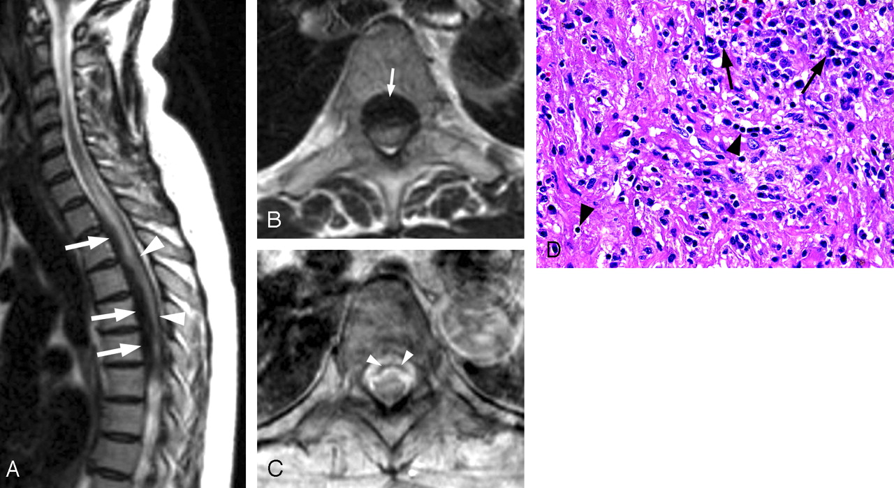
- Fig 1.
A, Sagittal fast spin-echo (FSE) T2-weighted image of the cervical and upper thoracic spine shows a mass of very low signal intensity (arrows) within the spinal canal located adjacent to the posterior aspect of the T1 through T6 vertebral bodies. High signal intensity is present centrally within the spinal cord, indicative of cord edema. There is also thickening and hypointensity of the dura posterior to the spinal cord (arrowheads).
B, Axial FSE T2-weighted image at T5 level shows a hypointense mass (arrow) in the anterior aspect of the spinal canal that appears to be arising from the dura. The lesion is displacing the spinal cord posteriorly and completely effacing the intradural subarachnoid spaces.
C, Axial postcontrast T1-weighted image at a level similar to that of B reveals thick enhancement of the anterior epidural mass with central nonenhancing area (arrowheads).
D, Photomicrograph shows fibrosis with plump reactive fibroblasts. Chronic inflammatory infiltrate is consisting chiefly of plasma cells (arrows) with additional lymphocytes (arrowheads) and scattered histiocytes (macrophages) (hematoxylin and eosin, original magnification 40×).
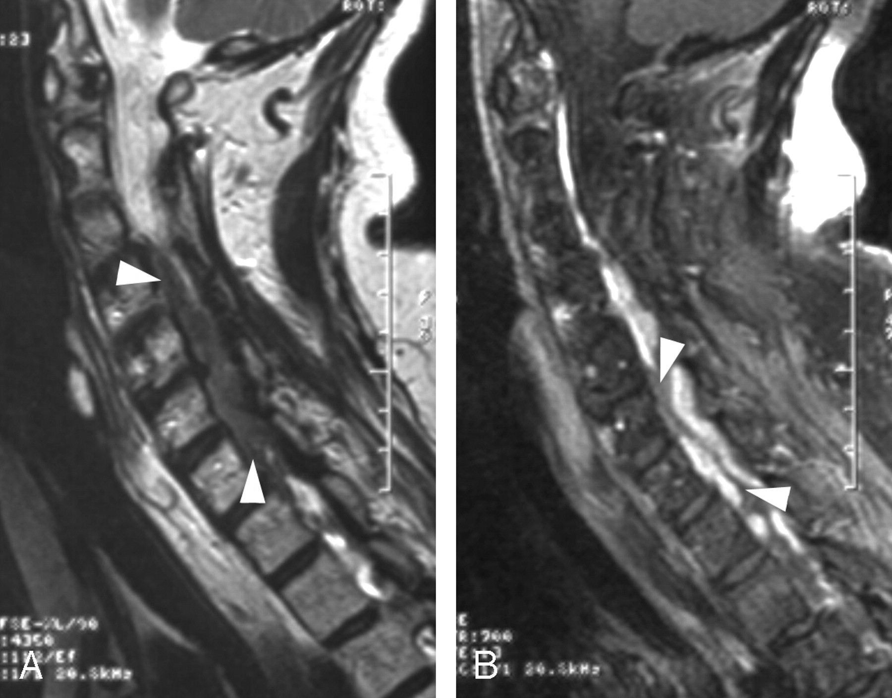
- Fig 2.
A, Sagittal fast spin-echo (FSE) T2-weighted image shows a hypointense mass (arrowheads) in the anterior aspect of the spinal canal that extends from C3 to T1 level.
B, Corresponding sagittal postcontrast T1-weighted image with fat suppression reveals attenuated enhancement of the mass, which is predominantly peripheral with central nonenhancing portions (arrowheads).


Tables
Characteristics of reported cases of idiopathic hypertrophic spinal pachymeningitis (IHSP) that included MR imaging findings
Study Patient Age (years) and Sex Spinal Levels Involved Lesion Length in Segments Location in Spinal Canal Relationship to Dura MRI Signal Intensity MRI Enhancement Pattern Ashkenazi et al8 65/F T1–T5 5 Dorsal Dural NA Homogenous* Botella et al10 55/F Cranial–C2 2 Dorsal & ventral Dural ↓T2 ≈T1 Peripheral Claus et al18 31/F T3–L1 11 Circumferential Intradural ↓T2 ≈T1 Homogenous Digman et al15 70/F All 25 Circumferential Dural NA Peripheral* Dumont et al14 30/F C4–T3 7 Dorsal Dural ↓T2 ≈T1 Peripheral Friedman and Flanders13 24/F Cranial–C7 7 Circumferential Dural ↓T2 ↓T1 Peripheral Friedman and Flanders13 51/M All 25 Dorsal Dural ↓T2 ↓T1 Peripheral Friedman and Flanders13 65/F Cranial–C2 2 Circumferential Dural ↓T2 ↓T1 Peripheral Kanamori et al11 28/M T5–L2 9 Circumferential Dural NA Peripheral Mikawa et al9 58/F C7–T11 12 Dorsal & ventral Dural ↓T1 Peripheral Park et al16 56/F C6–T8 10 Dorsal & ventral Dural ↓T2 ≈T1 Peripheral Pai et al** 47/F T1–T6 6 Dorsal & ventral Intra- and extradural ↓T2 ↓T1 Peripheral Pai et al** 68/F C3–T3 8 Dorsal & ventral Intradural ↓T2 ≈T1 Peripheral Sridhar et al12 48/F C7–T11 12 Circumferential Dural ↓T2 ↓T1 NA Voller et al17 54/M Cranial-C7 7 Dorsal Dural NA Peripheral Note:—The imaging features were assessed based on inspection of the provided images or on the description of findings, in cases when the respective images were not included in the reports. ↓ indicates hypointense; ≈ , isointense; *, poor image quality; **, this study; NA, not applicable (not performed or not mentioned).


