
Article Figures & Data
Figures
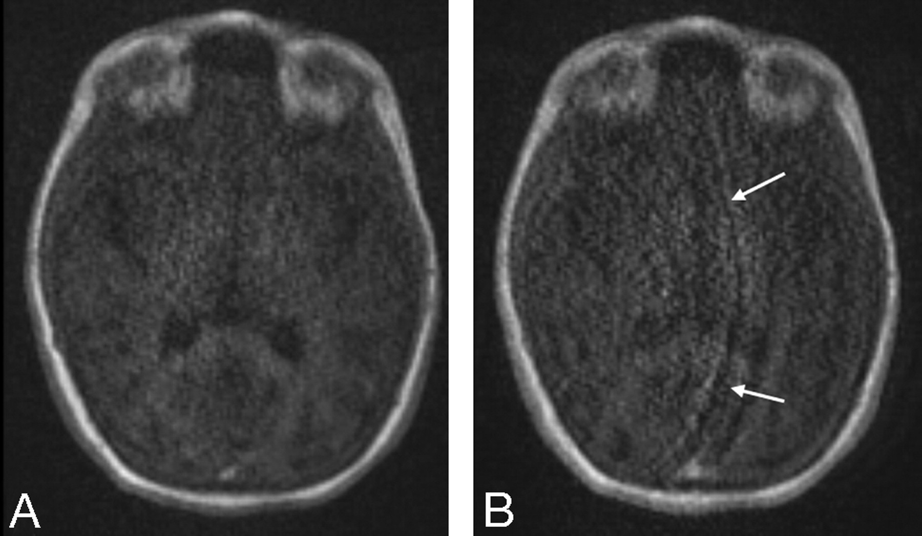
- Fig 1.
Wrap artifacts with IPAT 2 and 3. Note the wrap artifacts (arrows) due to the use of the higher IPAT 3 (B) reconstruction algorithm compared with the moderate IPAT 2 factor (A).
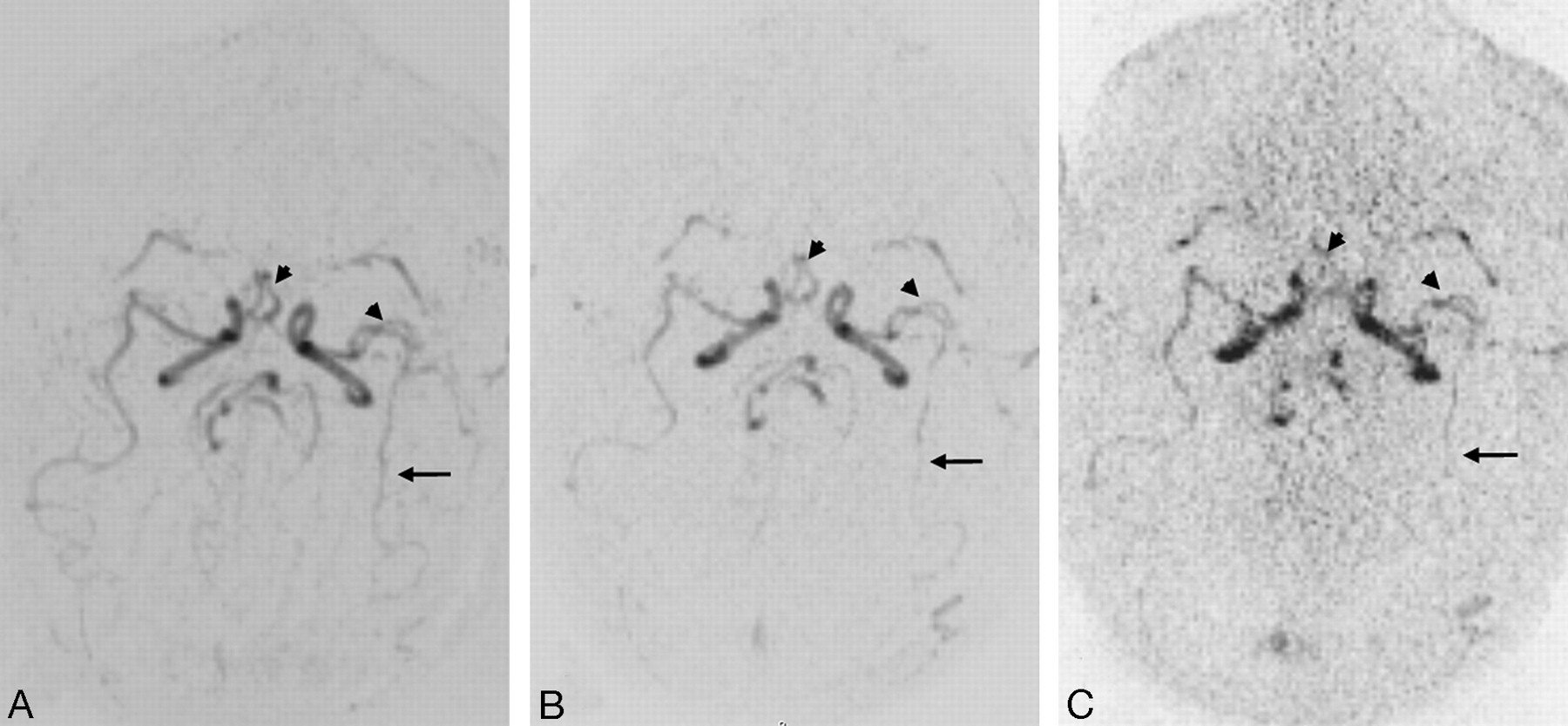
- Fig 2.
Axial TR MRA maximum intensity projection reconstructions without IPAT (A) and with IPAT 2 (B) and 3 (C). Note the better visualization of distal MCA (arrows) without IPAT and the similar conspicuity of proximal arterial branches (head arrows) without IPAT (A) compared with IPAT 2 (B). In contrast to the MRA without and with IPAT 2, the noise was slightly increased at IPAT 3 (C).
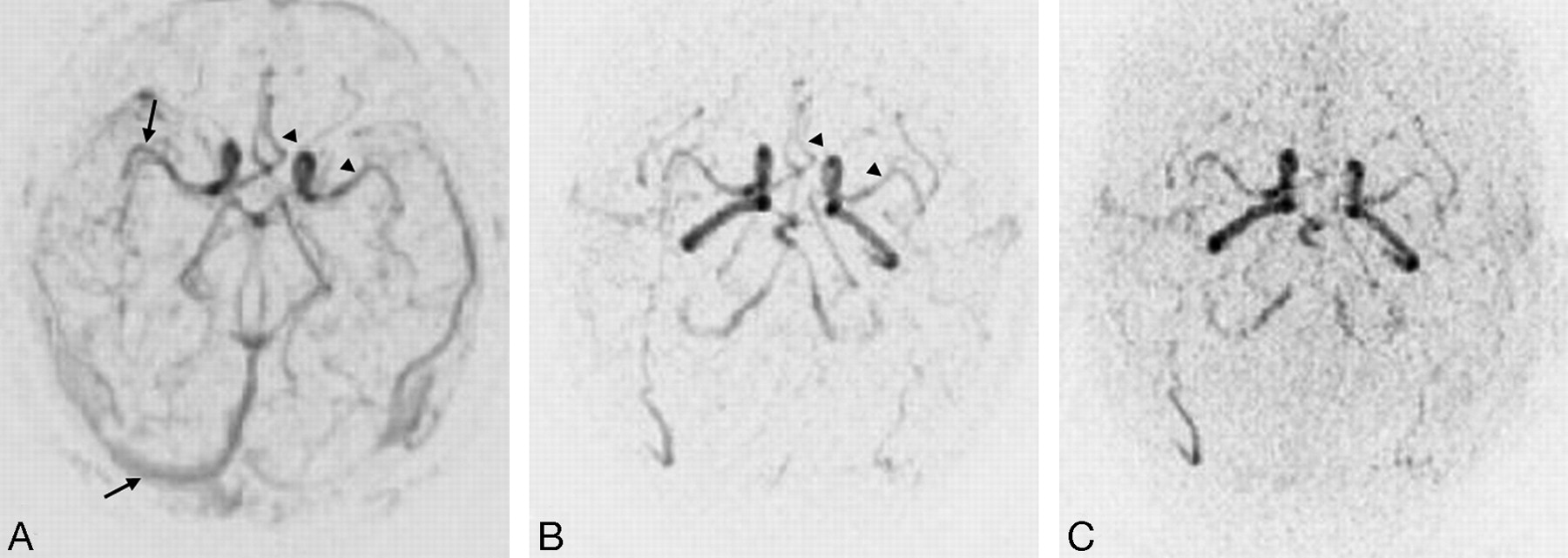
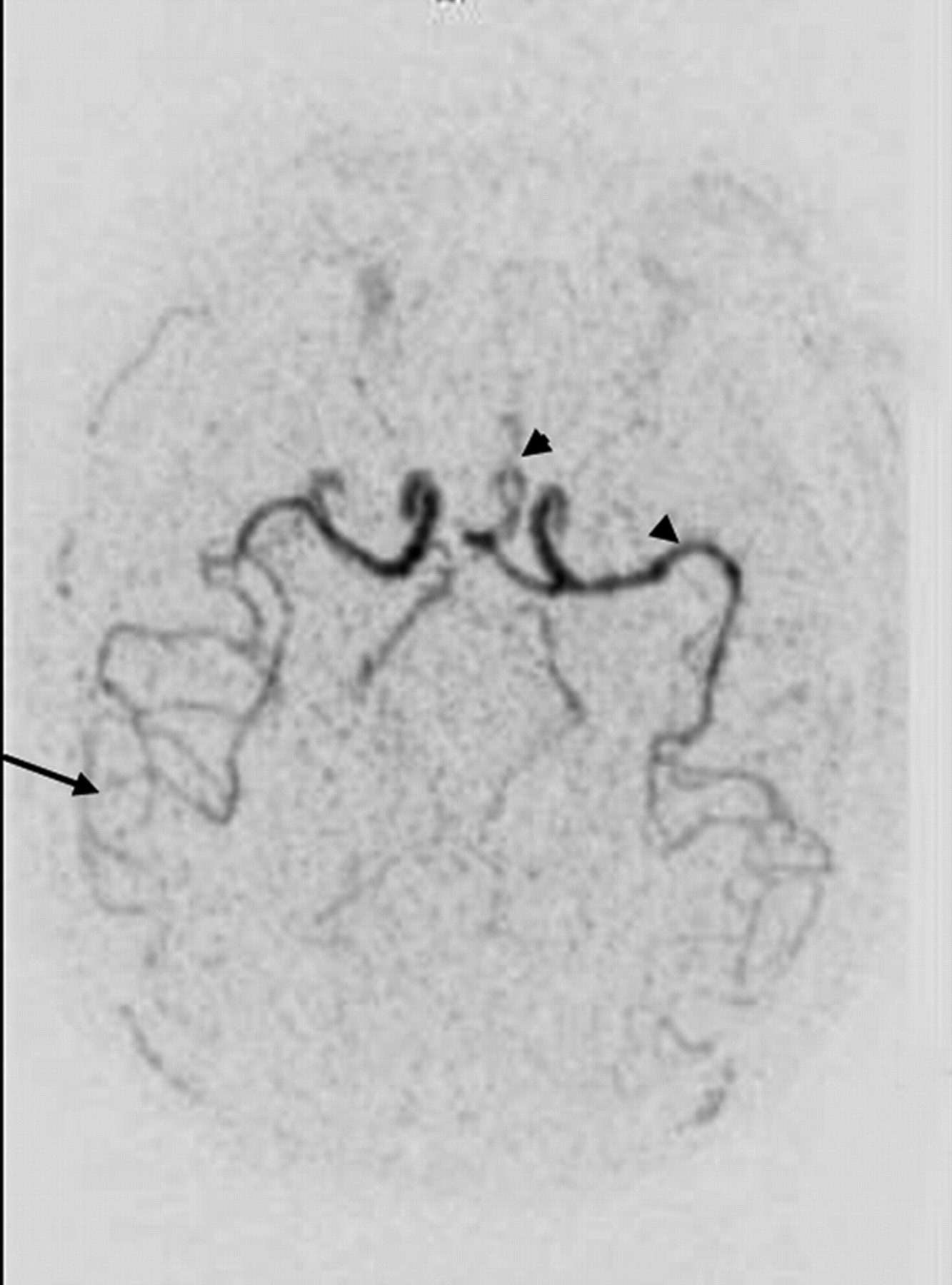
- Fig 3.
Axial TR MRA maximum intensity projection reconstructions without IPAT (A) and with IPAT 2 (B) and 3 (C). Note the simultaneous visualization of arterial and venous vessels (arrows) with TR MRA without IPAT (A), whereas the TR MRA with IPAT (B, C) provides the visualization of one arterial phase without venous opacification and the similar conspicuity of proximal arterial branches (head arrows) without IPAT (A) compared with IPAT 2 (B).
- Fig 4.
Axial TR MRA MIP reconstructions with IPAT 2 after an injection of 10 mL of contrast media. The increased volume of contrast media provides a better visualization of both proximal (head arrows) and distal (arrows) arterial vessels.


Tables
Parameter IPAT 0 IPAT 2 IPAT 3 TR (msec) 3.3 2.3 2.3 TE (msec) 1.1 0.8 0.8 BW (msec) 400 900 900 TA (seconds) 4.0 1.7 1.3 Total TA (seconds) 84 38 31 Section thickness (mm) 3 3 3 Flip angle (°) 20–25 20–25 25 FOV 167 × 250 167 × 250 167 × 250 Matrix (pixels) 128 × 192 128 × 192 128 × 192 Voxel size 1.3 × 1.3 × 3 1.3 × 1.3 × 3 1.3 × 1.3 × 3 Note:—BW indicates bandwidth; IPAT, integrated parallel acquisition technique; TA, acquisition time, FOV, field of view.
- Table 2:
The mean ± standard deviation of signal- (SNR) and contrast-to-noise ratios (CNR) at different IPAT factors
IPAT 0 IPAT 2 IPAT 3 SNR 108.6 ± 63.9 86.8 ± 49.8 77.1 ± 47.5 CNR 103.5 ± 61.8 80.8 ± 37.3 68.5 ± 35.8 Note:—IPAT indicates integrated parallel acquisition technique.
- Table 3:
The mean ± standard deviation of each ordinal assessment at different IPAT factors
IPAT Arterial Phases A1 A2 M1 M2 M3 BA P2 P3 0 0.8 ± 1.0 2.7 ± 0.7 2.9 ± 0.3 2.9 ± 0.3 2.6 ± 0.7 2.3 ± 0.9 3.0 ± 0.1 2.8 ± 0.6 2.1 ± 0.8 2 2.6 ± 0.7 2.2 ± 0.8 2.8 ± 0.5 2.8 ± 0.5 2.4 ± 0.8 1.6 ± 0.8 2.9 ± 0.3 2.3 ± 0.8 1.3 ± 0.8 3 3.7 ± 0.7 1.2 ± 0.4 1.3 ± 0.5 1.9 ± 0.7 1.7 ± 0.7 1.1 ± 0.3 2.0 ± 0.6 1.2 ± 0.4 1.0 ± 0.1 Note:—IPAT indicates integrated parallel acquisition technique; A1 and A2, A1 and A2 segments of anterior cerebral artery; M1, M2, and M3, M1, M2, and M3 segments of middle cerebral artery; P2 and P3, P2 and P3 segments of posterior cerebral artery; BA, basilar artery.
- Table 4:
Significance levels (P values) for the pairwise comparison of IPAT factor levels with respect to each end point
End Point IPAT Factor Levels Compared 0 vs 2 0 vs 3 2 vs 3 SNR .186 .042* .146 CNR .155 .020* .074 Score .004* .004* .014* A1 .059 .004* .009 A2 .371 .004* .004* M1 .371 .009* .009* M2 .371 .006* .009* M3 .022* .009* .100 AB .999 .006* .006* P2 .059 .004* .006* P3 .022* .009* .371 Note:—IPAT indicates integrated parallel acquisition technique; SNR, signal-to-noise ratio; CNR, contrast-to-noise ratio; A1 and A2, A1 and A2 segments of anterior cerebral artery; M1, M2, and M3, M1, M2, and M3 segments of middle cerebral artery; P2 and P3, P2 and P3 segments of posterior cerebral artery; BA, basilar artery. Statistically significant results are marked with asterisks.


