
Article Figures & Data
Figures
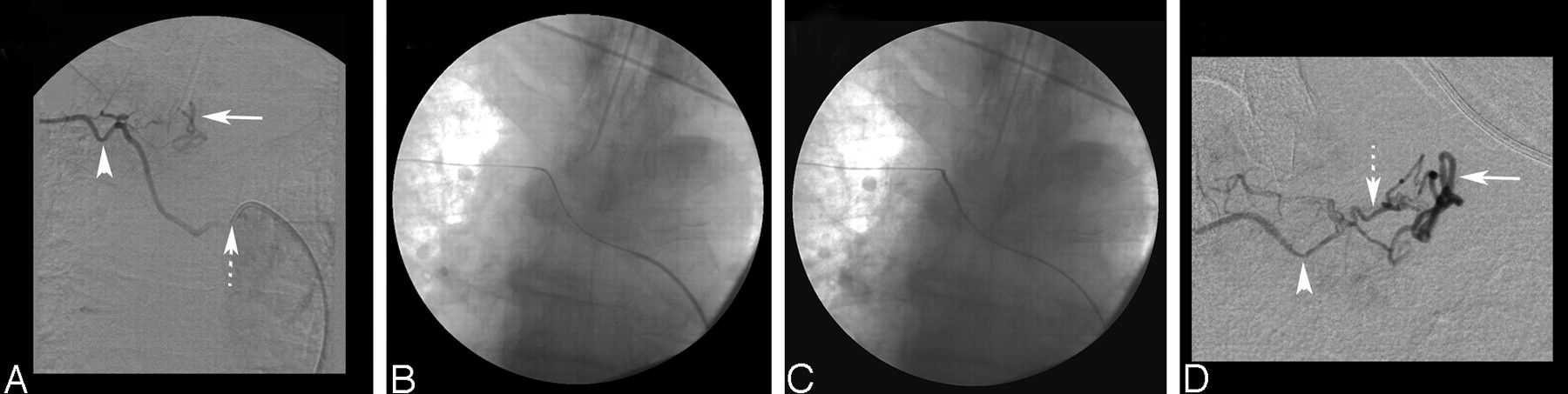
- Fig 1.
A, Selective right T6 intercostal artery digital subtraction angiogram (DSA) showing a DAVF. The dotted arrow points to the position of the 5F Cobra 2 catheter at the orifice of the right T6 intercostal artery. Note the size of the 5F catheter relative to the small size of the T6 intercostal artery (arrowhead). The solid arrow indicates the perimedullary vein. B, Nonsubtracted film shows the 0.035-inch Terumo exchange-length wire in the right T6 intercostal artery. C, Final distal position of the 4F guiding catheter with the exchange wire in situ. D, Superselective DSA through a Magic 1.2FM microcatheter shows the right T6 intercostal artery (arrowhead), the dural branch supplying the fistula (dotted arrow), and the draining perimedullary vein (solid arrow).
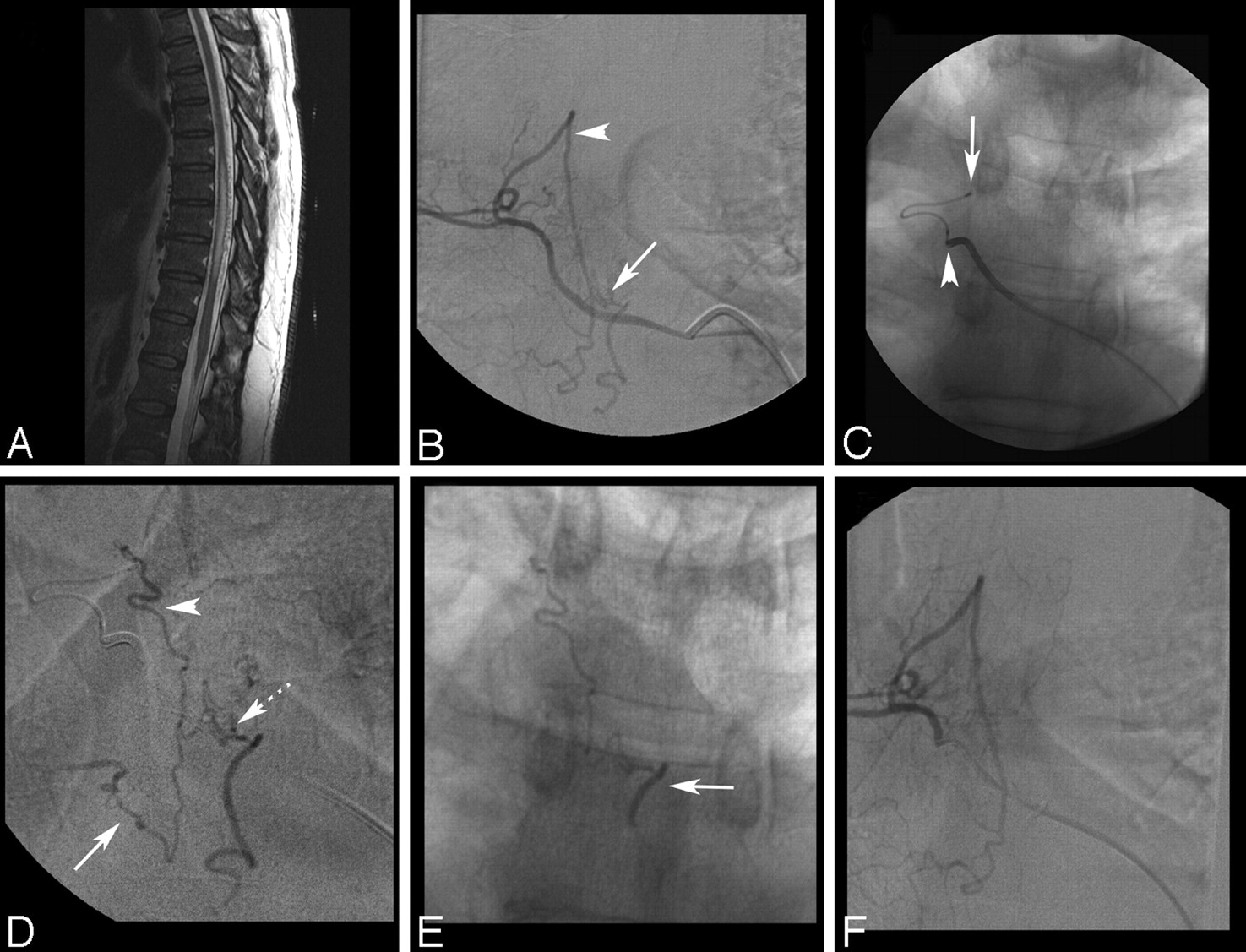
- Fig 2.
A, Sagittal T2-weighted MR image shows diffuse edema and mild expansion of the thoracic cord from T6 to T12. Extensive vascular flow voids are seen intradurally behind the spinal cord, suggesting a DAVF. B, Selective right T5 intercostal artery DSA shows the DAVF as it drains into a perimedullary vein (arrow). The arrowhead indicates a prominent retrolaminar muscular branch. C, Nonsubtracted film shows the distal position of the 4F catheter in the right T5 intercostal artery after the exchange procedure (arrowhead) and the selective position of a Prowler 10 microcatheter in the dural branch feeding the fistula (arrow). D, Superselective DSA through the microcatheter shows a bimetameric supply to the fistula from the right T5 (arrowhead) and T6 (solid arrow) dural branches. The dotted arrow marks the fistulous point. E, Nonsubtracted x-ray shows the glue cast reaching the draining perimedullary vein (arrow). F, Selective right T5 segmental artery DSA confirms disconnection of the dural AVF.


In this issue


Jump to section
Related Articles
Cited By...
- No citing articles found.