Advertisement

Main menu

  • Home
  • Content
    • Current Issue
    • Accepted Manuscripts
    • Article Preview
    • Past Issue Archive
    • AJNR Case Collection
    • Case of the Week Archive
    • Classic Case Archive
    • Case of the Month Archive
  • Special Collections
    • Spinal CSF Leak Articles (Jan 2020-June 2024)
    • 2024 AJNR Journal Awards
    • Most Impactful AJNR Articles
  • Multimedia
    • AJNR Podcast
    • AJNR Scantastics
    • Video Articles
  • For Authors
    • Submit a Manuscript
    • Author Policies
    • Fast publishing of Accepted Manuscripts
    • Graphical Abstract Preparation
    • Manuscript Submission Guidelines
    • Imaging Protocol Submission
    • Submit a Case for the Case Collection
  • About Us
    • About AJNR
    • Editorial Board
  • More
    • Become a Reviewer/Academy of Reviewers
    • Subscribers
    • Permissions
    • Alerts
    • Feedback
    • Advertisers
    • ASNR Home
  • Other Publications
    • ajnr

User menu

  • Alerts
  • Log in

Search

  • Advanced search
American Journal of Neuroradiology

American Journal of Neuroradiology

ASHNR American Society of Functional Neuroradiology ASHNR American Society of Pediatric Neuroradiology ASSR
  • Alerts
  • Log in

Advanced Search

  • Home
  • Content
    • Current Issue
    • Accepted Manuscripts
    • Article Preview
    • Past Issue Archive
    • AJNR Case Collection
    • Case of the Week Archive
    • Classic Case Archive
    • Case of the Month Archive
  • Special Collections
    • Spinal CSF Leak Articles (Jan 2020-June 2024)
    • 2024 AJNR Journal Awards
    • Most Impactful AJNR Articles
  • Multimedia
    • AJNR Podcast
    • AJNR Scantastics
    • Video Articles
  • For Authors
    • Submit a Manuscript
    • Author Policies
    • Fast publishing of Accepted Manuscripts
    • Graphical Abstract Preparation
    • Manuscript Submission Guidelines
    • Imaging Protocol Submission
    • Submit a Case for the Case Collection
  • About Us
    • About AJNR
    • Editorial Board
  • More
    • Become a Reviewer/Academy of Reviewers
    • Subscribers
    • Permissions
    • Alerts
    • Feedback
    • Advertisers
    • ASNR Home
  • Follow AJNR on Twitter
  • Visit AJNR on Facebook
  • Follow AJNR on Instagram
  • Join AJNR on LinkedIn
  • RSS Feeds

Welcome to the new AJNR, Updated Hall of Fame, and more. Read the full announcements.


AJNR is seeking candidates for the position of Associate Section Editor, AJNR Case Collection. Read the full announcement.


Getting new auth cookie, if you see this message a lot, tell someone!
Research ArticleINTERVENTIONAL

MR Angiography of Dural Arteriovenous Fistulas: Diagnosis and Follow-Up after Treatment Using a Time-Resolved 3D Contrast-Enhanced Technique

S. Meckel, M. Maier, D. San Millan Ruiz, H. Yilmaz, K. Scheffler, E.-W. Radue and S.G. Wetzel
American Journal of Neuroradiology May 2007, 28 (5) 877-884;
S. Meckel
  • Find this author on Google Scholar
  • Find this author on PubMed
  • Search for this author on this site
M. Maier
  • Find this author on Google Scholar
  • Find this author on PubMed
  • Search for this author on this site
D. San Millan Ruiz
  • Find this author on Google Scholar
  • Find this author on PubMed
  • Search for this author on this site
H. Yilmaz
  • Find this author on Google Scholar
  • Find this author on PubMed
  • Search for this author on this site
K. Scheffler
  • Find this author on Google Scholar
  • Find this author on PubMed
  • Search for this author on this site
E.-W. Radue
  • Find this author on Google Scholar
  • Find this author on PubMed
  • Search for this author on this site
S.G. Wetzel
  • Find this author on Google Scholar
  • Find this author on PubMed
  • Search for this author on this site
  • Article
  • Figures & Data
  • Info & Metrics
  • Responses
  • References
  • PDF
Loading

Article Figures & Data

Figures

  • Tables
  • Fig 1.
    • Download figure
    • Open in new tab
    • Download powerpoint
    Fig 1.

    Targeted sagittal MIP reconstructed image of a TR MRA dataset obtained during early arterial phase (A) shows minimal venous filling of right sigmoid sinus (black arrow) in a patient with a low flow DAVF (type 1). The corresponding lateral DSA image with a right common carotid artery injection (B) demonstrates the fistula with mild early filling of right sigmoid sinus (black arrow) that is surrounded by a network of small feeding artery branches from the right occipital artery and middle meningeal artery (white arrowheads). This fistula was missed on 3D TOF MRA images.

  • Fig 2.
    • Download figure
    • Open in new tab
    • Download powerpoint
    Fig 2.

    Preinterventional targeted MIP reconstructed sagittal image from an arterial phase of TR 3D MRA (A) shows a DAVF at the transverse/sigmoid sinus (short arrow) with strong early filling of the sinuses and feeding arterial branches from a prominent right occipital artery (long arrow). Small reflux into the proximal part of the vein of Labbé (arrowhead) is noted. The targeted MIP reconstructed image obtained from follow-up TR 3D MRA examination (B) after transvenous occlusion of the right transverse sinus clearly shows a residual fistula at the sigmoid sinus (short arrow) with downstaging of venous hypertension to anterograde sinus flow (type 1).

  • Fig 3.
    • Download figure
    • Open in new tab
    • Download powerpoint
    Fig 3.

    Sagittal targeted MIP reconstructed image obtained from an early arterial phase of TR 3D MRA shows a DAVF with strong and early filling of the torcular of Herophili, of the right transverse/sigmoid sinus (long arrow) and intense venous reflux into an enlarged superior vermian vein (short arrow) that follows the course of the tentorium in the midline. A large network of feeding branches from bilateral occipital arteries is observed (arrowheads).

  • Fig 4.
    • Download figure
    • Open in new tab
    • Download powerpoint
    Fig 4.

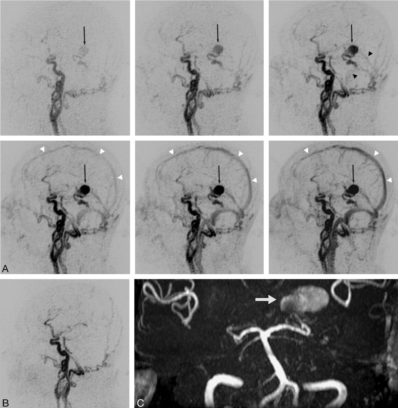
    TR 3D MRA is obtained before and after surgical exclusion of a high-grade DAVF (type 4) on a left medial tentorial sinus. The complete arteriovenous series of sagittal MIP-reconstructed images (A) shows early enhancement of dilated veins with a large venous varix (black arrow) on the left tentorium at early arterial inflow (top left). Consecutive filling of the straight sinus and the transverse sinuses from the fistula is noted at later arterial phase (top right, black arrowheads). Regular temporal enhancement of the superior sagittal sinus (white arrowheads) can be appreciated during venous phases (lower panel). Note that fistula occlusion is definitely demonstrated by the absence of early filling of tentorial veins and varix on the follow-up study after surgery (B). On 3D TOF MRA, however, the venous varix (white arrow) is still depicted with a high signal intensity imitating fistula flow (C).

  • Fig 5.
    • Download figure
    • Open in new tab
    • Download powerpoint
    Fig 5.

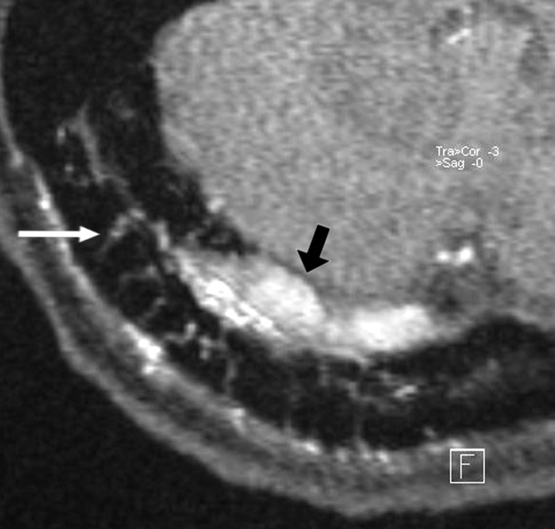
    In a patient with a DAVF involving the right transverse sinus, transvenous coil occlusion of the distal part of the transverse sinus and the sigmoid sinus has been performed. A residual type 2a fistula at proximal part of the right transverse sinus with arterial feeders from the right occipital artery is demonstrated at follow-up imaging. The magnified axial source image of 3D TOF MRA shows multiple hyperintense thin transosseous vessels (white arrow) in the vicinity of the arterialized right transverse sinus (black arrow).

Tables

  • Figures
    • View popup
    Table 1:

    Initial diagnosis of DAVFs on MR and MRA

    Presence of DAVFSide of DAVFVenous Segment of DAVF
    Reader 1Reader 2Reader 1Reader 2Reader 1Reader 2
    T2-weighted MR (n = 9)56% (21%–86%)56% (21%–86%)80% (28%–99%)80% (28%–99%)N/AN/A
    3D TOF MRA (n = 8*)88% (47%–100%)88% (47%–100%)100% (65%–100%)86% (42%–100%)43% (10%–82%)57% (18%–90%)
    TR 3D MRA (n = 9)100% (72%–100%)100% (72%–100%)100% (72%–100%)100% (72%–100%)78% (40%–97%)89% (52%–100%)
    • Note:—DAVF indicates dural arteriovenous fistula; MRA, MR angiography. Data are presented as percentage of correct detection with 95% CIs in parentheses. If signs suggestive of a DAVF were not encountered on an examination it was subsequently excluded from the location analysis.

    • * One examination was excluded because the fistulous point was located outside the scanned region.

    • View popup
    Table 2:

    Grading of patent DAVFs (Cognard) on TR 3D MRA

    Correct Grading (%)Weighted κ Values95% Confidence Intervals (%)
    Reader 1 vs. DSA11/13 (85%)0.9352%–100%
    Reader 2 vs. DSA10/13 (77%)0.8640%–100%
    Reader 1 vs. Reader 210/13 (77%)0.8640%–100%
    • Note:—DAVF indicates dural arteriovenous fistula; TR 3D MRA, time-resolved 3D MR angiography. Grading of DAVFs was performed according to the Cognard classification (6 types). All examinations that showed angiographically proved patent fistulas were included in this evaluation (n = 13). The interobserver agreement was determined by weighted κ values including 95% confidence intervals.

PreviousNext
Back to top

In this issue

American Journal of Neuroradiology: 28 (5)
American Journal of Neuroradiology
Vol. 28, Issue 5
May 2007
  • Table of Contents
  • Index by author
Advertisement
Print
Download PDF
Email Article

Thank you for your interest in spreading the word on American Journal of Neuroradiology.

NOTE: We only request your email address so that the person you are recommending the page to knows that you wanted them to see it, and that it is not junk mail. We do not capture any email address.

Enter multiple addresses on separate lines or separate them with commas.
MR Angiography of Dural Arteriovenous Fistulas: Diagnosis and Follow-Up after Treatment Using a Time-Resolved 3D Contrast-Enhanced Technique
(Your Name) has sent you a message from American Journal of Neuroradiology
(Your Name) thought you would like to see the American Journal of Neuroradiology web site.
CAPTCHA
This question is for testing whether or not you are a human visitor and to prevent automated spam submissions.
Cite this article
S. Meckel, M. Maier, D. San Millan Ruiz, H. Yilmaz, K. Scheffler, E.-W. Radue, S.G. Wetzel
MR Angiography of Dural Arteriovenous Fistulas: Diagnosis and Follow-Up after Treatment Using a Time-Resolved 3D Contrast-Enhanced Technique
American Journal of Neuroradiology May 2007, 28 (5) 877-884;

Citation Manager Formats

  • BibTeX
  • Bookends
  • EasyBib
  • EndNote (tagged)
  • EndNote 8 (xml)
  • Medlars
  • Mendeley
  • Papers
  • RefWorks Tagged
  • Ref Manager
  • RIS
  • Zotero
0 Responses
Respond to this article
Share
Bookmark this article
MR Angiography of Dural Arteriovenous Fistulas: Diagnosis and Follow-Up after Treatment Using a Time-Resolved 3D Contrast-Enhanced Technique
S. Meckel, M. Maier, D. San Millan Ruiz, H. Yilmaz, K. Scheffler, E.-W. Radue, S.G. Wetzel
American Journal of Neuroradiology May 2007, 28 (5) 877-884;
del.icio.us logo Twitter logo Facebook logo Mendeley logo
  • Tweet Widget
  • Facebook Like
  • Google Plus One

Jump to section

  • Article
    • Abstract
    • Materials and Methods
    • Results
    • Discussion
    • Conclusion
    • References
  • Figures & Data
  • Info & Metrics
  • Responses
  • References
  • PDF

Related Articles

  • No related articles found.
  • PubMed
  • Google Scholar

Cited By...

  • Four dimensional-flow magnetic resonance imaging analysis of carotid-cavernous fistula, dural arteriovenous fistula and spinal arteriovenous fistula: Detecting shunt point and diagnosing based on flow dynamics analysis
  • Spinal Dorsal Intradural Arteriovenous Fistulas: Natural History, Imaging, and Management
  • Vessel-Selective 4D-MRA Using Superselective Pseudocontinuous Arterial Spin-Labeling with Keyhole and View-Sharing for Visualizing Intracranial Dural AVFs
  • Assessment of 4D MR Angiography at 3T Compared with DSA for the Follow-up of Embolized Brain Dural Arteriovenous Fistula: A Dual-Center Study
  • Outcome of transarterial treatment of dural arteriovenous fistulas with direct or indirect cortical venous drainage
  • Arterial Spin-Labeling Improves Detection of Intracranial Dural Arteriovenous Fistulas with MRI
  • Intracranial Dural Arteriovenous Fistulae: Clinical Presentation and Management Strategies
  • Optimal MRI Sequence for Identifying Occlusion Location in Acute Stroke: Which Value of Time-Resolved Contrast-Enhanced MRA?
  • Identification of Venous Signal on Arterial Spin Labeling Improves Diagnosis of Dural Arteriovenous Fistulas and Small Arteriovenous Malformations
  • Quality-Evaluation Scheme for Cerebral Time-Resolved 3D Contrast-Enhanced MR Angiography Techniques
  • Cerebral Venous Thrombosis: Diagnostic Accuracy of Combined, Dynamic and Static, Contrast-Enhanced 4D MR Venography
  • Evaluation of Dural Arteriovenous Fistulas with 4D Contrast-Enhanced MR Angiography at 3T
  • Cranial Dural Arteriovenous Fistula: Diagnosis and Classification with Time-Resolved MR Angiography at 3T
  • Efficacy of DynaCT Digital Angiography in the Detection of the Fistulous Point of Dural Arteriovenous Fistulas
  • Crossref
  • Google Scholar

This article has not yet been cited by articles in journals that are participating in Crossref Cited-by Linking.

More in this TOC Section

  • SAVE vs. Solumbra Techniques for Thrombectomy
  • Contrast-Induced Encephalopathy after NeuroIR
  • CT Perfusion&Reperfusion in Acute Ischemic Stroke
Show more Interventional

Similar Articles

Advertisement

Indexed Content

  • Current Issue
  • Accepted Manuscripts
  • Article Preview
  • Past Issues
  • Editorials
  • Editors Choice
  • Fellow Journal Club
  • Letters to the Editor

Cases

  • Case Collection
  • Archive - Case of the Week
  • Archive - Case of the Month
  • Archive - Classic Case

Special Collections

  • Special Collections

Resources

  • News and Updates
  • Turn around Times
  • Submit a Manuscript
  • Author Policies
  • Manuscript Submission Guidelines
  • Evidence-Based Medicine Level Guide
  • Publishing Checklists
  • Graphical Abstract Preparation
  • Imaging Protocol Submission
  • Submit a Case
  • Become a Reviewer/Academy of Reviewers
  • Get Peer Review Credit from Publons

Multimedia

  • AJNR Podcast
  • AJNR SCANtastic
  • Video Articles

About Us

  • About AJNR
  • Editorial Board
  • Not an AJNR Subscriber? Join Now
  • Alerts
  • Feedback
  • Advertise with us
  • Librarian Resources
  • Permissions
  • Terms and Conditions

American Society of Neuroradiology

  • Not an ASNR Member? Join Now

© 2025 by the American Society of Neuroradiology All rights, including for text and data mining, AI training, and similar technologies, are reserved.
Print ISSN: 0195-6108 Online ISSN: 1936-959X

Powered by HighWire