Advertisement

Main menu

  • Home
  • Content
    • Current Issue
    • Accepted Manuscripts
    • Article Preview
    • Past Issue Archive
    • AJNR Case Collection
    • Case of the Week Archive
    • Classic Case Archive
    • Case of the Month Archive
  • Special Collections
    • Spinal CSF Leak Articles (Jan 2020-June 2024)
    • 2024 AJNR Journal Awards
    • Most Impactful AJNR Articles
  • Multimedia
    • AJNR Podcast
    • AJNR Scantastics
    • Video Articles
  • For Authors
    • Submit a Manuscript
    • Author Policies
    • Fast publishing of Accepted Manuscripts
    • Graphical Abstract Preparation
    • Manuscript Submission Guidelines
    • Imaging Protocol Submission
    • Submit a Case for the Case Collection
  • About Us
    • About AJNR
    • Editorial Board
  • More
    • Become a Reviewer/Academy of Reviewers
    • Subscribers
    • Permissions
    • Alerts
    • Feedback
    • Advertisers
    • ASNR Home
  • Other Publications
    • ajnr

User menu

  • Alerts
  • Log in

Search

  • Advanced search
American Journal of Neuroradiology

American Journal of Neuroradiology

ASHNR American Society of Functional Neuroradiology ASHNR American Society of Pediatric Neuroradiology ASSR
  • Alerts
  • Log in

Advanced Search

  • Home
  • Content
    • Current Issue
    • Accepted Manuscripts
    • Article Preview
    • Past Issue Archive
    • AJNR Case Collection
    • Case of the Week Archive
    • Classic Case Archive
    • Case of the Month Archive
  • Special Collections
    • Spinal CSF Leak Articles (Jan 2020-June 2024)
    • 2024 AJNR Journal Awards
    • Most Impactful AJNR Articles
  • Multimedia
    • AJNR Podcast
    • AJNR Scantastics
    • Video Articles
  • For Authors
    • Submit a Manuscript
    • Author Policies
    • Fast publishing of Accepted Manuscripts
    • Graphical Abstract Preparation
    • Manuscript Submission Guidelines
    • Imaging Protocol Submission
    • Submit a Case for the Case Collection
  • About Us
    • About AJNR
    • Editorial Board
  • More
    • Become a Reviewer/Academy of Reviewers
    • Subscribers
    • Permissions
    • Alerts
    • Feedback
    • Advertisers
    • ASNR Home
  • Follow AJNR on Twitter
  • Visit AJNR on Facebook
  • Follow AJNR on Instagram
  • Join AJNR on LinkedIn
  • RSS Feeds

Welcome to the new AJNR, Updated Hall of Fame, and more. Read the full announcements.


AJNR is seeking candidates for the position of Associate Section Editor, AJNR Case Collection. Read the full announcement.


OtherBRAIN

Decrease in the Volume of White Matter Lesions with Improvement of Hepatic Encephalopathy

B. Mínguez, A. Rovira, J. Alonso and J. Córdoba
American Journal of Neuroradiology September 2007, 28 (8) 1499-1500; DOI: https://doi.org/10.3174/ajnr.A0611
B. Mínguez
  • Find this author on Google Scholar
  • Find this author on PubMed
  • Search for this author on this site
A. Rovira
  • Find this author on Google Scholar
  • Find this author on PubMed
  • Search for this author on this site
J. Alonso
  • Find this author on Google Scholar
  • Find this author on PubMed
  • Search for this author on this site
J. Córdoba
  • Find this author on Google Scholar
  • Find this author on PubMed
  • Search for this author on this site
  • Article
  • Figures & Data
  • Info & Metrics
  • Responses
  • References
  • PDF
Loading

Abstract

SUMMARY: MR imaging has found abnormalities compatible with low-grade edema in the brain of patients with cirrhosis that have been related to hepatic encephalopathy. We present 3 patients with hepatic encephalopathy who exhibit supratentorial focal or diffuse white matter lesions compatible with small-vessel brain disease. The volume and number of white matter lesions reduced with the improvement of hepatic encephalopathy, suggesting the participation of the blood-brain barrier in the pathogenesis of brain edema in hepatic encephalopathy.

MR studies of the brain have described several abnormalities in cirrhosis.1–3 These abnormalities appear to be relevant, because it has been postulated that low-grade brain edema is involved in the pathogenesis of hepatic encephalopathy (HE).4 The previous studies and recent experimental evidence5 indicate that brain edema in liver failure is mainly located in white matter. White matter lesions (WMLs) are often seen on MR images of elderly people and have been related to cognitive decline.6 WMLs are attributed to degenerative small-vessel disease and are progressive. The development of WMLs can be prevented with adequate treatment of vascular risk factors,7 but once present, they are considered irreversible areas of demyelination, gliosis, and loss of axons.8 We observed 3 patients with HE and T2 hyperintense focal WMLs who experienced an important decrease in their volume with the improvement of HE.

Cases

Case 1

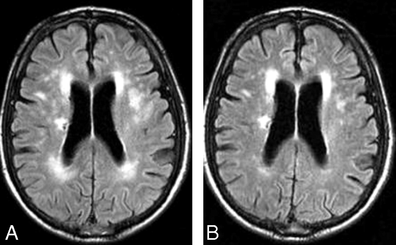
A 70-year-old male patient with hepatitis C cirrhosis and a history of myocardial infarction developed an episode of HE in 2003. After the episode of HE, he showed fluctuating manifestations of cognitive decline (apathy, inattention, and somnolence), compatible with persistent HE. In 2004, his liver function showed a Child Pugh Score of 7. At that time, the neurologic examination revealed that he was disoriented in time and showed bradykinesia. Brain MR imaging showed multifocal supratentorial WMLs (Fig 1A), and neuropsychologic assessment indicated severe impairment. The treatment of HE was optimized. Neomycin and branched chain amino acids were prescribed, and furosemide was withdrawn. Six months later, a follow-up MR imaging demonstrated a decrease in the number and size of WMLs (Fig 1B) associated with a significant improvement of the neuropsychologic function. Lesion volume from fast fluid-attenuated inversion recovery (FLAIR) images were outlined on hard copies. The lesion volumes were measured semiautomatically by a previously trained operator with the program DispImage9 (Dave Plummer, University College London Hospitals, London, UK. Available at: http://www.ucl.ac.uk/MedPhys/toast/misc/dispimg.htm) by using the hard copies as reference.

Fig 1.
  • Download figure
  • Open in new tab
  • Download powerpoint
Fig 1.

A, MR image (transverse T2-weighted fast-FLAIR images, 9900 ms/110 ms/2500 ms/1 [TR/TE/inversion time/acquisitions]) obtained in patient 1 at baseline, while showing signs of overt hepatic encephalopathy. The image shows multiple focal supratentorial WMLs (total lesion volume, 7,507 mm3). B, Patient 1 after improvement of hepatic encephalopathy: MR shows a clear reduction in size and number of these focal WMLs (final lesion volume of 2,033 mm3).

Case 2

A 65-year-old male patient with alcoholic cirrhosis and a history of insulin-dependent diabetes mellitus developed an episode of HE in 2004. After the episode of HE, he experienced manifestations of persistent HE. In May 2006, his Child Pugh Score was 13. At neurologic examination, he was oriented but exhibited flapping tremor, slurred speech, and bradykinesia, and neuropsychologic assessment indicated severe impairment. Brain MR imaging showed diffuse WMLs (Fig 2A). He was receiving diuretics that were withdrawn, and neomycin and branched chain amino acids were prescribed. After 1 month, the extension of WMLs (calculated in the same way as in case 1) decreased (Fig 2B). On physical examination, he appeared more rapid on mental processing, and the family reported an obvious improvement of daily activities.

Fig 2.
  • Download figure
  • Open in new tab
  • Download powerpoint
Fig 2.

A, MR image (transverse T2-weighted fast-FLAIR images, 9900 ms/110 ms/2500 ms/1 [TR/TE/inversion time/acquisitions ]) obtained in patient 1 at baseline, while showing signs of overt hepatic encephalopathy. The scan shows multiple diffuse supratentorial WMLs (total lesion volume, 25,901 mm3). B, Patient 2 after improvement of hepatic encephalopathy: MR shows a clear reduction in size and number of diffuse WMLs (final lesion volume, 16,586 mm3).

Case 3

A 73-year-old male patient with hepatitis C cirrhosis was referred with a history of arterial hypertension and a lacunar infarction in 1990 that caused left hemiparesis. In 2004, when his Child Pugh Score was 10, he developed gait disturbance accompanied by bradykinesia that was attributed to vascular Parkinsonism. In 2005, he experienced 2 episodes of confusion and worsening left hemiparesis of several days’ duration that were diagnosed as episodic HE. A brain MR imaging, performed during the second episode (Fig 3A) showed, in addition to a chronic lacunar infarct within the right centrum ovale, multifocal subcortical and periventricular WML. He was discharged and received treatment with neomycin. A follow-up MR imaging was obtained 6 months later (Fig 3B), which showed a decrease in size and volume of the WML, calculated in the same way as in case 1. At that time, neurologic examination was unremarkable except for mild left hemiparesis with pyramidal signs, and the neuropsychologic assessment showed only mild impairment on attention and speed of mental processing.

Fig 3.
  • Download figure
  • Open in new tab
  • Download powerpoint
Fig 3.

A, MR image (transverse T2-weighted fast-FLAIR images, 9900 ms/110 ms/2500 ms/1 [TR/TE/inversion time/acquisitions]) obtained in patient 3 at baseline, while showing signs of overt hepatic encephalopathy. MR shows multiple focal WMLs (total lesion volume, 18,601 mm3). B, Patient 3 after improvement of hepatic encephalopathy: MR shows clear reduction in size and number of focal WMLs (final lesion volume, 10,393 mm3).

Discussion

The present study shows supratentorial focal and diffuse WMLs compatible with small-vessel brain disease in 3 patients with cirrhosis and HE. These lesions, which are common in the general population, have probably been observed in patients with cirrhosis more frequently than reported.10 They are secondary to arteriolosclerosis and usually attributed to advanced age or hypertension.6 Thus, they have probably been ignored and interpreted as signs of normal involutive or chronic ischemic changes. In accordance with the interpretation of an ischemic source of their WMLs, the 3 patients of our study exhibited vascular risk factors.

The remarkable finding of our study is not the presence of WMLs but the observation that the volume and number of WMLs can be reduced with the improvement of HE. WMLs are considered irreversible consequences of small-vessel disease. The neuropathologic substrate of WMLs is a combination of perivascular demyelination, edema, gliosis, loss of axons, and glial cells accompanied by spongiosis. Of the different elements that constitute the neuropathologic substrate of WMLs, the one that can reverse in a short period of time is edema. The residual WML after improvement of HE probably corresponds with gliosis and other irreversible processes. MR studies in patients with minimal HE have demonstrated an increase in brain water, which may be involved in the pathogenesis of neurologic manifestations.11 The decrease of WMLs in parallel to the improvement of HE in the 3 patients of our study is in accordance with the association between brain edema and HE. Changes in the volume of edema in areas of small vessel-induced lesion support the participation of the blood-brain barrier in the pathogenesis of brain edema in liver failure.12

From a clinical perspective, the description that the volume of WMLs can decrease is important for the adequate interpretation of cognitive impairment in cirrhotic patients. This is especially important during the evaluation of candidates for liver transplantation. The presence of vascular risk factors and the finding of large WMLs may cause one to question the reversibility of the neurologic impairment after liver transplantation. According to our findings, in these patients, WMLs should be reassessed after therapy for HE to recognize to what extent the lesions can be reversible after liver transplantation.

In conclusion, we have observed a decrease in the volume and number of WMLs in relation to improvement of HE. These findings suggest that cognitive impairment in HE may be related to interstitial edema and dysfunction of cerebral microvasculature.

Footnotes

  • This study was supported by Instituto de Salud Carlos III (FIS PI03/0072). B.M. was the recipient of a grant FIS CM04/00044.

References

  1. ↵
    Spahr L, Butterworth RF, Fontaine S, et al. Increased blood manganese in cirrhotic patients: relationship to pallidal magnetic resonance signal hyperintensity and neurological symptoms. Hepatology 1996;24:1116–20
    CrossRefPubMed
  2. Rovira A, Cordoba J, Raguer N, et al. Magnetic resonance imaging measurement of brain edema in patients with liver disease: resolution after transplantation. Curr Opin Neurol 2002;15:731–37
    CrossRefPubMed
  3. ↵
    Kale RA, Gupta RK, Saraswat VA, et al. Demonstration of interstitial cerebral edema with diffusion tensor MR imaging in type C hepatic encephalopathy. Hepatology 2006;43:698–706
    CrossRefPubMed
  4. ↵
    Haussinger D, Kircheis G, Fischer R, et al. Hepatic encephalopathy in chronic liver disease: a clinical manifestation of astrocyte swelling and low-grade cerebral edema? J Hepatol 2000;32:1035–38
    CrossRefPubMed
  5. ↵
    Sen S, Rose C, Ytrebo LM, et al. Effect of albumin dialysis on intracranial pressure increase in pigs with acute liver failure: a randomized study. Crit Care Med 2006;34:158–64
    CrossRefPubMed
  6. ↵
    de Groot JC, de Leeuw FE, Oudkerk M, et al. Cerebral white matter lesions and subjective cognitive dysfunction: the Rotterdam Scan Study. Neurology 2001;56:1539–45
    Abstract/FREE Full Text
  7. ↵
    Dufouil C, Chalmers J, Coskun O, et al. Effects of blood pressure lowering on cerebral white matter hyperintensities in patients with stroke: the PROGRESS (Perindopril Protection Against Recurrent Stroke Study) Magnetic Resonance Imaging Substudy. Circulation 2005;112:1644–50
    Abstract/FREE Full Text
  8. ↵
    Pantoni L, Garcia JH. Pathogenesis of leukoaraiosis: a review. Stroke 1997;28:652–59
    Abstract/FREE Full Text
  9. ↵
    Plummer D. DispImage: a display and analysis tool for medical images. Rev Neuroradiol 1992;5:489–95
  10. ↵
    Matsusue E, Kinoshita T, Ohama E, et al. Cerebral cortical and white matter lesions in chronic hepatic encephalopathy: MR-pathologic correlations. AJNR Am J Neuroradiol 2005;26:347–51
    Abstract/FREE Full Text
  11. ↵
    Cordoba J, Alonso J, Rovira A, et al. The development of low-grade cerebral edema in cirrhosis is supported by the evolution of 1H-magnetic resonance abnormalities after liver transplantation. J Hepatol 2001;35:598–604
    CrossRefPubMed
  12. ↵
    Nguyen JH, Yamamoto S, Steers J, et al. Matrix metalloproteinase-9 contributes to brain extravasation and edema in fulminant hepatic failure mice. J Hepatol 2006;44:1105–14
    CrossRefPubMed
  • Received February 15, 2007.
  • Accepted after revision February 27, 2007.
  • Copyright © American Society of Neuroradiology
View Abstract
PreviousNext
Back to top

In this issue

American Journal of Neuroradiology: 28 (8)
American Journal of Neuroradiology
Vol. 28, Issue 8
September 2007
  • Table of Contents
  • Index by author
Advertisement
Print
Download PDF
Email Article

Thank you for your interest in spreading the word on American Journal of Neuroradiology.

NOTE: We only request your email address so that the person you are recommending the page to knows that you wanted them to see it, and that it is not junk mail. We do not capture any email address.

Enter multiple addresses on separate lines or separate them with commas.
Decrease in the Volume of White Matter Lesions with Improvement of Hepatic Encephalopathy
(Your Name) has sent you a message from American Journal of Neuroradiology
(Your Name) thought you would like to see the American Journal of Neuroradiology web site.
CAPTCHA
This question is for testing whether or not you are a human visitor and to prevent automated spam submissions.
Cite this article
B. Mínguez, A. Rovira, J. Alonso, J. Córdoba
Decrease in the Volume of White Matter Lesions with Improvement of Hepatic Encephalopathy
American Journal of Neuroradiology Sep 2007, 28 (8) 1499-1500; DOI: 10.3174/ajnr.A0611

Citation Manager Formats

  • BibTeX
  • Bookends
  • EasyBib
  • EndNote (tagged)
  • EndNote 8 (xml)
  • Medlars
  • Mendeley
  • Papers
  • RefWorks Tagged
  • Ref Manager
  • RIS
  • Zotero
0 Responses
Respond to this article
Share
Bookmark this article
Decrease in the Volume of White Matter Lesions with Improvement of Hepatic Encephalopathy
B. Mínguez, A. Rovira, J. Alonso, J. Córdoba
American Journal of Neuroradiology Sep 2007, 28 (8) 1499-1500; DOI: 10.3174/ajnr.A0611
del.icio.us logo Twitter logo Facebook logo Mendeley logo
  • Tweet Widget
  • Facebook Like
  • Google Plus One
Purchase

Jump to section

  • Article
    • Abstract
    • Cases
    • Discussion
    • Footnotes
    • References
  • Figures & Data
  • Info & Metrics
  • Responses
  • References
  • PDF

Related Articles

  • No related articles found.
  • PubMed
  • Google Scholar

Cited By...

  • Automatic Spatial Estimation of White Matter Hyperintensities Evolution in Brain MRI using Disease Evolution Predictor Deep Neural Networks
  • Enzyme replacement therapy and white matter hyperintensity progression in Fabry disease
  • Crossref (30)
  • Google Scholar

This article has been cited by the following articles in journals that are participating in Crossref Cited-by Linking.

  • MR Imaging Findings in Hepatic Encephalopathy
    A. Rovira, J. Alonso, J. Córdoba
    American Journal of Neuroradiology 2008 29 9
  • Hyponatremic and hepatic encephalopathies: similarities, differences and coexistence
    Juan Córdoba, Rita García-Martinez, Macarena Simón-Talero
    Metabolic Brain Disease 2010 25 1
  • Decreased white matter lesion volume and improved cognitive function after liver transplantation
    Alex Rovira, Beatriz Mínguez, Xavier F. Aymerich, Carlos Jacas, Elena Huerga, Juan Córdoba, Juli Alonso
    Hepatology 2007 46 5
  • Neurocognitive-neurological complications of liver transplantation: a review
    Francesca Campagna, A. Biancardi, U. Cillo, A. Gatta, P. Amodio
    Metabolic Brain Disease 2010 25 1
  • Neuroimaging Findings in Alcohol-Related Encephalopathies
    Giulio Zuccoli, Nasir Siddiqui, Isabel Cravo, Ariel Bailey, Massimo Gallucci, Clive G. Harper
    American Journal of Roentgenology 2010 195 6
  • White Matter Hyperintensity in Ischemic Stroke Patients: It May Regress Over Time
    A-Hyun Cho, Hyeong-Ryul Kim, Woojun Kim, Dong Won Yang
    Journal of Stroke 2015 17 1
  • Brain Magnetic Resonance in Hepatic Encephalopathy
    Juli Alonso, Juan Córdoba, Alex Rovira
    Seminars in Ultrasound, CT and MRI 2014 35 2
  • Brain Edema in Chronic Hepatic Encephalopathy
    Cristina Cudalbu, Simon D. Taylor-Robinson
    Journal of Clinical and Experimental Hepatology 2019 9 3
  • Dynamic Progression of White Matter Hyperintensities in Alzheimer’s Disease and Normal Aging: Results from the Sunnybrook Dementia Study
    Joel Ramirez, Alicia A. McNeely, Courtney Berezuk, Fuqiang Gao, Sandra E. Black
    Frontiers in Aging Neuroscience 2016 8
  • Brain Imaging and Hepatic Encephalopathy
    Mark J.W. McPhail, Neeral R. Patel, Simon D. Taylor-Robinson
    Clinics in Liver Disease 2012 16 1

More in this TOC Section

  • Fast Contrast-Enhanced 4D MRA and 4D Flow MRI Using Constrained Reconstruction (HYPRFlow): Potential Applications for Brain Arteriovenous Malformations
  • Quiet PROPELLER MRI Techniques Match the Quality of Conventional PROPELLER Brain Imaging Techniques
  • Predictors of Reperfusion in Patients with Acute Ischemic Stroke
Show more Brain

Similar Articles

Advertisement

Indexed Content

  • Current Issue
  • Accepted Manuscripts
  • Article Preview
  • Past Issues
  • Editorials
  • Editors Choice
  • Fellow Journal Club
  • Letters to the Editor

Cases

  • Case Collection
  • Archive - Case of the Week
  • Archive - Case of the Month
  • Archive - Classic Case

Special Collections

  • Special Collections

Resources

  • News and Updates
  • Turn around Times
  • Submit a Manuscript
  • Author Policies
  • Manuscript Submission Guidelines
  • Evidence-Based Medicine Level Guide
  • Publishing Checklists
  • Graphical Abstract Preparation
  • Imaging Protocol Submission
  • Submit a Case
  • Become a Reviewer/Academy of Reviewers
  • Get Peer Review Credit from Publons

Multimedia

  • AJNR Podcast
  • AJNR SCANtastic
  • Video Articles

About Us

  • About AJNR
  • Editorial Board
  • Not an AJNR Subscriber? Join Now
  • Alerts
  • Feedback
  • Advertise with us
  • Librarian Resources
  • Permissions
  • Terms and Conditions

American Society of Neuroradiology

  • Not an ASNR Member? Join Now

© 2025 by the American Society of Neuroradiology All rights, including for text and data mining, AI training, and similar technologies, are reserved.
Print ISSN: 0195-6108 Online ISSN: 1936-959X

Powered by HighWire