Advertisement

Main menu

  • Home
  • Content
    • Current Issue
    • Accepted Manuscripts
    • Article Preview
    • Past Issue Archive
    • AJNR Case Collection
    • Case of the Week Archive
    • Classic Case Archive
    • Case of the Month Archive
  • Special Collections
    • Spinal CSF Leak Articles (Jan 2020-June 2024)
    • 2024 AJNR Journal Awards
    • Most Impactful AJNR Articles
  • Multimedia
    • AJNR Podcast
    • AJNR Scantastics
    • Video Articles
  • For Authors
    • Submit a Manuscript
    • Author Policies
    • Fast publishing of Accepted Manuscripts
    • Graphical Abstract Preparation
    • Manuscript Submission Guidelines
    • Imaging Protocol Submission
    • Submit a Case for the Case Collection
  • About Us
    • About AJNR
    • Editorial Board
  • More
    • Become a Reviewer/Academy of Reviewers
    • Subscribers
    • Permissions
    • Alerts
    • Feedback
    • Advertisers
    • ASNR Home
  • Other Publications
    • ajnr

User menu

  • Alerts
  • Log in

Search

  • Advanced search
American Journal of Neuroradiology

American Journal of Neuroradiology

ASHNR American Society of Functional Neuroradiology ASHNR American Society of Pediatric Neuroradiology ASSR
  • Alerts
  • Log in

Advanced Search

  • Home
  • Content
    • Current Issue
    • Accepted Manuscripts
    • Article Preview
    • Past Issue Archive
    • AJNR Case Collection
    • Case of the Week Archive
    • Classic Case Archive
    • Case of the Month Archive
  • Special Collections
    • Spinal CSF Leak Articles (Jan 2020-June 2024)
    • 2024 AJNR Journal Awards
    • Most Impactful AJNR Articles
  • Multimedia
    • AJNR Podcast
    • AJNR Scantastics
    • Video Articles
  • For Authors
    • Submit a Manuscript
    • Author Policies
    • Fast publishing of Accepted Manuscripts
    • Graphical Abstract Preparation
    • Manuscript Submission Guidelines
    • Imaging Protocol Submission
    • Submit a Case for the Case Collection
  • About Us
    • About AJNR
    • Editorial Board
  • More
    • Become a Reviewer/Academy of Reviewers
    • Subscribers
    • Permissions
    • Alerts
    • Feedback
    • Advertisers
    • ASNR Home
  • Follow AJNR on Twitter
  • Visit AJNR on Facebook
  • Follow AJNR on Instagram
  • Join AJNR on LinkedIn
  • RSS Feeds

Welcome to the new AJNR, Updated Hall of Fame, and more. Read the full announcements.


AJNR is seeking candidates for the position of Associate Section Editor, AJNR Case Collection. Read the full announcement.


LetterLetter

Lymphomatous Meningosis Diagnosed with [18F]-Fluorodeoxyglucose–Positron-Emission Tomography/CT

S. Ceyssens, T. Van den Wyngaert, P. Blockx, L. van den Hauwe, P.M. Parizel, L. van den Hauwe and K. Geens
American Journal of Neuroradiology November 2009, 30 (10) E150-E151; DOI: https://doi.org/10.3174/ajnr.A1831
S. Ceyssens
  • Find this author on Google Scholar
  • Find this author on PubMed
  • Search for this author on this site
T. Van den Wyngaert
  • Find this author on Google Scholar
  • Find this author on PubMed
  • Search for this author on this site
P. Blockx
  • Find this author on Google Scholar
  • Find this author on PubMed
  • Search for this author on this site
L. van den Hauwe
  • Find this author on Google Scholar
  • Find this author on PubMed
  • Search for this author on this site
P.M. Parizel
  • Find this author on Google Scholar
  • Find this author on PubMed
  • Search for this author on this site
L. van den Hauwe
  • Find this author on Google Scholar
  • Find this author on PubMed
  • Search for this author on this site
K. Geens
  • Find this author on Google Scholar
  • Find this author on PubMed
  • Search for this author on this site
  • Article
  • Figures & Data
  • Info & Metrics
  • Responses
  • References
  • PDF
Loading

A primary manifestation of a highly malignant B-cell lymphoma is an important, albeit uncommon, cause of radiculopathy. Rarely, a primary central nervous system lymphoma (PCNSL) disseminates outside the central nervous system (CNS) as seen in the patient described in this case.

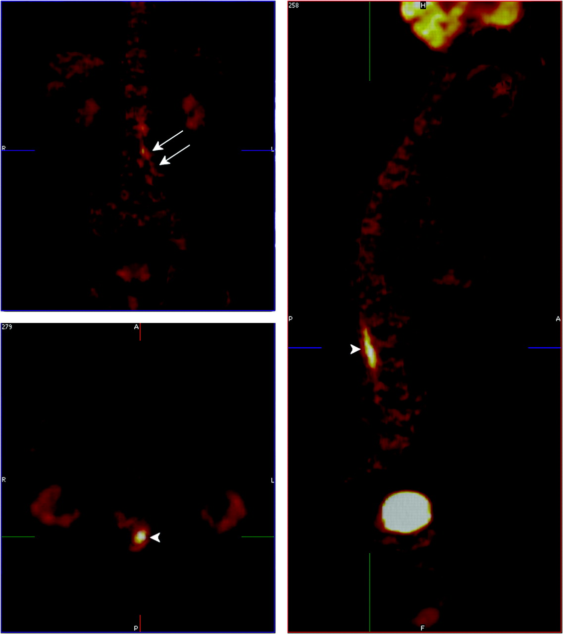
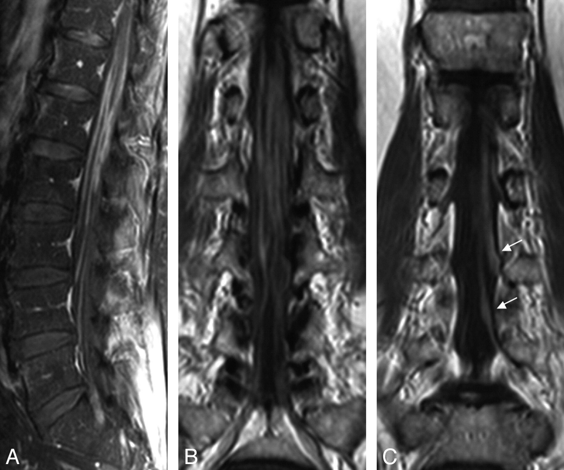
A previously healthy 79-year-old man presented with a progressive paresis and proximal paresthesia of the left lower extremity. Neurologic examination demonstrated paresis in the muscles innervated by the left L3, L4, and L5 nerve roots, absence of the Achilles tendon reflex, and sensory disturbances in the left dermatomes of L4 and L5. Other neurologic functions were intact. Except for a slightly increased erythrocyte sedimentation rate and a decreased absolute lymphocyte count, blood test results were negative or within normal limits. Initial examination of the CSF showed lymphocytic pleocytosis (0.143 × 10⁹ cells/L), an elevated protein level (1130 mg/L), and a mildly depressed glucose concentration (2.6 mmol/L). Bacterial and mycobacterial cultures were negative, as were the tests for Borrelia organisms, syphilis, herpes simplex virus, and Epstein-Barr virus. CSF cytology was negative for malignancy. Electromyography and nerve conduction studies revealed a sensorimotoric axonal polyneuropathy. To rule out underlying malignancy, we performed integrated [18F]-fluorodeoxyglucose–positron-emission tomography/CT (FDG-PET/CT), showing an intense 18F-FDG-uptake in the cauda equina, stretching out along the left nerve root on levels L3 and L4 (Fig 1). MR imaging of the lumbar spine confirmed thickening and contrast enhancement of the nerve roots of the cauda equina, more pronounced on the left side (Fig 2). Subsequent imaging of the brain, chest, and abdomen did not demonstrate any other lesions. A repeat CSF analysis with flow cytometric immunophenotyping confirmed the presence of B-cell lymphoma, which was treated with local radiation therapy (40 Gy). Follow-up FDG-PET/CT no longer demonstrated the hypermetabolic lesion in the spinal canal. Unfortunately, a new lesion was seen in the liver, considered a metastasis on the basis of imaging results, which responded well to chemotherapy (cyclophosphamide, rituximab, and prednisone). Vincristine was omitted due to the already prominent polyneuropathy. The neurologic condition of the patient remained stable.

Fig 1.
  • Download figure
  • Open in new tab
  • Download powerpoint
Fig 1.

Integrated [18F]-fluorodeoxyglucose (FDG)–PET/CT scan shows an intense FDG uptake in the cauda equina (arrowhead) and nerve roots L3 and L4 on the left side (arrows).

Fig 2.
  • Download figure
  • Open in new tab
  • Download powerpoint
Fig 2.

Gadolinium-enhanced T1-weighted images obtained in the sagittal plane with fat-suppression (FS) (A) and in the coronal plane without FS (B and C) show enhancement of the nerve roots of the cauda equina (A and B) and of the L3 and L4 dorsal root ganglia (arrows, C), the left more pronounced than the right.

Lymphomas in the CNS can occur in 3 settings. Most commonly, involvement of the CNS is a late manifestation of advanced lymphoma. A PCNSL is an unusual manifestation of extranodal non-Hodgkin lymphoma, usually consisting of a diffuse lymphomatous infiltration affecting the brain, leptomeninges, eyes, and, rarely, the spinal cord. Although considered a stage IE lymphoma, PCNSL is usually associated with a worse prognosis compared with other localized extranodal lymphomas with similar histologic characteristics. Rarely, a PCNSL disseminates outside the CNS as seen in our patient.1

Diagnosis of a PCNSL is often difficult. The presence of malignant lymphocytes distinguish it from infectious diseases, paraneoplastic or inflammatory neuropathies, and complications of medical interventions. The first objective of diagnostic work-up is to obtain tissue for pathologic diagnosis and immunophenotyping; and second, to evaluate the extent of the disease. Although a biopsy remains indispensable for providing tissue samples, a blind biopsy can be inconsequential because malignant infiltration in neurolymphomatosis affecting a peripheral nerve can be patchy, possibly resulting in sampling error.2 Currently, FDG-PET is increasingly being used for staging, restaging, and response assessment of lymphomas.3 Its role in the evaluation of PCNSL has not been established yet but seems promising.4

Our case highlights the diagnostic approach when evaluating a patient with a subacute sensorimotor loss. Diagnosis of PCNSL should be kept in mind. Although FDG-PET is currently mainly used for staging or restaging and response assessment of systemic lymphomas, our case illustrates the potential role in the diagnostic work-up of PCNSL.

References

  1. 1.↵
    1. Herrlinger U
    . Primary CNS lymphoma: findings outside the brain. J Neurooncol 1999;43:227–30
    CrossRefPubMed
  2. 2.↵
    1. van den Bent MJ,
    2. de Bruin HG,
    3. Bos GM,
    4. et al
    . Negative sural nerve biopsy in neurolymphomatosis. J Neurol 1999;246:1159–63
    CrossRefPubMed
  3. 3.↵
    1. Cheson BD,
    2. Pfistner B,
    3. Juweid ME,
    4. et al
    . Revised response criteria for malignant lymphoma. J Clin Oncol 2007;10;25:579–86
  4. 4.↵
    1. Lin M,
    2. Kilanowska J,
    3. Taper J,
    4. et al
    . Neurolymphomatosis: diagnosis and assessment of treatment response by FDG PET-CT. Hematol Oncol 2008;26:43–45
    CrossRefPubMed
  • Copyright © American Society of Neuroradiology
PreviousNext
Back to top

In this issue

American Journal of Neuroradiology: 30 (10)
American Journal of Neuroradiology
Vol. 30, Issue 10
1 Nov 2009
  • Table of Contents
  • Index by author
Advertisement
Print
Download PDF
Email Article

Thank you for your interest in spreading the word on American Journal of Neuroradiology.

NOTE: We only request your email address so that the person you are recommending the page to knows that you wanted them to see it, and that it is not junk mail. We do not capture any email address.

Enter multiple addresses on separate lines or separate them with commas.
Lymphomatous Meningosis Diagnosed with [18F]-Fluorodeoxyglucose–Positron-Emission Tomography/CT
(Your Name) has sent you a message from American Journal of Neuroradiology
(Your Name) thought you would like to see the American Journal of Neuroradiology web site.
CAPTCHA
This question is for testing whether or not you are a human visitor and to prevent automated spam submissions.
Cite this article
S. Ceyssens, T. Van den Wyngaert, P. Blockx, L. van den Hauwe, P.M. Parizel, L. van den Hauwe, K. Geens
Lymphomatous Meningosis Diagnosed with [18F]-Fluorodeoxyglucose–Positron-Emission Tomography/CT
American Journal of Neuroradiology Nov 2009, 30 (10) E150-E151; DOI: 10.3174/ajnr.A1831

Citation Manager Formats

  • BibTeX
  • Bookends
  • EasyBib
  • EndNote (tagged)
  • EndNote 8 (xml)
  • Medlars
  • Mendeley
  • Papers
  • RefWorks Tagged
  • Ref Manager
  • RIS
  • Zotero
0 Responses
Respond to this article
Share
Bookmark this article
Lymphomatous Meningosis Diagnosed with [18F]-Fluorodeoxyglucose–Positron-Emission Tomography/CT
S. Ceyssens, T. Van den Wyngaert, P. Blockx, L. van den Hauwe, P.M. Parizel, L. van den Hauwe, K. Geens
American Journal of Neuroradiology Nov 2009, 30 (10) E150-E151; DOI: 10.3174/ajnr.A1831
del.icio.us logo Twitter logo Facebook logo Mendeley logo
  • Tweet Widget
  • Facebook Like
  • Google Plus One
Purchase

Jump to section

  • Article
    • References
  • Figures & Data
  • Info & Metrics
  • Responses
  • References
  • PDF

Related Articles

  • No related articles found.
  • PubMed
  • Google Scholar

Cited By...

  • No citing articles found.
  • Crossref
  • Google Scholar

This article has not yet been cited by articles in journals that are participating in Crossref Cited-by Linking.

More in this TOC Section

  • Brain AVM’s Nidus: What if We Hadn’t Understood Anything?
  • Letter to the Editor regarding “Automated Volumetric Software in Dementia: Help or Hindrance to the Neuroradiologist?”
  • Reply:
Show more Letters

Similar Articles

Advertisement

Indexed Content

  • Current Issue
  • Accepted Manuscripts
  • Article Preview
  • Past Issues
  • Editorials
  • Editors Choice
  • Fellow Journal Club
  • Letters to the Editor

Cases

  • Case Collection
  • Archive - Case of the Week
  • Archive - Case of the Month
  • Archive - Classic Case

Special Collections

  • Special Collections

Resources

  • News and Updates
  • Turn around Times
  • Submit a Manuscript
  • Author Policies
  • Manuscript Submission Guidelines
  • Evidence-Based Medicine Level Guide
  • Publishing Checklists
  • Graphical Abstract Preparation
  • Imaging Protocol Submission
  • Submit a Case
  • Become a Reviewer/Academy of Reviewers
  • Get Peer Review Credit from Publons

Multimedia

  • AJNR Podcast
  • AJNR SCANtastic
  • Video Articles

About Us

  • About AJNR
  • Editorial Board
  • Not an AJNR Subscriber? Join Now
  • Alerts
  • Feedback
  • Advertise with us
  • Librarian Resources
  • Permissions
  • Terms and Conditions

American Society of Neuroradiology

  • Not an ASNR Member? Join Now

© 2025 by the American Society of Neuroradiology All rights, including for text and data mining, AI training, and similar technologies, are reserved.
Print ISSN: 0195-6108 Online ISSN: 1936-959X

Powered by HighWire