Advertisement

Main menu

  • Home
  • Content
    • Current Issue
    • Accepted Manuscripts
    • Article Preview
    • Past Issue Archive
    • AJNR Case Collection
    • Case of the Week Archive
    • Classic Case Archive
    • Case of the Month Archive
  • Special Collections
    • Spinal CSF Leak Articles (Jan 2020-June 2024)
    • 2024 AJNR Journal Awards
    • Most Impactful AJNR Articles
  • Multimedia
    • AJNR Podcast
    • AJNR Scantastics
    • Video Articles
  • For Authors
    • Submit a Manuscript
    • Author Policies
    • Fast publishing of Accepted Manuscripts
    • Graphical Abstract Preparation
    • Manuscript Submission Guidelines
    • Imaging Protocol Submission
    • Submit a Case for the Case Collection
  • About Us
    • About AJNR
    • Editorial Board
  • More
    • Become a Reviewer/Academy of Reviewers
    • Subscribers
    • Permissions
    • Alerts
    • Feedback
    • Advertisers
    • ASNR Home
  • Other Publications
    • ajnr

User menu

  • Alerts
  • Log in

Search

  • Advanced search
American Journal of Neuroradiology

American Journal of Neuroradiology

ASHNR American Society of Functional Neuroradiology ASHNR American Society of Pediatric Neuroradiology ASSR
  • Alerts
  • Log in

Advanced Search

  • Home
  • Content
    • Current Issue
    • Accepted Manuscripts
    • Article Preview
    • Past Issue Archive
    • AJNR Case Collection
    • Case of the Week Archive
    • Classic Case Archive
    • Case of the Month Archive
  • Special Collections
    • Spinal CSF Leak Articles (Jan 2020-June 2024)
    • 2024 AJNR Journal Awards
    • Most Impactful AJNR Articles
  • Multimedia
    • AJNR Podcast
    • AJNR Scantastics
    • Video Articles
  • For Authors
    • Submit a Manuscript
    • Author Policies
    • Fast publishing of Accepted Manuscripts
    • Graphical Abstract Preparation
    • Manuscript Submission Guidelines
    • Imaging Protocol Submission
    • Submit a Case for the Case Collection
  • About Us
    • About AJNR
    • Editorial Board
  • More
    • Become a Reviewer/Academy of Reviewers
    • Subscribers
    • Permissions
    • Alerts
    • Feedback
    • Advertisers
    • ASNR Home
  • Follow AJNR on Twitter
  • Visit AJNR on Facebook
  • Follow AJNR on Instagram
  • Join AJNR on LinkedIn
  • RSS Feeds

Welcome to the new AJNR, Updated Hall of Fame, and more. Read the full announcements.


AJNR is seeking candidates for the position of Associate Section Editor, AJNR Case Collection. Read the full announcement.


  • Getting new auth cookie, if you see this message a lot, tell someone!
  • Getting new auth cookie, if you see this message a lot, tell someone!
LetterLetter

Attenuation of Brain White Matter Hyperintensities after Cerebral Infarction

Y. Moriya, K. Kozaki, K. Nagai and K. Toba
American Journal of Neuroradiology March 2009, 30 (3) e43; DOI: https://doi.org/10.3174/ajnr.A1340
Y. Moriya
  • Find this author on Google Scholar
  • Find this author on PubMed
  • Search for this author on this site
K. Kozaki
  • Find this author on Google Scholar
  • Find this author on PubMed
  • Search for this author on this site
K. Nagai
  • Find this author on Google Scholar
  • Find this author on PubMed
  • Search for this author on this site
K. Toba
  • Find this author on Google Scholar
  • Find this author on PubMed
  • Search for this author on this site
  • Article
  • Figures & Data
  • Info & Metrics
  • Responses
  • References
  • PDF
Loading

Brain white matter hyperintensities (WMHs) on MR imaging are reported to increase with age1 and do not decrease,2 though their pathogenesis is not well understood. Here, we report an interesting case in which a patient showed attenuation of WMHs at 1 year after the incidence of cerebral infarction.

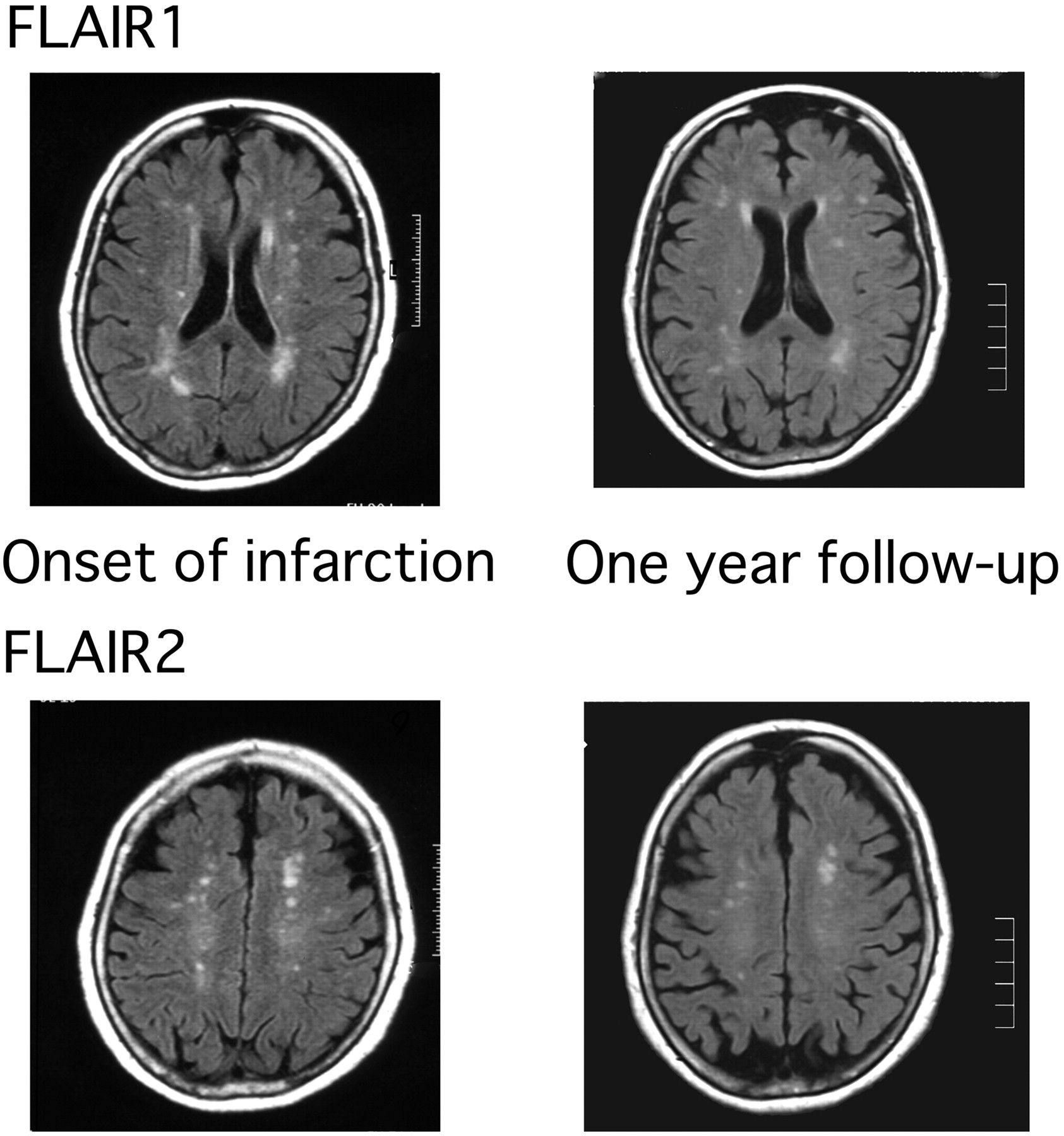
A 78-year-old female outpatient with hypertension and dyslipidemia presented with sudden onset of left homonymous hemianopia. She showed no other neurologic abnormality. The next day, brain MR imaging scans showed an acute infarct in the right occipital lobe. The symptom persisted thereafter with a little amelioration. When follow-up MR imaging scans were performed 1 year later, it was found that the WMHs attenuated (Fig 1). Measurement of the WMH area with MetaMorph 6.2 (Universal Imaging, Downingtown, Pa) confirmed this finding (Fig 2). Because the temporal decrease in WMHs was consistent in 2 different regions of the brain (fluid-attenuated inversion recovery [FLAIR] images 1 and 2), we could rule out a “chance appearance” of the attenuation of WMHs because of the difference in the levels of the 2 temporal images. Because the area of WMHs was apart from the infarct lesion, the change of WMHs was not considered to be from the direct effect of infarction.

Fig 1.
  • Download figure
  • Open in new tab
  • Download powerpoint
Fig 1.

Attenuation of WMHs at 2 different levels (FLAIR 1 and FLAIR 2).

Fig 2.
  • Download figure
  • Open in new tab
  • Download powerpoint
Fig 2.

WMH area was measured with use of MetaMorph 6.2. WMH 1 refers to the area of WMHs at the FLAIR 1 level (Fig 2), and WMH 2 refers to the area of WMHs at the FLAIR 2 level.

WMHs have not been reported to regress,2 though their pathogenesis is not well understood. The proposed mechanisms of their appearance are ischemia; blood-brain barrier dysfunction; and endothelial injury, which are not mutually exclusive.3 In this case, 1 possible explanation for the attenuation of WMHs could be that the blood-brain barrier was disturbed immediately after the onset of infarction, causing leakage of cerebral fluid into the white matter and this recovered afterward.

Image Analysis

The WMH area was quantified by a technician (who was not a physician) in a blind manner with use of MetaMorph 6.2. On acquired FLAIR images, 1) the scale was set; 2) unnecessary white areas such as bone were removed; 3) the average number of pixels in “normal white matter” fields in each image were measured (79 and 82 pixels), and the highest number of pixels in the 2 temporal images was set as the threshold for the ensuing measurement of WMHs; and 4) all of the areas above the threshold were selected, and their size was measured.

Sources of Funding

This study was supported by the Mitsui Sumitomo Insurance Welfare Foundation (2006) and the Japan Health Foundation.

References

  1. ↵
    Sachdev P, Wen W, Chen X, et al. Progression of white matter hyperintensities in elderly individuals over 3 years. Neurology 2007;68:214–22
    Abstract/FREE Full Text
  2. ↵
    Schmidt R, Fazekas F, Kapeller P, et al. MRI white matter hyperintensities: three-year follow-up of the Austrian Stroke Prevention Study. Neurology 1999;53:132–39
    Abstract/FREE Full Text
  3. ↵
    O'Sullivan M. Leukoaraiosis. Pract Neurol 2008;8:26–38
    Abstract/FREE Full Text
  • Copyright © American Society of Neuroradiology
PreviousNext
Back to top

In this issue

American Journal of Neuroradiology: 30 (3)
American Journal of Neuroradiology
Vol. 30, Issue 3
March 2009
  • Table of Contents
  • Index by author
Advertisement
Print
Download PDF
Email Article

Thank you for your interest in spreading the word on American Journal of Neuroradiology.

NOTE: We only request your email address so that the person you are recommending the page to knows that you wanted them to see it, and that it is not junk mail. We do not capture any email address.

Enter multiple addresses on separate lines or separate them with commas.
Attenuation of Brain White Matter Hyperintensities after Cerebral Infarction
(Your Name) has sent you a message from American Journal of Neuroradiology
(Your Name) thought you would like to see the American Journal of Neuroradiology web site.
CAPTCHA
This question is for testing whether or not you are a human visitor and to prevent automated spam submissions.
Cite this article
Y. Moriya, K. Kozaki, K. Nagai, K. Toba
Attenuation of Brain White Matter Hyperintensities after Cerebral Infarction
American Journal of Neuroradiology Mar 2009, 30 (3) e43; DOI: 10.3174/ajnr.A1340

Citation Manager Formats

  • BibTeX
  • Bookends
  • EasyBib
  • EndNote (tagged)
  • EndNote 8 (xml)
  • Medlars
  • Mendeley
  • Papers
  • RefWorks Tagged
  • Ref Manager
  • RIS
  • Zotero
0 Responses
Respond to this article
Share
Bookmark this article
Attenuation of Brain White Matter Hyperintensities after Cerebral Infarction
Y. Moriya, K. Kozaki, K. Nagai, K. Toba
American Journal of Neuroradiology Mar 2009, 30 (3) e43; DOI: 10.3174/ajnr.A1340
del.icio.us logo Twitter logo Facebook logo Mendeley logo
  • Tweet Widget
  • Facebook Like
  • Google Plus One
Purchase

Jump to section

  • Article
    • Image Analysis
    • Sources of Funding
    • References
  • Figures & Data
  • Info & Metrics
  • Responses
  • References
  • PDF

Related Articles

  • No related articles found.
  • PubMed
  • Google Scholar

Cited By...

  • Prediction of white matter hyperintensities evolution one-year post-stroke from a single-point brain MRI and stroke lesions information
  • Automatic Spatial Estimation of White Matter Hyperintensities Evolution in Brain MRI using Disease Evolution Predictor Deep Neural Networks
  • Disease progression and regression in sporadic small vessel disease-insights from neuroimaging
  • Crossref (10)
  • Google Scholar

This article has been cited by the following articles in journals that are participating in Crossref Cited-by Linking.

  • What are White Matter Hyperintensities Made of?
    Joanna M. Wardlaw, Maria C. Valdés Hernández, Susana Muñoz‐Maniega
    Journal of the American Heart Association 2015 4 6
  • White matter hyperintensity reduction and outcomes after minor stroke
    Joanna M. Wardlaw, Francesca M. Chappell, Maria del Carmen Valdés Hernández, Stephen D.J. Makin, Julie Staals, Kirsten Shuler, Michael J. Thrippleton, Paul A. Armitage, Susana Muñoz-Maniega, Anna K. Heye, Eleni Sakka, Martin S. Dennis
    Neurology 2017 89 10
  • Comparison of cerebral blood flow and structural penumbras in relation to white matter hyperintensities: A multi-modal magnetic resonance imaging study
    Nutta-on Promjunyakul, David L Lahna, Jeffrey A Kaye, Hiroko H Dodge, Deniz Erten-Lyons, William D Rooney, Lisa C Silbert
    Journal of Cerebral Blood Flow & Metabolism 2016 36 9
  • Disease progression and regression in sporadic small vessel disease–insights from neuroimaging
    Esther M.C. van Leijsen, Frank-Erik de Leeuw, Anil M. Tuladhar
    Clinical Science 2017 131 12
  • Dynamic Progression of White Matter Hyperintensities in Alzheimer’s Disease and Normal Aging: Results from the Sunnybrook Dementia Study
    Joel Ramirez, Alicia A. McNeely, Courtney Berezuk, Fuqiang Gao, Sandra E. Black
    Frontiers in Aging Neuroscience 2016 8
  • Quantitative and qualitative MRI evaluation of cerebral small vessel disease in an elderly population: a longitudinal study
    Ruta Nylander, Markus Fahlström, Egill Rostrup, Joel Kullberg, Soheil Damangir, Håkan Ahlström, Lars Lind, Elna-Marie Larsson
    Acta Radiologica 2018 59 5
  • Automatic spatial estimation of white matter hyperintensities evolution in brain MRI using disease evolution predictor deep neural networks
    Muhammad Febrian Rachmadi, Maria del C. Valdés-Hernández, Stephen Makin, Joanna Wardlaw, Taku Komura
    Medical Image Analysis 2020 63
  • Cerebral White Matter Lesions May Be Partially Reversible in Patients with Carotid Artery Stenosis
    K. Yamada, K. Sakai, K. Owada, K. Mineura, T. Nishimura
    American Journal of Neuroradiology 2010 31 7
  • Greater accelerometer-measured physical activity is associated with better cognition and cerebrovascular health in older adults
    Katherine J. Bangen, Amanda T. Calcetas, Kelsey R. Thomas, Christina Wierenga, Christine N. Smith, Maria Bordyug, Einat K. Brenner, David Wing, Conan Chen, Thomas T. Liu, Zvinka Z. Zlatar
    Journal of the International Neuropsychological Society 2023 29 9
  • Prediction of white matter hyperintensities evolution one-year post-stroke from a single-point brain MRI and stroke lesions information
    Muhammad Febrian Rachmadi, Maria del C. Valdés-Hernández, Stephen Makin, Joanna Wardlaw, Henrik Skibbe
    Scientific Reports 2025 15 1

More in this TOC Section

  • Reply:
  • Brain AVM’s Nidus: What if We Hadn’t Understood Anything?
  • Letter to the Editor regarding “Automated Volumetric Software in Dementia: Help or Hindrance to the Neuroradiologist?”
Show more Letters

Similar Articles

Advertisement

Indexed Content

  • Current Issue
  • Accepted Manuscripts
  • Article Preview
  • Past Issues
  • Editorials
  • Editors Choice
  • Fellow Journal Club
  • Letters to the Editor

Cases

  • Case Collection
  • Archive - Case of the Week
  • Archive - Case of the Month
  • Archive - Classic Case

Special Collections

  • Special Collections

Resources

  • News and Updates
  • Turn around Times
  • Submit a Manuscript
  • Author Policies
  • Manuscript Submission Guidelines
  • Evidence-Based Medicine Level Guide
  • Publishing Checklists
  • Graphical Abstract Preparation
  • Imaging Protocol Submission
  • Submit a Case
  • Become a Reviewer/Academy of Reviewers
  • Get Peer Review Credit from Publons

Multimedia

  • AJNR Podcast
  • AJNR SCANtastic
  • Video Articles

About Us

  • About AJNR
  • Editorial Board
  • Not an AJNR Subscriber? Join Now
  • Alerts
  • Feedback
  • Advertise with us
  • Librarian Resources
  • Permissions
  • Terms and Conditions

American Society of Neuroradiology

  • Not an ASNR Member? Join Now

© 2025 by the American Society of Neuroradiology All rights, including for text and data mining, AI training, and similar technologies, are reserved.
Print ISSN: 0195-6108 Online ISSN: 1936-959X

Powered by HighWire