
Article Figures & Data
Figures
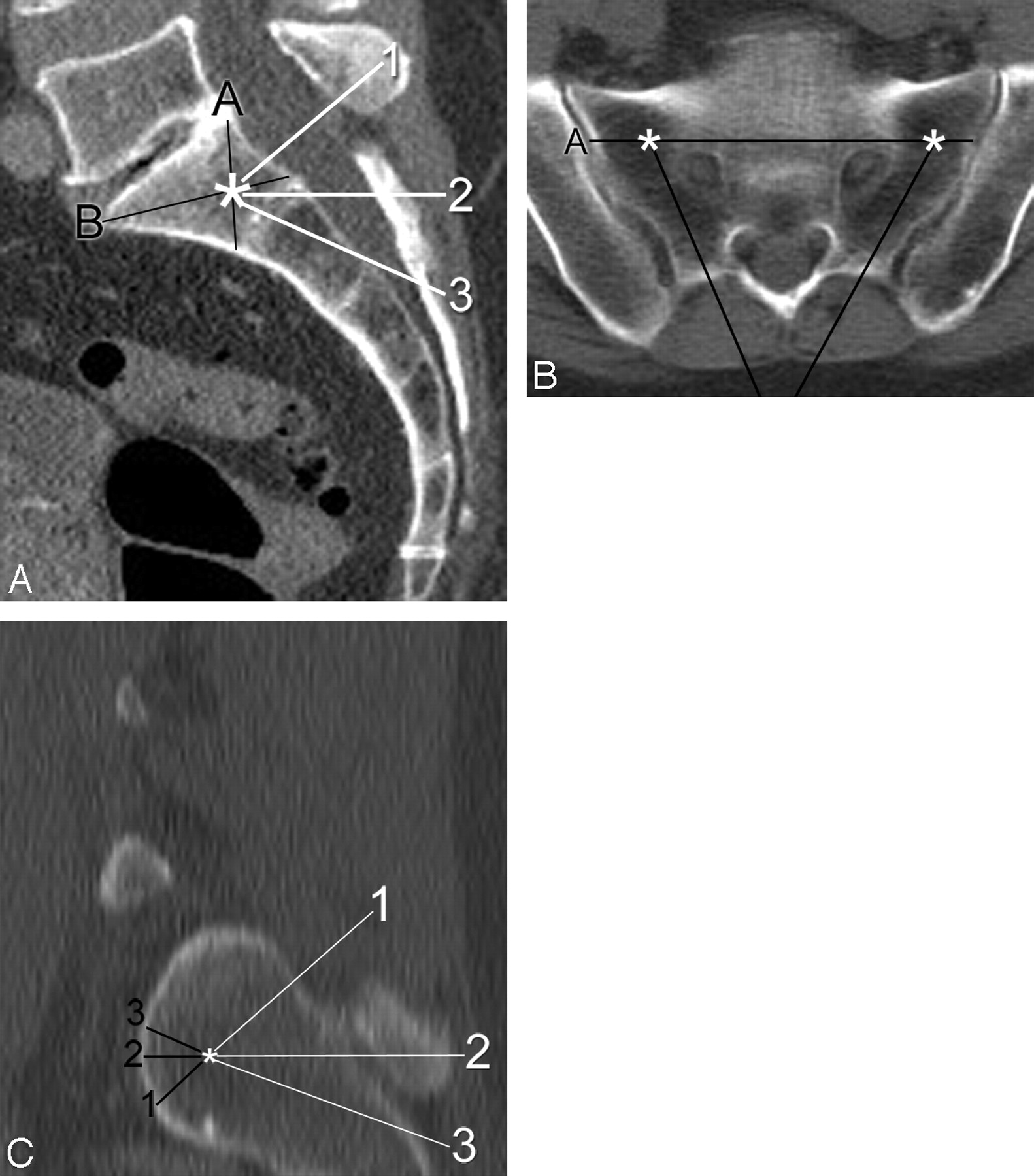
- Fig 1.
Demonstration of measurements made during virtual needle placement on a CT scan of the pelvis in a 65-year-old woman. A, On a sagittal midline reconstructed image, note the target (asterisk) by the intersection of line A (from the posterosuperior corner of S1 to the anteroinferior corner of S1) with line B (from the anterosuperior corner of S1 to the posteroinferior corner of S1). Three possible needle trajectories have been described and are indicated by the numbered white lines: 1) parallel to the L5-S1 disk space, 2) neutral or axial with respect to the patient, and 3) along the sacral long axis. B, Axial image taken along line 2 (axial plane) from the same patient demonstrates the target zones (asterisks) for each sacral ala. Line A represents the projection of line A from the sagittal image (A). Note how the 2 needle trajectories (black lines) are both parallel to their respective sacroiliac joints. C, Sagittal oblique image obtained along a line parallel to the sacroiliac joint shows the relative location of the target (asterisk) within the lateral sacrum. The distance from the target point to the anterior sacral cortex was obtained for the 3 needle trajectories described previously.
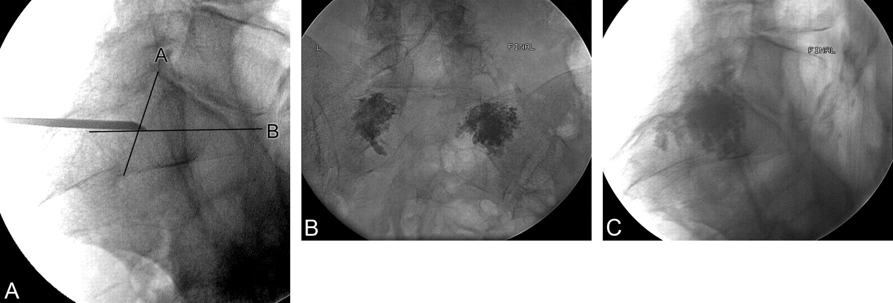
- Fig 2.
Use of a fluoroscopic landmark in performing sacroplasty in a 76-year-old woman. A, Initial lateral image shows the needle in position at the intersection of lines as defined in Fig 1. B and C, Posttreatment frontal (B) and lateral (C) projections demonstrate polymethylmethacrylate cement in both sacral alas with no presacral extravasation.
- Fig 3.
Example of a potential breach of the anterior sacral cortex in an 88-year-old woman with lumbarization of S1. A, Midline sagittal reformatted image from a CT scan shows the target zone (arrow) within S1 as described in Fig 1. B, Left parasagittal reformatted image shows that needle placement (arrow) would be anterior to the sacral cortex.


Tables
- Table 1:
Distance measurements from a predefined target point to the anterior sacral cortex for each of the 3 trajectories described (Fig 1)*
Distance to Anterior Sacral Cortex from Target Point Trajectory 1 Trajectory 2 Trajectory 3 Mean ± SD 11.3 ± 4.6 mm 11.1 ± 9.2 mm 12.8 ± 5.2 mm <3 mm 7 (3.5%) 8 (4%) 7 (3.5%) 3–10 mm 123 (61.5%) 138 (69%) 117 (58.5%) >10 mm 70 (35%) 54 (27%) 76 (38%) * Data from both the left and right hemisacrum were pooled, resulting in 200 measurements total.
- Table 2:
Demographics of 13 consecutive patients treated with percutaneous sacroplasty using fluoroscopic guidance only
Age (yr) Sex Sides Treated Needle Size (Gauge) Extraosseous Cement Seen on Imaging? Clinical Complication or Worsening of Symptoms? 73 M Bilateral 13 No No 65 F Bilateral 11 No No 65 F Bilateral 11 No No 68 F Bilateral 11 No No 61 F Bilateral 11 No No 74 F Bilateral 11 No No 76 F Bilateral 11 No No 83 F Right 11 No No 81 F Bilateral 13 No No 67 F Right 13 No No 78 F Bilateral 11 No No 81 F Bilateral 13 No No 76 F Bilateral 11 No No


