Advertisement

Main menu

  • Home
  • Content
    • Current Issue
    • Accepted Manuscripts
    • Article Preview
    • Past Issue Archive
    • AJNR Case Collection
    • Case of the Week Archive
    • Classic Case Archive
    • Case of the Month Archive
  • Special Collections
    • Spinal CSF Leak Articles (Jan 2020-June 2024)
    • 2024 AJNR Journal Awards
    • Most Impactful AJNR Articles
  • Multimedia
    • AJNR Podcast
    • AJNR Scantastics
    • Video Articles
  • For Authors
    • Submit a Manuscript
    • Author Policies
    • Fast publishing of Accepted Manuscripts
    • Graphical Abstract Preparation
    • Manuscript Submission Guidelines
    • Imaging Protocol Submission
    • Submit a Case for the Case Collection
  • About Us
    • About AJNR
    • Editorial Board
  • More
    • Become a Reviewer/Academy of Reviewers
    • Subscribers
    • Permissions
    • Alerts
    • Feedback
    • Advertisers
    • ASNR Home
  • Other Publications
    • ajnr

User menu

  • Alerts
  • Log in

Search

  • Advanced search
American Journal of Neuroradiology

American Journal of Neuroradiology

ASHNR American Society of Functional Neuroradiology ASHNR American Society of Pediatric Neuroradiology ASSR
  • Alerts
  • Log in

Advanced Search

  • Home
  • Content
    • Current Issue
    • Accepted Manuscripts
    • Article Preview
    • Past Issue Archive
    • AJNR Case Collection
    • Case of the Week Archive
    • Classic Case Archive
    • Case of the Month Archive
  • Special Collections
    • Spinal CSF Leak Articles (Jan 2020-June 2024)
    • 2024 AJNR Journal Awards
    • Most Impactful AJNR Articles
  • Multimedia
    • AJNR Podcast
    • AJNR Scantastics
    • Video Articles
  • For Authors
    • Submit a Manuscript
    • Author Policies
    • Fast publishing of Accepted Manuscripts
    • Graphical Abstract Preparation
    • Manuscript Submission Guidelines
    • Imaging Protocol Submission
    • Submit a Case for the Case Collection
  • About Us
    • About AJNR
    • Editorial Board
  • More
    • Become a Reviewer/Academy of Reviewers
    • Subscribers
    • Permissions
    • Alerts
    • Feedback
    • Advertisers
    • ASNR Home
  • Follow AJNR on Twitter
  • Visit AJNR on Facebook
  • Follow AJNR on Instagram
  • Join AJNR on LinkedIn
  • RSS Feeds

Welcome to the new AJNR, Updated Hall of Fame, and more. Read the full announcements.


AJNR is seeking candidates for the position of Associate Section Editor, AJNR Case Collection. Read the full announcement.


LetterLetter

Imaging Cerebral Malaria with a Susceptibility-Weighted MR Sequence

J.P. Nickerson, K.A. Tong and R. Raghavan
American Journal of Neuroradiology June 2009, 30 (6) e85-e86; DOI: https://doi.org/10.3174/ajnr.A1568
J.P. Nickerson
  • Find this author on Google Scholar
  • Find this author on PubMed
  • Search for this author on this site
K.A. Tong
  • Find this author on Google Scholar
  • Find this author on PubMed
  • Search for this author on this site
R. Raghavan
  • Find this author on Google Scholar
  • Find this author on PubMed
  • Search for this author on this site
  • Article
  • Figures & Data
  • Info & Metrics
  • Responses
  • References
  • PDF
Loading

Cerebral malaria is a severe complication seen in approximately 2% of patients with acute Plasmodium falciparum infection. Characteristic diffuse petechial hemorrhages that were previously only seen by gross pathologic or microscopic examination, and virtually undetectable by conventional imaging modalities, were well demonstrated in a patient with cerebral malaria, with use of susceptibility-weighted imaging (SWI). This case highlights the usefulness of SWI imaging to accelerate the diagnosis of cerebral malaria.

A 31-year-old woman was seen in the emergency department within 1 week of returning from a trip to the Philippines, complaining of cyclic fever and chills, headache, nausea, and vomiting. She admitted to inconsistently taking her malaria prophylaxis medication while out of the country. Her initial admission blood smear result was positive for P falciparum. A head CT scan was obtained; results were unremarkable. She was admitted to the intensive care unit, where she required intubation for respiratory failure. Head MR imaging was obtained 3 days after admission and demonstrated diffuse petechial hemorrhage throughout the gray-white matter junction, corpus callosum, and internal capsules of both hemispheres on SWI as well as edema in the posterior limbs of the bilateral internal capsules on fluid-attenuated inversion recovery imaging (Fig 1). The patient was extubated on hospital day 10 and was discharged home on day 12. Follow-up MR imaging at 6 weeks continued to demonstrate diffuse small hemorrhages with some interval improvement and resolution of the previously noted edema (Fig 2).

Cerebral malaria is a rare but potentially fatal complication of infection with P falciparum. Diffuse petechial hemorrhage occurs in these cases when cerebral capillaries and small veins are occluded by infected erythrocytes. Although the mortality rate of cerebral malaria has been estimated between 20% and 50%, those patients who survive often have a full recovery with no long-term sequelae.

Imaging findings in cerebral malaria have been limited in the past. Significant hemorrhage or infarction with resultant edema is required for CT detection. Millan et al1 described MR imaging findings in cerebral malaria by using conventional T1-weighted and T2-weighted imaging to demonstrate hemorrhage and infarction in a single case, and Cordoliani et al2 reported a series of 12 patients with cerebral malaria who received MR imaging, only 3 of whom demonstrated any findings suggestive of a pathologic process on conventional T1 and T2 sequences. The SWI sequence described by Tong et al3 is much more sensitive in the detection of small hemorrhages than is conventional MR imaging, including standard 2D gradient-echo T2* sequences. This high spatial-resolution technique is extremely sensitive to susceptibility changes. SWI has been shown to be helpful in the detection of small hemorrhages in various conditions including diffuse axonal injury, coagulopathy, neoplasms, and neurodegenerative disorders.4 Imaging findings with use of this sequence seem to correlate much more closely with the expected pathologic findings of diffuse petechial hemorrhage. In the future, SWI may play a significant role in the detection of small hemorrhagic lesions that are not detectable by conventional MR images or CT scan.

Fig 1.
  • Download figure
  • Open in new tab
  • Download powerpoint
Fig 1.

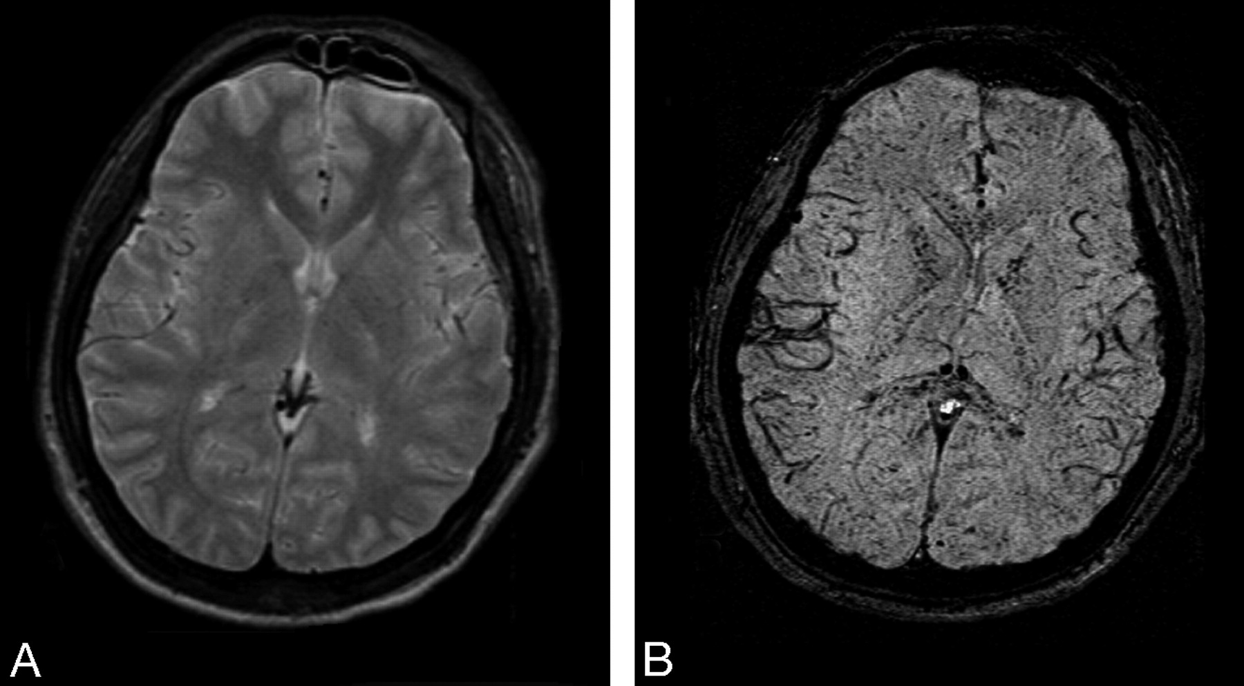
Admission MR images (A and B): T2 and SWI. Diffuse petechial hemorrhages present throughout the brain (particularly the gray-white junction, corpus callosum, and internal capsule) are seen only on the SWI sequence.

Fig 2.
  • Download figure
  • Open in new tab
  • Download powerpoint
Fig 2.

Six-week follow-up study (A and B): T2 and SWI. SWI reveals residual hemorrhages, though there has been some interval improvement.

References

  1. ↵
    Millan J, San Millan J, Munoz M, et al. CNS complications in acute malaria: MR findings. AJNR Am J Neuroradiol 1993;14:493–94
    Abstract/FREE Full Text
  2. ↵
    Cordoliani YS, Sarrazin JL, Felten D, et al. MR of cerebral malaria. AJNR Am J Neuroradiol 1998;19:871–74
    Abstract
  3. ↵
    Tong KA, Ashwal S, Holshouser BA, et al. Hemorrhagic shearing lesions in children and adolescents with posttraumatic diffuse axonal injury: improved detection and initial results. Radiology 2003;227:332
    CrossRefPubMed
  4. ↵
    Tong KA, Ashwal S, Obenaus A, et al. Susceptibility-weighted MR imaging: a review of clinical applications in children. AJNR Am J Neuroradiol 2008;29:9–17.
    Abstract/FREE Full Text
  • Copyright © American Society of Neuroradiology
PreviousNext
Back to top

In this issue

American Journal of Neuroradiology: 30 (6)
American Journal of Neuroradiology
Vol. 30, Issue 6
June 2009
  • Table of Contents
  • Index by author
Advertisement
Print
Download PDF
Email Article

Thank you for your interest in spreading the word on American Journal of Neuroradiology.

NOTE: We only request your email address so that the person you are recommending the page to knows that you wanted them to see it, and that it is not junk mail. We do not capture any email address.

Enter multiple addresses on separate lines or separate them with commas.
Imaging Cerebral Malaria with a Susceptibility-Weighted MR Sequence
(Your Name) has sent you a message from American Journal of Neuroradiology
(Your Name) thought you would like to see the American Journal of Neuroradiology web site.
CAPTCHA
This question is for testing whether or not you are a human visitor and to prevent automated spam submissions.
Cite this article
J.P. Nickerson, K.A. Tong, R. Raghavan
Imaging Cerebral Malaria with a Susceptibility-Weighted MR Sequence
American Journal of Neuroradiology Jun 2009, 30 (6) e85-e86; DOI: 10.3174/ajnr.A1568

Citation Manager Formats

  • BibTeX
  • Bookends
  • EasyBib
  • EndNote (tagged)
  • EndNote 8 (xml)
  • Medlars
  • Mendeley
  • Papers
  • RefWorks Tagged
  • Ref Manager
  • RIS
  • Zotero
0 Responses
Respond to this article
Share
Bookmark this article
Imaging Cerebral Malaria with a Susceptibility-Weighted MR Sequence
J.P. Nickerson, K.A. Tong, R. Raghavan
American Journal of Neuroradiology Jun 2009, 30 (6) e85-e86; DOI: 10.3174/ajnr.A1568
del.icio.us logo Twitter logo Facebook logo Mendeley logo
  • Tweet Widget
  • Facebook Like
  • Google Plus One
Purchase

Jump to section

  • Article
    • References
  • Figures & Data
  • Info & Metrics
  • Responses
  • References
  • PDF

Related Articles

  • No related articles found.
  • PubMed
  • Google Scholar

Cited By...

  • Cerebral malaria is regulated by host mediated changes in Plasmodium gene expression
  • Transcellular blood-brain barrier disruption in malaria-induced reversible brain edema
  • Reversible brain edema in experimental cerebral malaria is associated with transcellular blood-brain barrier disruption and delayed microhemorrhages
  • Magnetic resonance imaging in cerebral malaria: a report of four cases
  • Crossref (38)
  • Google Scholar

This article has been cited by the following articles in journals that are participating in Crossref Cited-by Linking.

  • Infectious causes of stroke
    Jennifer E Fugate, Jennifer L Lyons, Kiran T Thakur, Bryan R Smith, E Tessa Hedley-Whyte, Farrah J Mateen
    The Lancet Infectious Diseases 2014 14 9
  • Bacterial, Fungal, and Parasitic Infections of the Central Nervous System: Radiologic-Pathologic Correlation and Historical Perspectives:From the Radiologic Pathology Archives
    Robert Y. Shih, Kelly K. Koeller
    RadioGraphics 2015 35 4
  • Nervous System Involvement in Coronavirus Disease 2019: Results from a Retrospective Consecutive Neuroimaging Cohort
    Stefanos Klironomos, Antonios Tzortzakakis, Annika Kits, Claes Öhberg, Evangelia Kollia, Amir Ahoromazdae, Håkan Almqvist, Åsa Aspelin, Heather Martin, Russell Ouellette, Jonathan Al-Saadi, Mikael Hasselberg, Mansour Haghgou, Mitra Pedersen, Sven Petersson, Johannes Finnsson, Johan Lundberg, Anna Falk Delgado, Tobias Granberg
    Radiology 2020 297 3
  • Parasitic Diseases of the Central Nervous System
    Ahmed Abdel Khalek Abdel Razek, Arvemas Watcharakorn, Mauricio Castillo
    Neuroimaging Clinics of North America 2011 21 4
  • CNS Hypoxia Is More Pronounced in Murine Cerebral than Noncerebral Malaria and Is Reversed by Erythropoietin
    Casper Hempel, Valery Combes, Nicholas Henry Hunt, Jørgen Anders Lindholm Kurtzhals, Georges Emile Raymond Grau
    The American Journal of Pathology 2011 179 4
  • Cerebral amyloid angiopathy: Review of clinico‐radiological features and mimics
    Rohit Sharma, Stephanie Dearaugo, Bernard Infeld, Richard O'Sullivan, Richard P Gerraty
    Journal of Medical Imaging and Radiation Oncology 2018 62 4
  • Rethinking cerebral malaria pathology
    Danny A Milner
    Current Opinion in Infectious Diseases 2010 23 5
  • Susceptibility-weighted imaging in pediatric neuroimaging
    Thangamadhan Bosemani, Andrea Poretti, Thierry A.G.M. Huisman
    Journal of Magnetic Resonance Imaging 2014 40 3
  • Imaging Plasmodium immunobiology in the liver, brain, and lung
    Ute Frevert, Adéla Nacer, Mynthia Cabrera, Alexandru Movila, Maike Leberl
    Parasitology International 2014 63 1
  • Magnetic resonance imaging in cerebral malaria: a report of four cases
    D D Rasalkar, B K Paunipagar, D Sanghvi, B D Sonawane, P Loniker
    The British Journal of Radiology 2011 84 1000

More in this TOC Section

  • Letter to the Editor regarding “Automated Volumetric Software in Dementia: Help or Hindrance to the Neuroradiologist?”
  • Reply:
  • Brain AVM’s Nidus: What if We Hadn’t Understood Anything?
Show more Letters

Similar Articles

Advertisement

Indexed Content

  • Current Issue
  • Accepted Manuscripts
  • Article Preview
  • Past Issues
  • Editorials
  • Editors Choice
  • Fellow Journal Club
  • Letters to the Editor

Cases

  • Case Collection
  • Archive - Case of the Week
  • Archive - Case of the Month
  • Archive - Classic Case

Special Collections

  • Special Collections

Resources

  • News and Updates
  • Turn around Times
  • Submit a Manuscript
  • Author Policies
  • Manuscript Submission Guidelines
  • Evidence-Based Medicine Level Guide
  • Publishing Checklists
  • Graphical Abstract Preparation
  • Imaging Protocol Submission
  • Submit a Case
  • Become a Reviewer/Academy of Reviewers
  • Get Peer Review Credit from Publons

Multimedia

  • AJNR Podcast
  • AJNR SCANtastic
  • Video Articles

About Us

  • About AJNR
  • Editorial Board
  • Not an AJNR Subscriber? Join Now
  • Alerts
  • Feedback
  • Advertise with us
  • Librarian Resources
  • Permissions
  • Terms and Conditions

American Society of Neuroradiology

  • Not an ASNR Member? Join Now

© 2025 by the American Society of Neuroradiology All rights, including for text and data mining, AI training, and similar technologies, are reserved.
Print ISSN: 0195-6108 Online ISSN: 1936-959X

Powered by HighWire