
Article Figures & Data
Figures
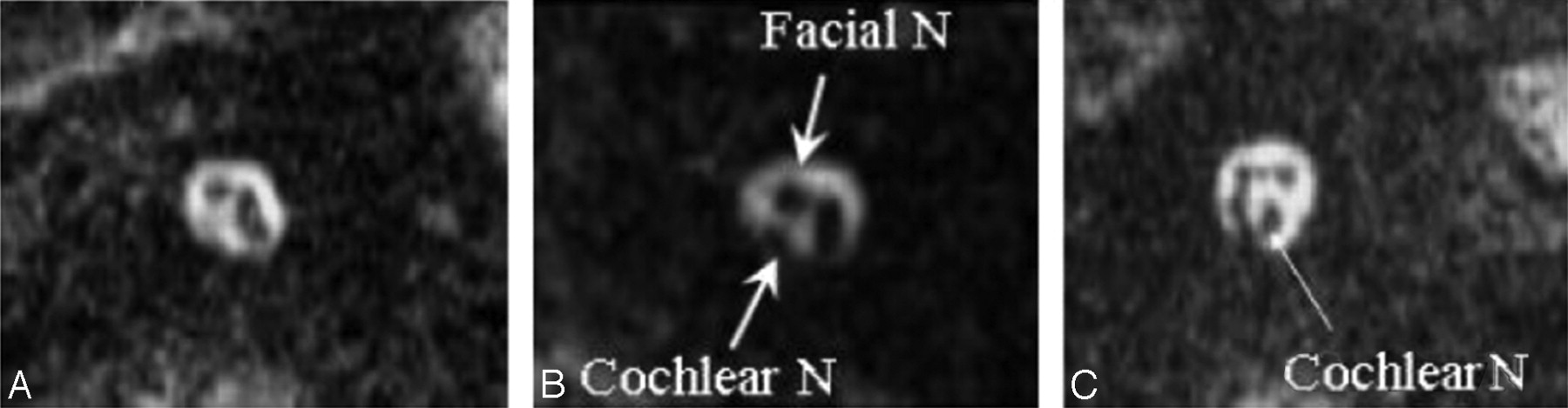
- Fig 1.
Representative 3D MR images of cochlear nerve (N) aplasia (A), hypoplasia (B), and the normal cochlear nerve (C). Arrows indicate the cochlear nerve and facial nerve in the internal auditory canal. B, The caliber of the cochlear nerve is smaller than that of the facial nerve, which is the diagnostic criterion for cochlear nerve hypoplasia.
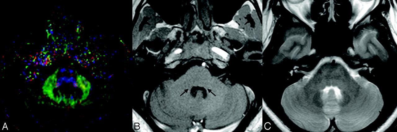
- Fig 2.
A, A sample DTI at the level of the LL and the selected region of interest (3 × 3 × 2 mm rectangular box). Note that in the DTIs, the most basic red-green-blue color-coded scheme attributes a color to each orientation of the fibers: Fibers crossing from left to the right are shown in red, fibers crossing anteroposteriorly are visualized in green, and fibers crossing inferosuperiorly are visualized in blue.30 B, In the corresponding T1-weighted MR image at the same sectional level, long arrows pinpoint the anatomic sites of lateral lemniscus. C, The corresponding T2-weighted image at the same section level.
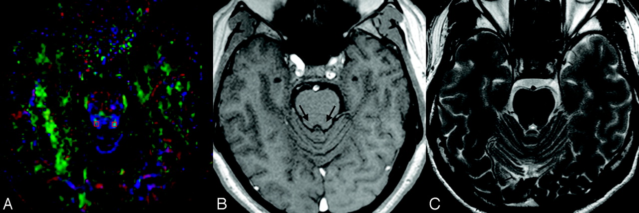
- Fig 3.
A, A sample DTI at the level of the IC and the selected area of region of interest. B, In the corresponding T1-weighted MR image at the same sectional level, long arrows pinpoint the anatomic sites of the IC. C, The corresponding T2-weighted image at the same sectional level.


Tables
- Table 1:
Basic clinical data of all subjects with unilateral cochlear nerve deficiency (aplasia and hypoplasia)
Case Age (yr) Sex Cochlear Nerve Deficiency Hearing Threshold (PTA) dB HL Right Left Right Left 1 12 M Aplasia Normal 100 10 2 10 F Aplasia Normal >100 20 3 8 F Aplasia Normal >100 10 4 9 F Aplasia Normal >110 15 5 14 F Hypoplasia Normal 100 5 6 8 M Hypoplasia Normal >110 10 7 12 M Normal Aplasia 5 >110 8 25 F Aplasia Normal 100 10 9 9 M Normal Aplasia 10 >110 10 11 M Normal Aplasia 10 >110 11 29 M Normal Aplasia 15 >110 12 9 F Hypoplasia Normal 90 10 Note:—PTA indicates pure tone average; HL, hearing level.
LL IC Contralateral Mean (SEM) Control Mean (SEM) P Value Contralateral Mean (SEM) Control Mean (SEM) P Value λ‖ 1.12 (0.22) 1.22 (0.19) .2817 1.50 (0.37) 1.37 (0.11) .2558 l⊥ 0.58 (0.16) 0.34 (0.05) .0003† 0.45 (0.13) 0.26 (0.06) .0004† MD 0.76 (0.18) 0.62 (0.06) .0263† 0.80 (0.20) 0.64 (0.09) .0231† FA 0.43 (0.07) 0.67 (0.08) .0001† 0.68 (0.06) 0.79 (0.04) .0001† Note:—LL indicates lateral lemniscus; IC, inferior colliculus; CND, cochlear nerve deficiency; SEM, standard error of the mean; λ‖, axial diffusivity; l⊥ , radial diffusivity; MD, mean diffusivity; FA, fractional anisotropy.
* The contralateral side of the patients was compared with that in the control group.
† Statistically significant.
- Table 3:
Summary of the diffusion indices (mean and SEM) at the LL and IC for the unilateral CND group*
LL IC Ipsilateral Mean (SEM) Control Mean (SEM) P Value Ipsilateral Mean (SEM) Control Mean (SEM) P Value λ‖ 1.18 (0.26) 1.22 (0.19) .6907 1.56 (0.38) 1.37 (0.11) .1236 l⊥ 0.59 (0.17) 0.34 (0.05) .0002† 0.43 (0.15) 0.26 (0.06) .0026† MD 0.79 (0.19) 0.62 (0.06) .0151† 0.81 (0.21) 0.64 (0.09) .0228† FA 0.44 (0.06) 0.67 (0.08) .0001† 0.70 (0.09) 0.79 (0.04) .0081† * The ipsilateral side of the patients was compared with that in the control group.
† Statistically significant.
In this issue


Jump to section
Related Articles
Cited By...
- Greater working memory and speech perception scores in cochlear implant users predict better subjective quality of life and hearing
- Probabilistic Fiber-Tracking Reveals Degeneration of the Contralateral Auditory Pathway in Patients with Vestibular Schwannoma
- Altered White Matter Integrity in Adolescents with Prelingual Deafness: A High-Resolution Tract-Based Spatial Statistics Imaging Study
- Brain Stem and Inner Ear Abnormalities in Children with Auditory Neuropathy Spectrum Disorder and Cochlear Nerve Deficiency