
Article Figures & Data
Figures
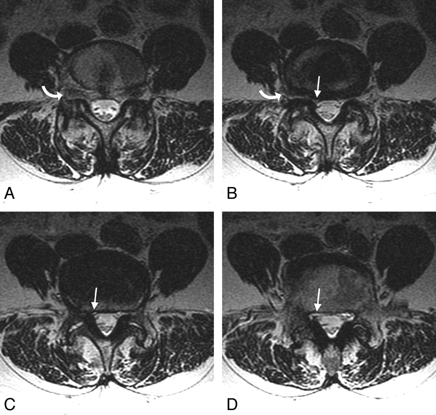
- Fig 1.
A 69-year-old woman with right-sided radiating leg pain that projected to her posterolateral thigh, knee, calf, and ankle, referred for right-sided nerve root block and steroid administration. Preprocedural MR imaging demonstrated an abnormality at L4–5, with right-sided L4 involvement in the foramen and L5 involvement in the lateral recess. Injection of the L4 root reproduced the patient's thigh and knee pain, and injection of the L5 root reproduced her right calf and ankle pain. She was completely pain-free after the injection. A−C, Axial T2-weighted MR images at L4–5 demonstrate uplifting of the right L4 root in the foramen and far lateral region due to disk (curved arrows), with compression of the L5 root in the lateral recess (arrows). D, At the lowest extent of the lateral recess (arrow), the lateral canal widens and the root reappears in the narrowed niche.
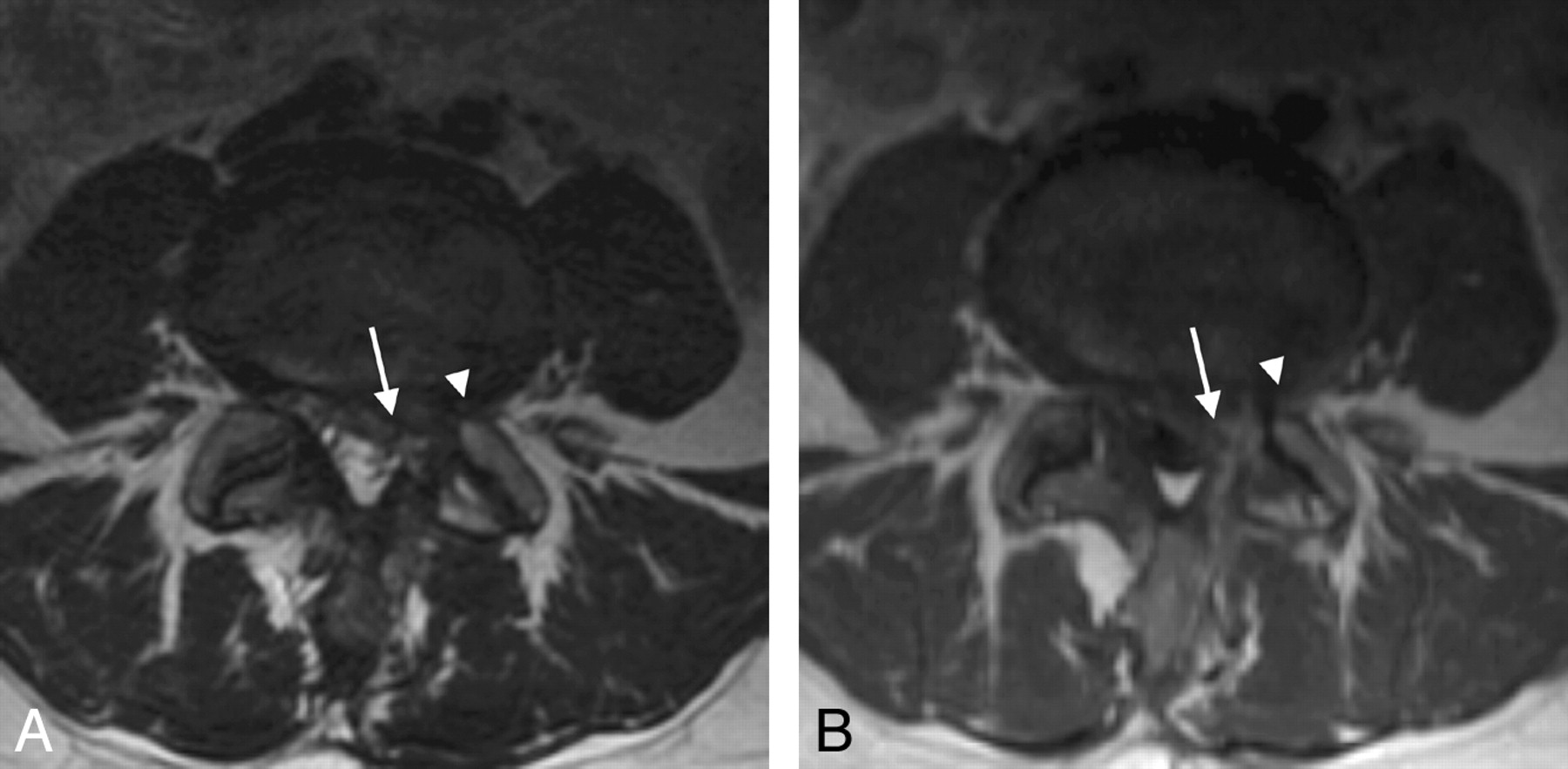
- Fig 2.
A 46-year-old woman with prior left L4–5 diskectomy 9 months prior, who presented with recurrent left-sided gluteal, posterior thigh, knee, and shin pain and was referred for left-sided nerve root block and steroid administration. Preprocedural MR imaging demonstrated postoperative changes on the left at L4–5, with a scar and root distortion involving the left L4 and L5. Injection of the L4 root reproduced the patient's gluteal and thigh pain, like sciatica, consistent with prominent furcal nerve contribution from L4 to L5. Injection of the L5 root reproduced the patient's shin pain only. She was completely pain-free after the procedure. A, Axial T2-weighted MR image demonstrates postoperative changes on the left, with distortion of the corner of the canal (arrow) affecting the L5 root, and the foraminal region (arrowhead) affecting the left L4. B, Axial T1-weighted image after contrast administration demonstrates a postoperative scar in the canal (arrow) and neural foramen (arrowhead) on the left, involving the L5 and L4 roots.
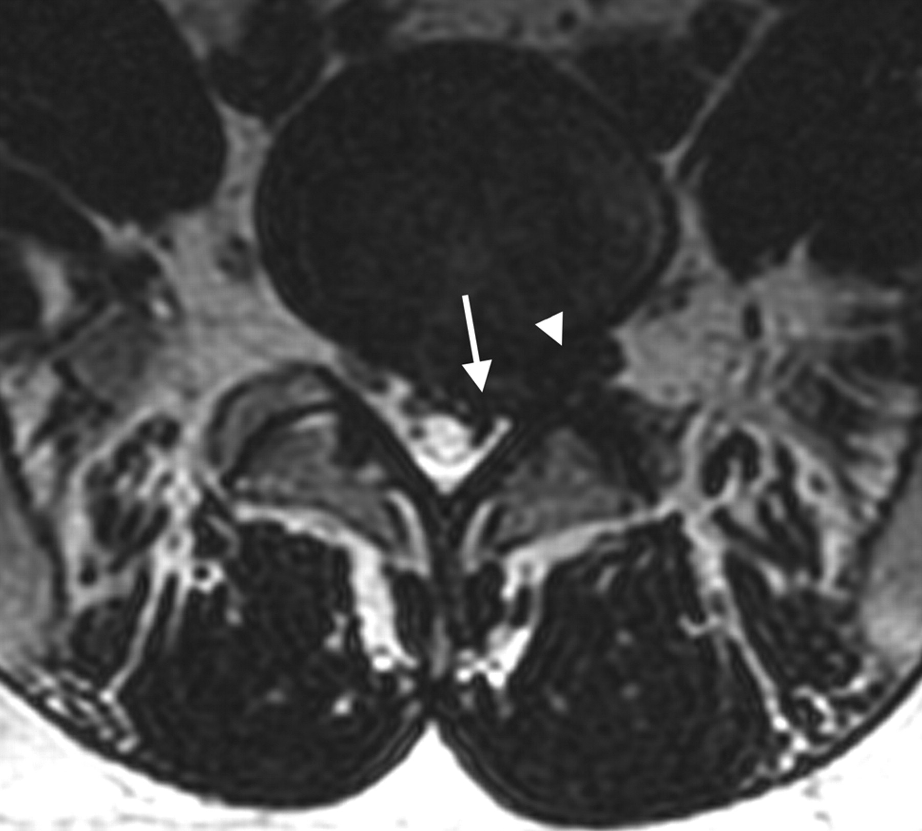
- Fig 3.
A 58-year-old man with left-sided leg pain that projected to the thigh, knee, calf, ankle, and foot, referred for nerve root block and steroid administration. Preprocedural MR imaging demonstrated a prominent disk bulge/protrusion at L3–4 and L4–5, suggesting a possible L4 and L5 root involvement. Injection at L4 reproduced the patient's calf and ankle pain, consistent with a significant furcal nerve contribution to L4 from L5, with injection at L5 reproducing a component of the patient's foot and toe pain. A, Axial T2-weighted image at L3–4 demonstrates a prominent asymmetric diffuse disk bulge, with distortion of the left lateral recess (arrow), likely affecting the L4 root. B, Axial T2-weighted image at L4–5 demonstrates a prominent diffuse disk bulge and asymmetric central disk protrusion, with distortion of the left lateral recess (arrow), very likely affecting the left L5 root.
- Fig 4.
A 35-year-old man with left-sided sciatica radiating down his posterior thigh to the level of the knee, referred for nerve root block and steroid administration. Preprocedural MR imaging demonstrates a moderately large disk protrusion at L5–S1 on the right, with involvement of the S1 root (arrow) in the canal and L5 root (arrowhead) in the neural foramen. Injection of both L5 and S1 reproduced the patient's sciatic pain, with slightly greater pain reproduction from the S1 root.


Tables
Clinical Presentation No. Pts Patient's Response to Injection L4, L5, Symptomatic Single Root, Symptomatic Neither Root, Symptomatic Proximal sciatica 15 7 5 3 Knee (± anterior thigh) and calf, ankle, or foot pain 32 21 8 3 Total 47 28 13 6 Clinical Presentation No. Pts Patient's Response to Injection L5, S1, Symptomatic Single Root, Symptomatic Neither Root, Symptomatic Proximal sciatica 23 14 6 3 Sciatica and calf, ankle, or foot pain 49 33 13 3 Total 72 47 19 6 - Table 3:
Patient's sense of radiating/radicular pain during double-root injection: L3+L4 and L2+L3
Roots Injected No. Pts Patient's Response to Injection Both Roots, Symptomatic Single Root, Symptomatic Neither Root, Symptomatic L3+L4 11 6 5 0 L2+L3 2 1 0 1 Total 13 7 5 1
In this issue


Jump to section
Related Articles
Cited By...
- No citing articles found.