Advertisement

Main menu

  • Home
  • Content
    • Current Issue
    • Accepted Manuscripts
    • Article Preview
    • Past Issue Archive
    • AJNR Case Collection
    • Case of the Week Archive
    • Classic Case Archive
    • Case of the Month Archive
  • Special Collections
    • Spinal CSF Leak Articles (Jan 2020-June 2024)
    • 2024 AJNR Journal Awards
    • Most Impactful AJNR Articles
  • Multimedia
    • AJNR Podcast
    • AJNR Scantastics
    • Video Articles
  • For Authors
    • Submit a Manuscript
    • Author Policies
    • Fast publishing of Accepted Manuscripts
    • Graphical Abstract Preparation
    • Manuscript Submission Guidelines
    • Imaging Protocol Submission
    • Submit a Case for the Case Collection
  • About Us
    • About AJNR
    • Editorial Board
  • More
    • Become a Reviewer/Academy of Reviewers
    • Subscribers
    • Permissions
    • Alerts
    • Feedback
    • Advertisers
    • ASNR Home
  • Other Publications
    • ajnr

User menu

  • Alerts
  • Log in

Search

  • Advanced search
American Journal of Neuroradiology

American Journal of Neuroradiology

ASHNR American Society of Functional Neuroradiology ASHNR American Society of Pediatric Neuroradiology ASSR
  • Alerts
  • Log in

Advanced Search

  • Home
  • Content
    • Current Issue
    • Accepted Manuscripts
    • Article Preview
    • Past Issue Archive
    • AJNR Case Collection
    • Case of the Week Archive
    • Classic Case Archive
    • Case of the Month Archive
  • Special Collections
    • Spinal CSF Leak Articles (Jan 2020-June 2024)
    • 2024 AJNR Journal Awards
    • Most Impactful AJNR Articles
  • Multimedia
    • AJNR Podcast
    • AJNR Scantastics
    • Video Articles
  • For Authors
    • Submit a Manuscript
    • Author Policies
    • Fast publishing of Accepted Manuscripts
    • Graphical Abstract Preparation
    • Manuscript Submission Guidelines
    • Imaging Protocol Submission
    • Submit a Case for the Case Collection
  • About Us
    • About AJNR
    • Editorial Board
  • More
    • Become a Reviewer/Academy of Reviewers
    • Subscribers
    • Permissions
    • Alerts
    • Feedback
    • Advertisers
    • ASNR Home
  • Follow AJNR on Twitter
  • Visit AJNR on Facebook
  • Follow AJNR on Instagram
  • Join AJNR on LinkedIn
  • RSS Feeds

Welcome to the new AJNR, Updated Hall of Fame, and more. Read the full announcements.


AJNR is seeking candidates for the position of Associate Section Editor, AJNR Case Collection. Read the full announcement.


Case ReportExpedited Publication
Open Access

MR Imaging in Novel Influenza A(H1N1)-Associated Meningoencephalitis

A. Haktanir
American Journal of Neuroradiology March 2010, 31 (3) 394-395; DOI: https://doi.org/10.3174/ajnr.A2037
A. Haktanir
  • Find this author on Google Scholar
  • Find this author on PubMed
  • Search for this author on this site
  • Article
  • Figures & Data
  • Info & Metrics
  • Responses
  • References
  • PDF
Loading

Abstract

SUMMARY: MR imaging manifestations of influenza A−associated encephalitis have been described previously. However, there is very limited information about the central nervous system complications of the novel influenza A(H1N1) virus. MR imaging findings of novel influenza A−associated meningoencephalitis in a child are presented.

Abbreviations

ADC
apparent diffusion coefficient
FFE
fast-field echo
WHO
World Health Organization

Since the first report of novel influenza A(H1N1) virus in Mexico in April 2009,1 a rapid spread to many countries around the world has occurred. In June 2009, the WHO raised the alert level to phase 6 (pandemic phase) on the basis of documented human-to-human spread of infection in at least 3 countries in 2 WHO regions.

Awareness of imaging features of the disease and imminent complications of the virus is important. MR imaging findings of encephalitis associated with influenza A virus have been described previously.2–5 However, to my knowledge, there are very limited reports about H1N1 virus−associated central nervous system lesions. Lyon et al6 presented radiologic findings of acute necrotizing encephalopathy in a child with H1N1 influenza infection. I present conventional and diffusion MR imaging findings of H1N1-associated meningoencephalitis in a child.

Case Report

A previously healthy 3-year-old girl presented to our emergency department with high fever (39°C) and focal convulsion. Her routine immunizations were up-to-date. She did not receive the seasonal influenza or H1N1 vaccine. The following day she experienced focal convulsions and lost consciousness. The girl was given anticonvulsive and antiviral (acyclovir) medication; however, no acute improvement was noticed. Lumbar puncture showed a normal biochemical structure of the CSF. Contrast-enhanced MR imaging was performed for suspected encephalitis and complications. Antiviral therapy for H1N1 (oseltamivir) was administered after the nasopharyngeal swabs confirmed H1N1 virus infection.

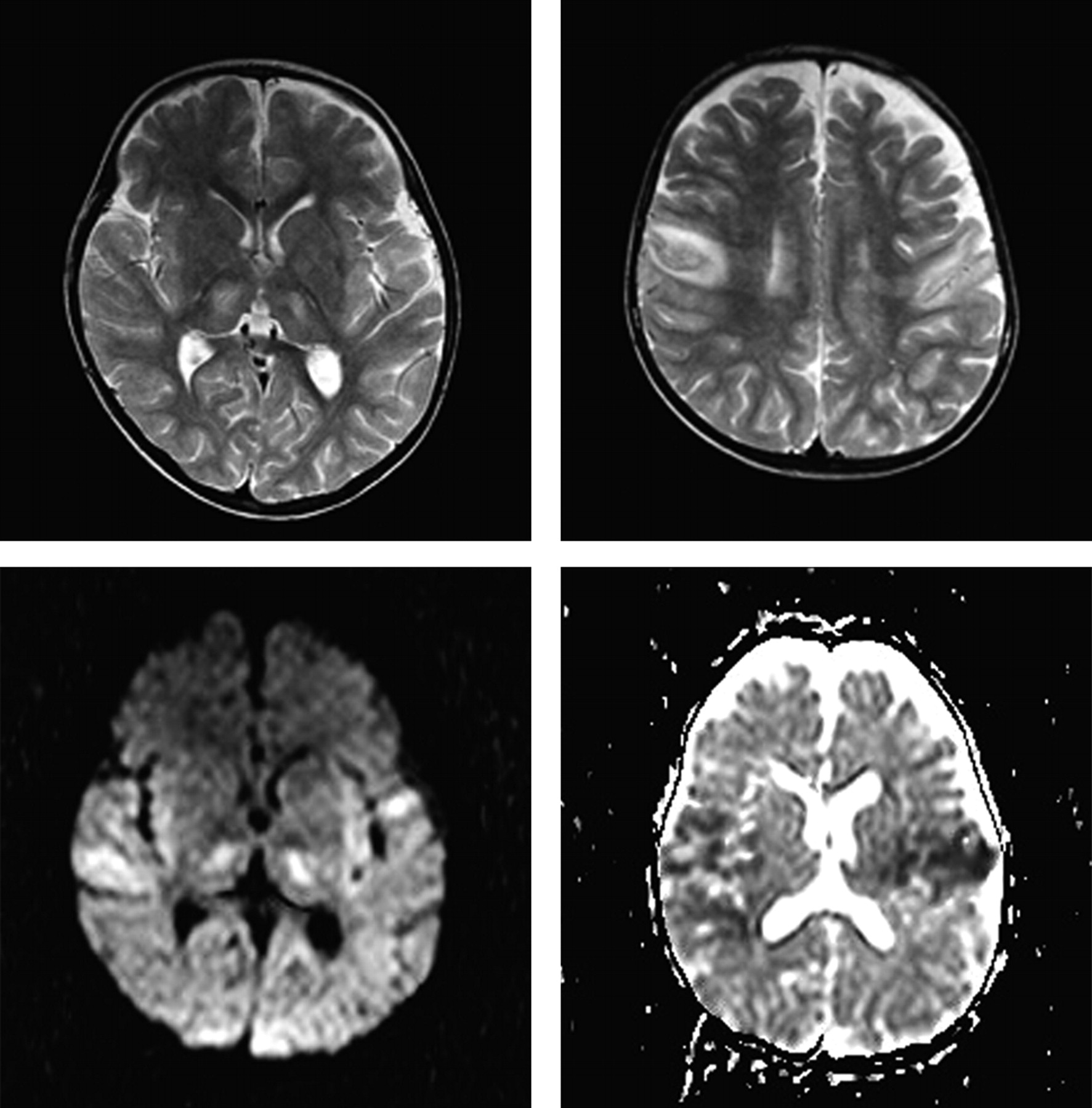
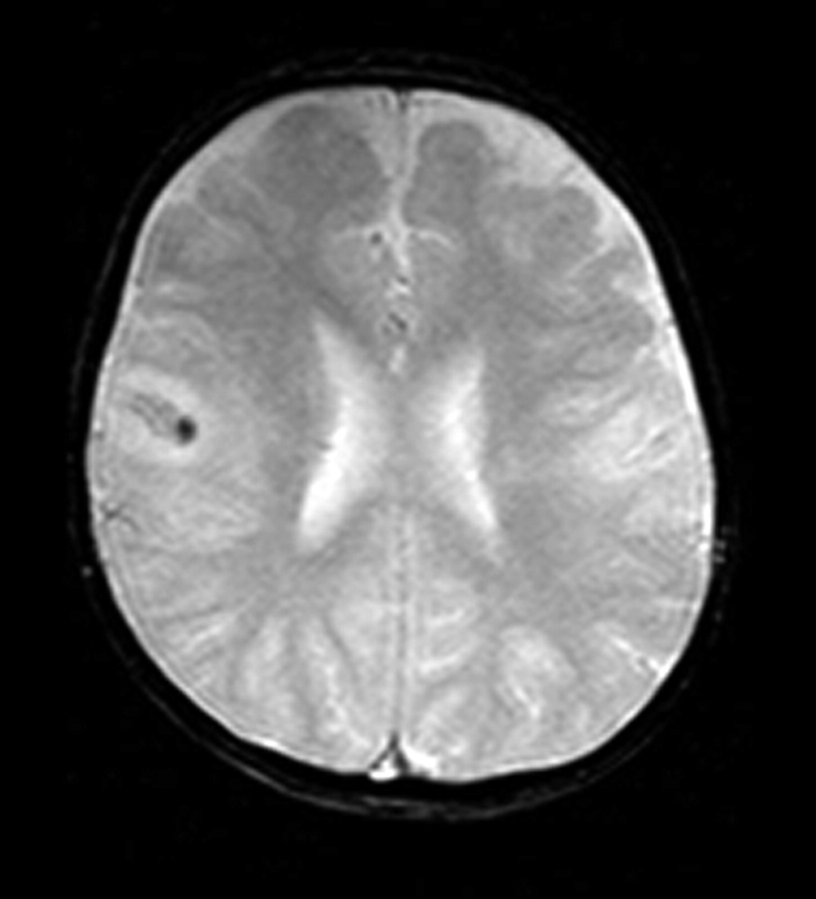
MR imaging demonstrated bilateral perirolandic T2 hyperintensities with restricted diffusion and round T2 hyperintense lesions in both thalami (Fig 1). Following intravenous contrast agent administration, diffuse meningeal enhancement was seen in both cerebral hemispheres. Enhancement was more pronounced in the right perirolandic area (Fig 2). T2 hyperintensity was more prominent in the right perirolandic area as well, along with magnetic susceptibility on T2 FFE images (Fig 3). There was no sign of cerebral edema.

Fig 1.
  • Download figure
  • Open in new tab
  • Download powerpoint
Fig 1.

T2-weighted transverse MR images demonstrate bilateral thalamic (upper right) and perirolandic (upper left) hyperintensities with restricted diffusion in the isotropic b = 1000 image (lower right) and the ADC map (lower left).

Fig 2.
  • Download figure
  • Open in new tab
  • Download powerpoint
Fig 2.

Contrast-enhanced transverse T1-weighted images show diffuse meningeal enhancement in both cerebral hemispheres, which is more pronounced in the right perirolandic area.

Fig 3.
  • Download figure
  • Open in new tab
  • Download powerpoint
Fig 3.

Magnetic susceptibility on a T2 FFE image in the right perirolandic area.

On the fifth hospital day, she recovered consciousness, and oral nourishment was begun.

Discussion

Association of influenza virus infection with central nervous system lesions was reported previously and named “influenza-associated encephalitis/encephalopathy.”7 The beginning of neurologic symptoms is usually within a few days to a week after the first signs of influenza A infection, and complete recovery occurs within a month.4 Symmetric splenial lesions (restricted diffusion and T2 hyperintensities) of the corpus callosum have been frequently reported in these patients.3,4,8 In addition, symmetric localized hypoattenuated lesions within the thalami and pons on CT and ringlike areas of high signal intensity in the pons on MR imaging were reported.2

Lyon et al6 reported CT and MR imaging findings in a 12-year-old girl infected with influenza A(H1N1) whose clinical course was complicated by acute necrotizing encephalopathy. The authors reported T2 hyperintensity and restricted diffusion in the thalami, cerebellar hemispheres, brain stem, and centrum semiovale bilaterally; enlarged heterogeneous thalami with ring-enhancing lesions; and abnormal enhancement in the centrum semiovale and brain stem. In addition, magnetic susceptibility in the bilateral thalamus was demonstrated in T2* gradient-recalled echo images. CT showed diffuse hypoattenuation in the thalamus and brain stem with sulcal effacement and brain stem edema. Lyon et al did not report any meningeal abnormality. In my case, bilateral thalamic T2 hyperintensities seem similar to the findings of Lyon et al. I observed magnetic susceptibility artifacts, too; however, they were in the right perirolandic area, not in the thalami. Also, bilateral perirolandic changes and diffuse meningeal enhancement are the differential imaging characteristics of this case.

Kimura et al5 divided influenza-related brain changes into 5 categories based on the MR imaging and CT findings: normal (category 1), diffuse involvement of the cerebral cortex (category 2), diffuse brain edema (category 3), symmetric involvement of the thalamus (category 4), and postinfectious focal encephalitis (category 5). Because of the bilateral thalamic lesions, my case may be classified as category 4; however, accompanying meningeal and perirolandic changes do not appear to be simply categorized. These varied findings can be explained by the unknown pathogenesis of brain lesions associated with influenza infection. Alternatively, the novelty of the influenza A(H1N1) virus infection may have caused these changes.

To the best of my knowledge, MR imaging findings of novel influenza A(H1N1) meningoencephalitis have not been reported before. In this regard, this report is the first in the literature. H1N1 encephalitis should be added to the differential diagnosis of bilateral lesions in the thalami and perirolandic area with meningeal enhancement. Neuroradiologists should be aware of this entity and its MR imaging manifestations, particularly during the pandemic phase.

Indicates open access to non-subscribers at www.ajnr.org

References

  1. 1.↵
    1. Perez-Padilla R,
    2. de la Rosa-Zamboni D,
    3. Ponce de Leon S,
    4. et al
    . Pneumonia and respiratory failure from swine-origin influenza A (H1N1) in Mexico. N Engl J Med 2009 8 13;361:680–89
    CrossRefPubMed
  2. 2.↵
    1. Protheroe SM,
    2. Mellor DH
    . Imaging in influenza A encephalitis. Arch Dis Child 1991;66:702–05
    Abstract/FREE Full Text
  3. 3.↵
    1. Takanashi J,
    2. Barkovich AJ,
    3. Yamaguchi K,
    4. et al
    . Influenza-associated encephalitis/encephalopathy with a reversible lesion in the splenium of the corpus callosum: a case report and literature review. AJNR Am J Neuroradiol 2004;25:798–802
    Abstract/FREE Full Text
  4. 4.↵
    1. Bulakbasi N,
    2. Kocaoglu M,
    3. Tayfun C,
    4. et al
    . Transient splenial lesion of the corpus callosum in clinically mild influenza-associated encephalitis/encephalopathy. AJNR Am J Neuroradiol 2006;27:1983–86
    Abstract/FREE Full Text
  5. 5.↵
    1. Kimura S,
    2. Ohtuki N,
    3. Nezu A,
    4. et al
    . Clinical and radiological variability of influenza-related encephalopathy or encephalitis. Acta Paediatr Jpn 1998;40:264–70
    PubMed
  6. 6.↵
    1. Lyon JB,
    2. Remigio C,
    3. Milligan T,
    4. et al
    . Acute necrotizing encephalopathy in a child with H1N1 influenza infection. Pediatr Radiol 2010;40:200–05
    CrossRefPubMed
  7. 7.↵
    1. Yoshikawa H,
    2. Yamazaki S,
    3. Watanabe T,
    4. et al
    . Study of influenza-associated encephalitis/encephalopathy in children during the 1997 to 2001 influenza seasons. J Child Neurol 2001;16:885–90
    Abstract/FREE Full Text
  8. 8.↵
    1. Tada H,
    2. Takanashi J,
    3. Barkovich AJ,
    4. et al
    . Clinically mild encephalitis/encephalopathy with a reversible splenial lesion. Neurology 2004;63:1854–58
    Abstract/FREE Full Text
  • Received January 4, 2010.
  • Accepted after revision January 7, 2010.
  • Copyright © American Society of Neuroradiology
View Abstract
PreviousNext
Back to top

In this issue

American Journal of Neuroradiology: 31 (3)
American Journal of Neuroradiology
Vol. 31, Issue 3
1 Mar 2010
  • Table of Contents
  • Index by author
Advertisement
Print
Download PDF
Email Article

Thank you for your interest in spreading the word on American Journal of Neuroradiology.

NOTE: We only request your email address so that the person you are recommending the page to knows that you wanted them to see it, and that it is not junk mail. We do not capture any email address.

Enter multiple addresses on separate lines or separate them with commas.
MR Imaging in Novel Influenza A(H1N1)-Associated Meningoencephalitis
(Your Name) has sent you a message from American Journal of Neuroradiology
(Your Name) thought you would like to see the American Journal of Neuroradiology web site.
CAPTCHA
This question is for testing whether or not you are a human visitor and to prevent automated spam submissions.
Cite this article
A. Haktanir
MR Imaging in Novel Influenza A(H1N1)-Associated Meningoencephalitis
American Journal of Neuroradiology Mar 2010, 31 (3) 394-395; DOI: 10.3174/ajnr.A2037

Citation Manager Formats

  • BibTeX
  • Bookends
  • EasyBib
  • EndNote (tagged)
  • EndNote 8 (xml)
  • Medlars
  • Mendeley
  • Papers
  • RefWorks Tagged
  • Ref Manager
  • RIS
  • Zotero
0 Responses
Respond to this article
Share
Bookmark this article
MR Imaging in Novel Influenza A(H1N1)-Associated Meningoencephalitis
A. Haktanir
American Journal of Neuroradiology Mar 2010, 31 (3) 394-395; DOI: 10.3174/ajnr.A2037
del.icio.us logo Twitter logo Facebook logo Mendeley logo
  • Tweet Widget
  • Facebook Like
  • Google Plus One
Purchase

Jump to section

  • Article
    • Abstract
    • Abbreviations
    • Case Report
    • Discussion
    • References
  • Figures & Data
  • Info & Metrics
  • Responses
  • References
  • PDF

Related Articles

  • No related articles found.
  • PubMed
  • Google Scholar

Cited By...

  • MRI Brain Findings in 126 Patients with COVID-19: Initial Observations from a Descriptive Literature Review
  • Neurologic complications of influenza A(H1N1)pdm09: Surveillance in 6 pediatric hospitals
  • Are Neuroimaging Findings in Novel Influenza A(H1N1) Infection Really Novel?
  • Crossref (26)
  • Google Scholar

This article has been cited by the following articles in journals that are participating in Crossref Cited-by Linking.

  • H1N1 Encephalitis with Malignant Edema and Review of Neurologic Complications from Influenza
    Paul Taylor Akins, John Belko, Timothy M. Uyeki, Yekaterina Axelrod, Kenneth K. Lee, James Silverthorn
    Neurocritical Care 2010 13 3
  • A Population-Based Study of Neurologic Manifestations of Severe Influenza A(H1N1)pdm09 in California
    C. A. Glaser, K. Winter, K. DuBray, K. Harriman, T. M. Uyeki, J. Sejvar, S. Gilliam, J. K. Louie
    Clinical Infectious Diseases 2012 55 4
  • Neurologic complications of influenza A(H1N1)pdm09
    Gulam Khandaker, Yvonne Zurynski, Jim Buttery, Helen Marshall, Peter C. Richmond, Russell C. Dale, Jenny Royle, Michael Gold, Tom Snelling, Bruce Whitehead, Cheryl Jones, Leon Heron, Mary McCaskill, Kristine Macartney, Elizabeth J. Elliott, Robert Booy
    Neurology 2012 79 14
  • MRI Brain Findings in 126 Patients with COVID-19: Initial Observations from a Descriptive Literature Review
    E. Gulko, M.L. Oleksk, W. Gomes, S. Ali, H. Mehta, P. Overby, F. Al-Mufti, A. Rozenshtein
    American Journal of Neuroradiology 2020 41 12
  • Neurological complications of pandemic influenza A H1N1 2009 infection: European case series and review
    Pinki Surana, Shan Tang, Marilyn McDougall, Cheuk Yan William Tong, Esse Menson, Ming Lim
    European Journal of Pediatrics 2011 170 8
  • Neurological events related to influenza A (H1N1) pdm09
    Graciela Cárdenas, José Luis Soto‐Hernández, Alexandra Díaz‐Alba, Yair Ugalde, Jorge Mérida‐Puga, Marcos Rosetti, Edda Sciutto
    Influenza and Other Respiratory Viruses 2014 8 3
  • Post-Marketing Assessment of Neuropsychiatric Adverse Events in Influenza Patients Treated with Oseltamivir: An Updated Review
    Stephen Toovey, Eric P. Prinssen, Craig R. Rayner, Bharat T. Thakrar, Regina Dutkowski, Annette Koerner, Tom Chu, Alexandra Sirzen-Zelenskaya, Markus Britschgi, Sudhir Bansod, Barbara Donner
    Advances in Therapy 2012 29 10
  • Neuroimaging of Emergent and Reemergent Infections
    Rafael Lourenço do Carmo, Aylla Keiner Alves Simão, Lázaro Luís Faria do Amaral, Bruno Shigueo Yonekura Inada, Camila Filardi Silveira, Christiane Monteiro de Siqueira Campos, Leonardo Furtado Freitas, Victor Bonadio, Victor Hugo Rocha Marussi
    RadioGraphics 2019 39 6
  • Clinical and MRI features of neurological complications after influenza A (H1N1) infection in critically ill children
    Hongwu Zeng, Stephen Quinet, Wenxian Huang, Yungen Gan, Chunxi Han, Yanxia He, Yonker Wang
    Pediatric Radiology 2013 43 9
  • Novel influenza A (H1N1)-associated encephalopathy/encephalitis with severe neurological sequelae and unique Image features — A case report
    Ying-Chu Chen, Chung-Ping Lo, Tzu-Pu Chang
    Journal of the Neurological Sciences 2010 298 1-2

More in this TOC Section

  • MRI Findings in Children with Acute Flaccid Paralysis and Cranial Nerve Dysfunction Occurring during the 2014 Enterovirus D68 Outbreak
  • MRI Surrogates for Molecular Subgroups of Medulloblastoma
  • WEB-DL Endovascular Treatment of Wide-Neck Bifurcation Aneurysms: Short- and Midterm Results in a European Study
Show more Expedited Publication

Similar Articles

Advertisement

Indexed Content

  • Current Issue
  • Accepted Manuscripts
  • Article Preview
  • Past Issues
  • Editorials
  • Editors Choice
  • Fellow Journal Club
  • Letters to the Editor

Cases

  • Case Collection
  • Archive - Case of the Week
  • Archive - Case of the Month
  • Archive - Classic Case

Special Collections

  • Special Collections

Resources

  • News and Updates
  • Turn around Times
  • Submit a Manuscript
  • Author Policies
  • Manuscript Submission Guidelines
  • Evidence-Based Medicine Level Guide
  • Publishing Checklists
  • Graphical Abstract Preparation
  • Imaging Protocol Submission
  • Submit a Case
  • Become a Reviewer/Academy of Reviewers
  • Get Peer Review Credit from Publons

Multimedia

  • AJNR Podcast
  • AJNR SCANtastic
  • Video Articles

About Us

  • About AJNR
  • Editorial Board
  • Not an AJNR Subscriber? Join Now
  • Alerts
  • Feedback
  • Advertise with us
  • Librarian Resources
  • Permissions
  • Terms and Conditions

American Society of Neuroradiology

  • Not an ASNR Member? Join Now

© 2025 by the American Society of Neuroradiology All rights, including for text and data mining, AI training, and similar technologies, are reserved.
Print ISSN: 0195-6108 Online ISSN: 1936-959X

Powered by HighWire