Advertisement

Main menu

  • Home
  • Content
    • Current Issue
    • Accepted Manuscripts
    • Article Preview
    • Past Issue Archive
    • AJNR Case Collection
    • Case of the Week Archive
    • Classic Case Archive
    • Case of the Month Archive
  • Special Collections
    • Spinal CSF Leak Articles (Jan 2020-June 2024)
    • 2024 AJNR Journal Awards
    • Most Impactful AJNR Articles
  • Multimedia
    • AJNR Podcast
    • AJNR Scantastics
    • Video Articles
  • For Authors
    • Submit a Manuscript
    • Author Policies
    • Fast publishing of Accepted Manuscripts
    • Graphical Abstract Preparation
    • Manuscript Submission Guidelines
    • Imaging Protocol Submission
    • Submit a Case for the Case Collection
  • About Us
    • About AJNR
    • Editorial Board
  • More
    • Become a Reviewer/Academy of Reviewers
    • Subscribers
    • Permissions
    • Alerts
    • Feedback
    • Advertisers
    • ASNR Home
  • Other Publications
    • ajnr

User menu

  • Alerts
  • Log in

Search

  • Advanced search
American Journal of Neuroradiology

American Journal of Neuroradiology

ASHNR American Society of Functional Neuroradiology ASHNR American Society of Pediatric Neuroradiology ASSR
  • Alerts
  • Log in

Advanced Search

  • Home
  • Content
    • Current Issue
    • Accepted Manuscripts
    • Article Preview
    • Past Issue Archive
    • AJNR Case Collection
    • Case of the Week Archive
    • Classic Case Archive
    • Case of the Month Archive
  • Special Collections
    • Spinal CSF Leak Articles (Jan 2020-June 2024)
    • 2024 AJNR Journal Awards
    • Most Impactful AJNR Articles
  • Multimedia
    • AJNR Podcast
    • AJNR Scantastics
    • Video Articles
  • For Authors
    • Submit a Manuscript
    • Author Policies
    • Fast publishing of Accepted Manuscripts
    • Graphical Abstract Preparation
    • Manuscript Submission Guidelines
    • Imaging Protocol Submission
    • Submit a Case for the Case Collection
  • About Us
    • About AJNR
    • Editorial Board
  • More
    • Become a Reviewer/Academy of Reviewers
    • Subscribers
    • Permissions
    • Alerts
    • Feedback
    • Advertisers
    • ASNR Home
  • Follow AJNR on Twitter
  • Visit AJNR on Facebook
  • Follow AJNR on Instagram
  • Join AJNR on LinkedIn
  • RSS Feeds

Welcome to the new AJNR, Updated Hall of Fame, and more. Read the full announcements.


AJNR is seeking candidates for the position of Associate Section Editor, AJNR Case Collection. Read the full announcement.


Research ArticleSpine
Open Access

Erythropoietin Promotes Functional Recovery and Enhances Nerve Regeneration after Peripheral Nerve Injury in Rats

Z.-S. Yin, H. Zhang and W. Gao
American Journal of Neuroradiology March 2010, 31 (3) 509-515; DOI: https://doi.org/10.3174/ajnr.A1820
Z.-S. Yin
  • Find this author on Google Scholar
  • Find this author on PubMed
  • Search for this author on this site
H. Zhang
  • Find this author on Google Scholar
  • Find this author on PubMed
  • Search for this author on this site
W. Gao
  • Find this author on Google Scholar
  • Find this author on PubMed
  • Search for this author on this site
  • Article
  • Figures & Data
  • Info & Metrics
  • Responses
  • References
  • PDF
Loading

Article Figures & Data

Figures

  • Tables
  • Fig 1.
    • Download figure
    • Open in new tab
    • Download powerpoint
    Fig 1.

    Photograph shows tubulation with a 7-mm-long interstump gap. The proximal and distal nerve stumps were joined to a 10-mm-long silicone rubber tube, leaving a 7-mm interstump gap. The ruler is graduated in millimeters.

  • Fig 2.
    • Download figure
    • Open in new tab
    • Download powerpoint
    Fig 2.

    Comparison of the mean functional recovery of each group after sciatic nerve transection and repair. Functional analysis of neural regeneration was assessed at 4 and 8 weeks after surgery by using walking-track analysis. Measurements made from walking-track prints were then submitted to an SFI. Data are the mean ± SEM. The asterisk indicates P < .05 compared with the NGF group significance.
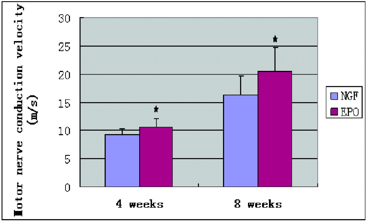
  • Fig 3.
    • Download figure
    • Open in new tab
    • Download powerpoint
    Fig 3.

    Effect of EPO administration on MNCV. At weeks 4 and 8 after surgery, MNCV was significantly lower in the NGF group than in the EPO group. There are statistically significant differences in MNCV between the 2 groups. Data are mean ± SEM. The asterisk indicates P < .05 compared with the NGF group.

  • Fig 4.
    • Download figure
    • Open in new tab
    • Download powerpoint
    Fig 4.

    The arrows demonstrate the thickness of scar tissue of nerve fiber bundles 8 weeks after surgery. A, A rat treated with NGF shows a very thick band of scar tissue surrounding fiber bundles. B, A rat treated with EPO shows very thin bands of scar tissue surrounding fiber bundles (hematoxylin-eosin stain, original magnification ×100).
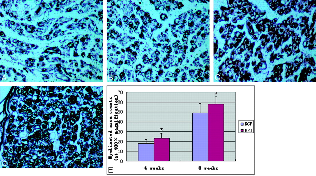
  • Fig 5.
    • Download figure
    • Open in new tab
    • Download powerpoint
    Fig 5.

    Representative photomicrographs of regenerated sciatic nerve cross-sections. The modified Bielschowsky silver stain was used to visualize myelinated axons. A and B, Regeneration of myelinated axons at 4 weeks. C and D, Regeneration at 8 weeks. A−D, These photomicrographs demonstrated significantly better regeneration in the EPO group (B and D) compared with the NGF group (A and C). E, Quantitation of myelinated axon counts in regenerated sciatic nerve cross-sections is shown. There are statistically significant differences in myelinated axon counts between the 2 groups at 4 and 8 weeks after surgery. Data are the mean ± SEM. The asterisk indicates P < .05 compared with the NGF group significance (uranyl acetate-lead stain, original magnification ×400).
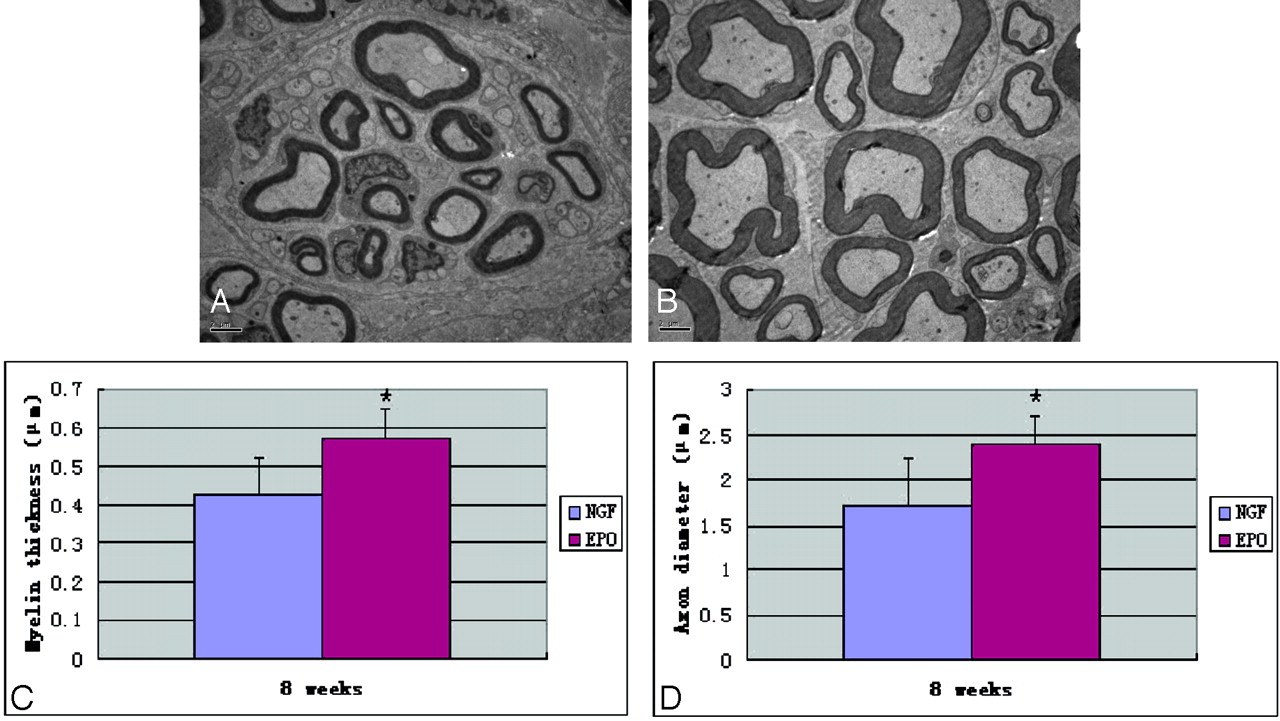
  • Fig 6.
    • Download figure
    • Open in new tab
    • Download powerpoint
    Fig 6.

    A and B, Representative ultrastructural imaging of cross-sections of nerve originally located 10-mm distal to the silicone rubber tube in the NGF and EPO groups respectively at week 8 after surgery. A, The NGF group shows lower myelin thickness and axonal diameter and fewer myelinated fibers. B, The EPO group shows higher myelin thickness and axonal diameter and more myelinated fibers. C and D, There are statistically significant differences in myelin thickness (C) and axonal diameter (D) between the 2 groups at 8 weeks after surgery. Data are the mean ± SEM. The asterisk indicates P < .05 compared with the NGF group significance.
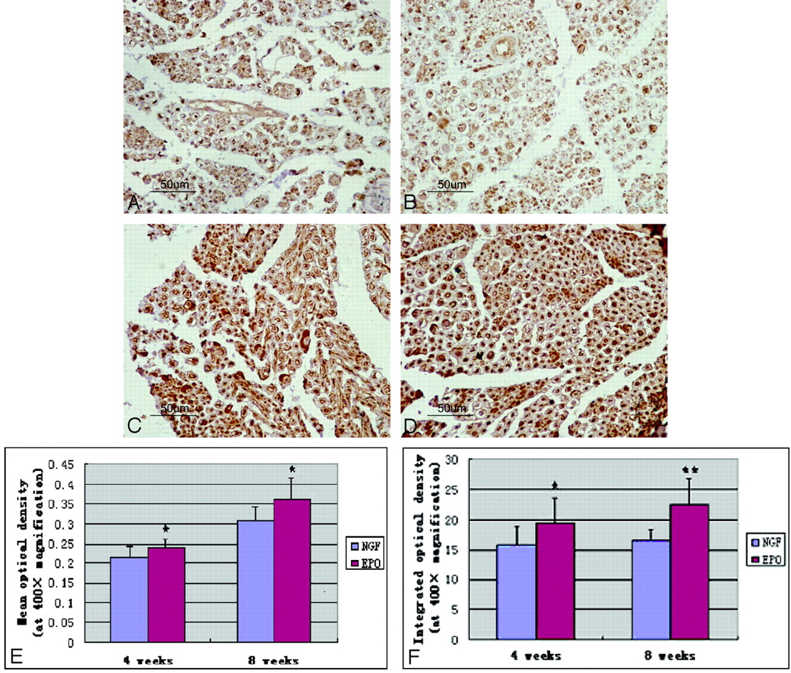
  • Fig 7.
    • Download figure
    • Open in new tab
    • Download powerpoint
    Fig 7.

    Representative photomicrographs of immunohistochemical staining of PGP 9.5 in regenerated sciatic nerve cross-sections. Axons and the periphery of the axons show strong immunoreactivity. A and B, Expression of PGP 9.5 in immunopositive nerve fibers at 4 weeks. C and D, Expression of PGP 9.5 at week 8. A−F, Quantitative analysis by using MOD and IOD reveals that nerves treated with EPO (B and D) have significantly higher MOD (E) and IOD (F) compared with those in the NGF group (A and C), and there are statistically significant differences between the 2 groups at the 2 time points. Data are the mean ± SEM. The asterisk indicates P < .05. Double asterisks indicate P < .01 compared with the NGF group significance.

Tables

  • Figures
    • View popup
    Table 1:

    Results of functional and electrophysiologic evaluation (mean ± SEM)

    4 Weeks8 Weeks
    SFIa
        NGF−79.98 ± 4.58−64.65 ± 4.11
        EPO−78.85 ± 3.87, P > .05−60.26 ± 2.91, P < .05
    MNCV (m/s)b
        NGF9.20 ± 1.0716.37 ± 3.40
        EPO10.60 ± 1.36, P < .0520.56 ± 4.18, P < .05
    • a The SFI studies at 8 weeks postsurgery demonstrated better improvement in nerve function in rats that received EPO.

    • b Rats treated with EPO showed better results than those of the NGF group at the 2 time points.

    • View popup
    Table 2:

    Results of histomorphometry and electron microscopy and immunohistochemistry (mean ± SEM)

    4 Weeks8 Weeks
    Myelin thickness (μm)a
        NGF0.43 ± 0.09
        EPO0.57 ± 0.08, P < .05
    Axon diameter (μm)a
        NGF1.73 ± 0.51
        EPO2.39 ± 0.32, P < .05
    Myelinated axon counts (at original magnification ×400)b
        NGF17.56 ± 4.1949.30 ± 9.56
        EPO22.90 ± 5.24, P < .0558.00 ± 7.73, P < .05
    MOD (at original magnification ×400)b
        NGF0.2173 ± 0.02570.3089 ± 0.0343
        EPO0.2419 ± 0.0204, P < .050.3611 ± 0.0547, P < .05
    IOD (at original magnification ×400)b
        NGF15.8494 ± 3.082516.6742 ± 1.7156
        EPO19.4382 ± 4.1861, P < .0522.4111 ± 4.3418, P < .01
    • a At 8 weeks after surgery, nerves treated with EPO had significantly larger myelin thickness and axon diameter compared with those in the NGF group, and there were statistically significant differences between the 2 groups.

    • b In addition, there were also statistically significant differences in MOD and IOD as well as myelinated axon counts between the 2 groups at the 2 time points.

PreviousNext
Back to top

In this issue

American Journal of Neuroradiology: 31 (3)
American Journal of Neuroradiology
Vol. 31, Issue 3
1 Mar 2010
  • Table of Contents
  • Index by author
Advertisement
Print
Download PDF
Email Article

Thank you for your interest in spreading the word on American Journal of Neuroradiology.

NOTE: We only request your email address so that the person you are recommending the page to knows that you wanted them to see it, and that it is not junk mail. We do not capture any email address.

Enter multiple addresses on separate lines or separate them with commas.
Erythropoietin Promotes Functional Recovery and Enhances Nerve Regeneration after Peripheral Nerve Injury in Rats
(Your Name) has sent you a message from American Journal of Neuroradiology
(Your Name) thought you would like to see the American Journal of Neuroradiology web site.
CAPTCHA
This question is for testing whether or not you are a human visitor and to prevent automated spam submissions.
Cite this article
Z.-S. Yin, H. Zhang, W. Gao
Erythropoietin Promotes Functional Recovery and Enhances Nerve Regeneration after Peripheral Nerve Injury in Rats
American Journal of Neuroradiology Mar 2010, 31 (3) 509-515; DOI: 10.3174/ajnr.A1820

Citation Manager Formats

  • BibTeX
  • Bookends
  • EasyBib
  • EndNote (tagged)
  • EndNote 8 (xml)
  • Medlars
  • Mendeley
  • Papers
  • RefWorks Tagged
  • Ref Manager
  • RIS
  • Zotero
0 Responses
Respond to this article
Share
Bookmark this article
Erythropoietin Promotes Functional Recovery and Enhances Nerve Regeneration after Peripheral Nerve Injury in Rats
Z.-S. Yin, H. Zhang, W. Gao
American Journal of Neuroradiology Mar 2010, 31 (3) 509-515; DOI: 10.3174/ajnr.A1820
del.icio.us logo Twitter logo Facebook logo Mendeley logo
  • Tweet Widget
  • Facebook Like
  • Google Plus One
Purchase

Jump to section

  • Article
    • Abstract
    • Abbreviations
    • Materials and Methods
    • Results
    • Discussion
    • Acknowledgments
    • Footnotes
    • References
  • Figures & Data
  • Info & Metrics
  • Responses
  • References
  • PDF

Related Articles

  • Erratum
  • PubMed
  • Google Scholar

Cited By...

  • Predictors of vibration perception threshold in type 2 diabetic patients with proliferative retinopathy
  • Crossref (56)
  • Google Scholar

This article has been cited by the following articles in journals that are participating in Crossref Cited-by Linking.

  • Erythropoietin as neuroprotective and neuroregenerative treatment strategy: Comprehensive overview of 12 years of preclinical and clinical research
    Derya Sargin, Heidi Friedrichs, Ahmed El-Kordi, Hannelore Ehrenreich
    Best Practice & Research Clinical Anaesthesiology 2010 24 4
  • Prevention and Treatment for Chemotherapy-Induced Peripheral Neuropathy: Therapies Based on CIPN Mechanisms
    Lang-Yue Hu, Wen-Li Mi, Gen-Cheng Wu, Yan-Qing Wang, Qi-Liang Mao-Ying
    Current Neuropharmacology 2019 17 2
  • Sciatic nerve regeneration by transplantation of Schwann cells via erythropoietin controlled‐releasing polylactic acid/multiwalled carbon nanotubes/gelatin nanofibrils neural guidance conduit
    Majid Salehi, Mahdi Naseri‐Nosar, Somayeh Ebrahimi‐Barough, Mohammdreza Nourani, Arash Khojasteh, Amir‐Ali Hamidieh, Amir Amani, Saeed Farzamfar, Jafar Ai
    Journal of Biomedical Materials Research Part B: Applied Biomaterials 2018 106 4
  • Alternative Erythropoietin Receptors in the Nervous System
    Daniela Ostrowski, Ralf Heinrich
    Journal of Clinical Medicine 2018 7 2
  • Erythropoietin and Nonhematopoietic Effects
    Alireza Nekoui, Gilbert Blaise
    The American Journal of the Medical Sciences 2017 353 1
  • Non-surgical therapies for peripheral nerve injury
    P. Martinez de Albornoz, P. J. Delgado, F. Forriol, N. Maffulli
    British Medical Bulletin 2011 100 1
  • Treatment and diagnosis of chemotherapy-induced peripheral neuropathy: An update
    Allison D. Desforges, Chance M. Hebert, Allyson L. Spence, Bailey Reid, Hemangini A. Dhaibar, Diana Cruz-Topete, Elyse M. Cornett, Alan David Kaye, Ivan Urits, Omar Viswanath
    Biomedicine & Pharmacotherapy 2022 147
  • Sprouting of corticospinal tract axons from the contralateral hemisphere into the denervated side of the spinal cord is associated with functional recovery in adult rat after traumatic brain injury and erythropoietin treatment
    Yanlu Zhang, Ye Xiong, Asim Mahmood, Yuling Meng, Zhongwu Liu, Changsheng Qu, Michael Chopp
    Brain Research 2010 1353
  • Repairing sciatic nerve injury with an EPO-loaded nerve conduit and sandwiched-in strategy of transplanting mesenchymal stem cells
    Wei Zhang, Lihai Zhang, Jianheng Liu, Licheng Zhang, Jian Zhang, Peifu Tang
    Biomaterials 2017 142
  • Erythropoietin promotes M2 macrophage phagocytosis of Schwann cells in peripheral nerve injury
    Prem Kumar Govindappa, John C. Elfar
    Cell Death & Disease 2022 13 3

More in this TOC Section

  • Bern Score Validity for SIH
  • MP2RAGE 7T in MS Lesions of the Cervical Spine
  • Deep Learning for STIR Spine MRI Quality
Show more Spine

Similar Articles

Advertisement

Indexed Content

  • Current Issue
  • Accepted Manuscripts
  • Article Preview
  • Past Issues
  • Editorials
  • Editors Choice
  • Fellow Journal Club
  • Letters to the Editor

Cases

  • Case Collection
  • Archive - Case of the Week
  • Archive - Case of the Month
  • Archive - Classic Case

Special Collections

  • Special Collections

Resources

  • News and Updates
  • Turn around Times
  • Submit a Manuscript
  • Author Policies
  • Manuscript Submission Guidelines
  • Evidence-Based Medicine Level Guide
  • Publishing Checklists
  • Graphical Abstract Preparation
  • Imaging Protocol Submission
  • Submit a Case
  • Become a Reviewer/Academy of Reviewers
  • Get Peer Review Credit from Publons

Multimedia

  • AJNR Podcast
  • AJNR SCANtastic
  • Video Articles

About Us

  • About AJNR
  • Editorial Board
  • Not an AJNR Subscriber? Join Now
  • Alerts
  • Feedback
  • Advertise with us
  • Librarian Resources
  • Permissions
  • Terms and Conditions

American Society of Neuroradiology

  • Not an ASNR Member? Join Now

© 2025 by the American Society of Neuroradiology All rights, including for text and data mining, AI training, and similar technologies, are reserved.
Print ISSN: 0195-6108 Online ISSN: 1936-959X

Powered by HighWire