Advertisement

Main menu

  • Home
  • Content
    • Current Issue
    • Accepted Manuscripts
    • Article Preview
    • Past Issue Archive
    • AJNR Case Collection
    • Case of the Week Archive
    • Classic Case Archive
    • Case of the Month Archive
  • Special Collections
    • Spinal CSF Leak Articles (Jan 2020-June 2024)
    • 2024 AJNR Journal Awards
    • Most Impactful AJNR Articles
  • Multimedia
    • AJNR Podcast
    • AJNR Scantastics
    • Video Articles
  • For Authors
    • Submit a Manuscript
    • Author Policies
    • Fast publishing of Accepted Manuscripts
    • Graphical Abstract Preparation
    • Manuscript Submission Guidelines
    • Imaging Protocol Submission
    • Submit a Case for the Case Collection
  • About Us
    • About AJNR
    • Editorial Board
  • More
    • Become a Reviewer/Academy of Reviewers
    • Subscribers
    • Permissions
    • Alerts
    • Feedback
    • Advertisers
    • ASNR Home
  • Other Publications
    • ajnr

User menu

  • Alerts
  • Log in

Search

  • Advanced search
American Journal of Neuroradiology

American Journal of Neuroradiology

ASHNR American Society of Functional Neuroradiology ASHNR American Society of Pediatric Neuroradiology ASSR
  • Alerts
  • Log in

Advanced Search

  • Home
  • Content
    • Current Issue
    • Accepted Manuscripts
    • Article Preview
    • Past Issue Archive
    • AJNR Case Collection
    • Case of the Week Archive
    • Classic Case Archive
    • Case of the Month Archive
  • Special Collections
    • Spinal CSF Leak Articles (Jan 2020-June 2024)
    • 2024 AJNR Journal Awards
    • Most Impactful AJNR Articles
  • Multimedia
    • AJNR Podcast
    • AJNR Scantastics
    • Video Articles
  • For Authors
    • Submit a Manuscript
    • Author Policies
    • Fast publishing of Accepted Manuscripts
    • Graphical Abstract Preparation
    • Manuscript Submission Guidelines
    • Imaging Protocol Submission
    • Submit a Case for the Case Collection
  • About Us
    • About AJNR
    • Editorial Board
  • More
    • Become a Reviewer/Academy of Reviewers
    • Subscribers
    • Permissions
    • Alerts
    • Feedback
    • Advertisers
    • ASNR Home
  • Follow AJNR on Twitter
  • Visit AJNR on Facebook
  • Follow AJNR on Instagram
  • Join AJNR on LinkedIn
  • RSS Feeds

Welcome to the new AJNR, Updated Hall of Fame, and more. Read the full announcements.


AJNR is seeking candidates for the position of Associate Section Editor, AJNR Case Collection. Read the full announcement.


ReplyLetter

Reply:

D. Dai and D.F. Kallmes
American Journal of Neuroradiology March 2010, 31 (3) E37; DOI: https://doi.org/10.3174/ajnr.A2019
D. Dai
  • Find this author on Google Scholar
  • Find this author on PubMed
  • Search for this author on this site
D.F. Kallmes
  • Find this author on Google Scholar
  • Find this author on PubMed
  • Search for this author on this site
  • Article
  • Figures & Data
  • Info & Metrics
  • Responses
  • References
  • PDF
Loading

We greatly appreciate the correspondence from Meng et al regarding our recent article about the basilar bifurcation in rabbits following carotid ligation.1 We remain absolutely confident that the focal excrescences along the basilar apex in our model represent branch arteries and not aneurysms. Dr. Meng and colleagues raised concern that de- and re-staining our histologic sections might have compromised the ability to identify the internal elastic lamellae. However, the article they cite regarding such loss of accuracy was for immunohistochemical techniques,2 not the histochemical staining, Verhoeff Elastic-Van Gieson (VVG), used in our study. We have re-reviewed all of our slides in detail. There are no aneurysms.

We are grateful to Dr. Meng and colleagues for providing new histologic images in their letter, different from those published previously.3 We note with interest that in Fig 1A in their letter, the artery denoted as the basilar artery is much much smaller than the P1 segment (the first segment of the posterior cerebral artery). From our experience, we have found that it is very easy to lose track during histologic processing of which branch is the basilar and which are the P1s; as such, simple rotation of the slide by 90° would place the bifurcation of the superior cerebellar artery/P1 at the “basilar tip.” Thus, the “aneurysm” noted in their Fig 1 looks similar to the origins of superior cerebellar arteries in a subject from our lab (Fig 1).

Fig 1.
  • Download figure
  • Open in new tab
  • Download powerpoint
Fig 1.

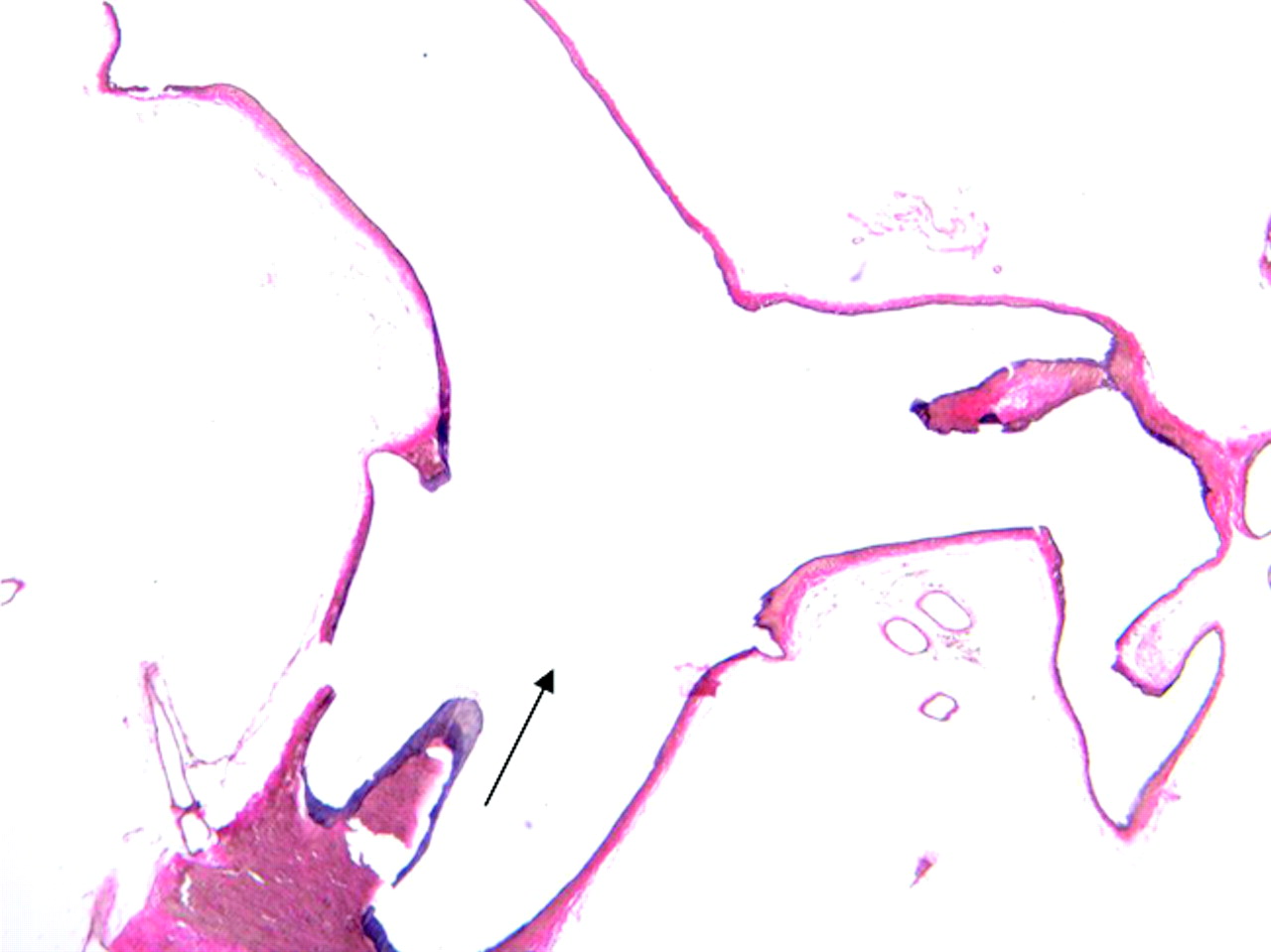
A subject at 8 weeks after right common carotid artery ligation in the elastase aneurysm model. Photomicrograph shows the basilar artery, superior cerebellar artery bifurcation, and the posterior cerebral artery at a low magnification. Arrow indicates the blood flow direction (VVG, original magnification ×40).

Given the disparate results between our study and their previous work, we speculated in our article that the apparent aneurysms noted by them might in fact have been branch arteries. Certainly there may be other potential explanations for different results between our study and theirs. In any event, because they identify aneurysms in 100% of cases, it should be relatively easy for them to confirm, by using complementary methods in addition to serial histologic sections, that they have induced aneurysms. These complementary and potentially confirmatory techniques could include angiographic or micro-CT imaging,4,5 or casting techniques such Microfil perfusion (MTS Medication Technologies, St. Petersburg, Florida).6–9 In addition, we would assume that the microaneurysms induced in their model might, with time, grow to become large and thus intuitively obvious to all observers, including us.

Perhaps the most efficient way to move forward in clarifying the apparent disparity in conclusions between our 2 research groups would be to provide the histologic data to an independent vascular pathologist. We would be delighted to supply all of our serial histologic slides to such an expert in hopes of advancing this important field.

References

  1. 1.↵
    1. Dai D,
    2. Ding YH,
    3. Kadirvel R,
    4. et al
    . Experience with microaneurysm formation at the basilar terminus in the rabbit elastase aneurysm model. AJNR Am J Neuroradiol 2010;31:300–03. Epub 2009 Oct 1
    Abstract/FREE Full Text
  2. 2.↵
    1. Dardik M,
    2. Epstein J
    . Efficacy of restaining prostate needle biopsies with high-molecular weight cytokeratin. Hum Pathol 2000;31:1155–61
    CrossRefPubMed
  3. 3.↵
    1. Gao L,
    2. Hoi Y,
    3. Swartz DD,
    4. et al
    . Nascent aneurysm formation at the basilar terminus induced by hemodynamics. Stroke 2008;39:2085–90
    Abstract/FREE Full Text
  4. 4.↵
    1. Dorr A,
    2. Sled JG,
    3. Kabani N
    . Three-dimensional cerebral vasculature of the CBA mouse brain: a magnetic resonance imaging and micro computed tomography study. Neuroimage 2007;35:1409–23. Epub 2007 Jan 23
    CrossRefPubMed
  5. 5.↵
    1. Schambach SJ,
    2. Bag S,
    3. Groden C,
    4. et al
    . Vascular imaging in small rodents using micro-CT. Methods 2010;50:26–35. Epub 2009 Sep 20
    CrossRefPubMed
  6. 6.↵
    1. Krucker T,
    2. Schuler A,
    3. Meyer EP,
    4. et al
    . Magnetic resonance angiography and vascular corrosion casting as tools in biomedical research: application to transgenic mice modeling Alzheimer's disease. Neurol Res 2004;26:507–16
    CrossRefPubMed
  7. 7.↵
    1. Heinzer S,
    2. Krucker T,
    3. Stampanoni M,
    4. et al
    . Hierarchical microimaging for multiscale analysis of large vascular networks. Neuroimage 2006;32:626–36
    CrossRefPubMed
  8. 8.↵
    1. Krucker T,
    2. Lang A,
    3. Meyer EP
    . New polyurethane-based material for vascular corrosion casting with improved physical and imaging characteristics. Microsc Res Tech 2006;69:138–47
    CrossRefPubMed
  9. 9.↵
    1. Cruise GM,
    2. Rivera EA,
    3. Jones RM,
    4. et al
    . A comparison of experimental aneurysm occlusion determination by angiography, scanning electron microscopy, MICROFIL perfusion, and histology. J Biomed Mater Res B Appl Biomater 2009;91:669–78
    PubMed
  • Copyright © American Society of Neuroradiology
PreviousNext
Back to top

In this issue

American Journal of Neuroradiology: 31 (3)
American Journal of Neuroradiology
Vol. 31, Issue 3
1 Mar 2010
  • Table of Contents
  • Index by author
Advertisement
Print
Download PDF
Email Article

Thank you for your interest in spreading the word on American Journal of Neuroradiology.

NOTE: We only request your email address so that the person you are recommending the page to knows that you wanted them to see it, and that it is not junk mail. We do not capture any email address.

Enter multiple addresses on separate lines or separate them with commas.
Reply:
(Your Name) has sent you a message from American Journal of Neuroradiology
(Your Name) thought you would like to see the American Journal of Neuroradiology web site.
CAPTCHA
This question is for testing whether or not you are a human visitor and to prevent automated spam submissions.
Cite this article
D. Dai, D.F. Kallmes
Reply:
American Journal of Neuroradiology Mar 2010, 31 (3) E37; DOI: 10.3174/ajnr.A2019

Citation Manager Formats

  • BibTeX
  • Bookends
  • EasyBib
  • EndNote (tagged)
  • EndNote 8 (xml)
  • Medlars
  • Mendeley
  • Papers
  • RefWorks Tagged
  • Ref Manager
  • RIS
  • Zotero
0 Responses
Respond to this article
Share
Bookmark this article
Reply:
D. Dai, D.F. Kallmes
American Journal of Neuroradiology Mar 2010, 31 (3) E37; DOI: 10.3174/ajnr.A2019
del.icio.us logo Twitter logo Facebook logo Mendeley logo
  • Tweet Widget
  • Facebook Like
  • Google Plus One
Purchase

Jump to section

  • Article
    • References
  • Figures & Data
  • Info & Metrics
  • Responses
  • References
  • PDF

Related Articles

  • No related articles found.
  • Google Scholar

Cited By...

  • No citing articles found.
  • Crossref (1)
  • Google Scholar

This article has been cited by the following articles in journals that are participating in Crossref Cited-by Linking.

  • Lack of aneurysm formation after carotid artery ligation in rabbits: a polymer MICROFIL® study
    Daying Dai, Yong Hong Ding, Ramanathan Kadirvel, Arash Ehteshami Rad, Debra A. Lewis, David F. Kallmes
    Neuroradiology 2013 55 1

More in this TOC Section

  • Brain AVM’s Nidus: What if We Hadn’t Understood Anything?
  • Letter to the Editor regarding “Automated Volumetric Software in Dementia: Help or Hindrance to the Neuroradiologist?”
  • Reply:
Show more Letters

Similar Articles

Advertisement

Indexed Content

  • Current Issue
  • Accepted Manuscripts
  • Article Preview
  • Past Issues
  • Editorials
  • Editors Choice
  • Fellow Journal Club
  • Letters to the Editor

Cases

  • Case Collection
  • Archive - Case of the Week
  • Archive - Case of the Month
  • Archive - Classic Case

Special Collections

  • Special Collections

Resources

  • News and Updates
  • Turn around Times
  • Submit a Manuscript
  • Author Policies
  • Manuscript Submission Guidelines
  • Evidence-Based Medicine Level Guide
  • Publishing Checklists
  • Graphical Abstract Preparation
  • Imaging Protocol Submission
  • Submit a Case
  • Become a Reviewer/Academy of Reviewers
  • Get Peer Review Credit from Publons

Multimedia

  • AJNR Podcast
  • AJNR SCANtastic
  • Video Articles

About Us

  • About AJNR
  • Editorial Board
  • Not an AJNR Subscriber? Join Now
  • Alerts
  • Feedback
  • Advertise with us
  • Librarian Resources
  • Permissions
  • Terms and Conditions

American Society of Neuroradiology

  • Not an ASNR Member? Join Now

© 2025 by the American Society of Neuroradiology All rights, including for text and data mining, AI training, and similar technologies, are reserved.
Print ISSN: 0195-6108 Online ISSN: 1936-959X

Powered by HighWire