Advertisement

Main menu

  • Home
  • Content
    • Current Issue
    • Accepted Manuscripts
    • Article Preview
    • Past Issue Archive
    • AJNR Case Collection
    • Case of the Week Archive
    • Classic Case Archive
    • Case of the Month Archive
  • Special Collections
    • Spinal CSF Leak Articles (Jan 2020-June 2024)
    • 2024 AJNR Journal Awards
    • Most Impactful AJNR Articles
  • Multimedia
    • AJNR Podcast
    • AJNR Scantastics
    • Video Articles
  • For Authors
    • Submit a Manuscript
    • Author Policies
    • Fast publishing of Accepted Manuscripts
    • Graphical Abstract Preparation
    • Manuscript Submission Guidelines
    • Imaging Protocol Submission
    • Submit a Case for the Case Collection
  • About Us
    • About AJNR
    • Editorial Board
  • More
    • Become a Reviewer/Academy of Reviewers
    • Subscribers
    • Permissions
    • Alerts
    • Feedback
    • Advertisers
    • ASNR Home
  • Other Publications
    • ajnr

User menu

  • Alerts
  • Log in

Search

  • Advanced search
American Journal of Neuroradiology

American Journal of Neuroradiology

ASHNR American Society of Functional Neuroradiology ASHNR American Society of Pediatric Neuroradiology ASSR
  • Alerts
  • Log in

Advanced Search

  • Home
  • Content
    • Current Issue
    • Accepted Manuscripts
    • Article Preview
    • Past Issue Archive
    • AJNR Case Collection
    • Case of the Week Archive
    • Classic Case Archive
    • Case of the Month Archive
  • Special Collections
    • Spinal CSF Leak Articles (Jan 2020-June 2024)
    • 2024 AJNR Journal Awards
    • Most Impactful AJNR Articles
  • Multimedia
    • AJNR Podcast
    • AJNR Scantastics
    • Video Articles
  • For Authors
    • Submit a Manuscript
    • Author Policies
    • Fast publishing of Accepted Manuscripts
    • Graphical Abstract Preparation
    • Manuscript Submission Guidelines
    • Imaging Protocol Submission
    • Submit a Case for the Case Collection
  • About Us
    • About AJNR
    • Editorial Board
  • More
    • Become a Reviewer/Academy of Reviewers
    • Subscribers
    • Permissions
    • Alerts
    • Feedback
    • Advertisers
    • ASNR Home
  • Follow AJNR on Twitter
  • Visit AJNR on Facebook
  • Follow AJNR on Instagram
  • Join AJNR on LinkedIn
  • RSS Feeds

Welcome to the new AJNR, Updated Hall of Fame, and more. Read the full announcements.


AJNR is seeking candidates for the position of Associate Section Editor, AJNR Case Collection. Read the full announcement.


LetterLetter

A Meningioma Exclusively Located inside the Superior Sagittal Sinus Responsible for Intracranial Hypertension

B. Szitkar
American Journal of Neuroradiology June 2010, 31 (6) E57-E58; DOI: https://doi.org/10.3174/ajnr.A2130
B. Szitkar
  • Find this author on Google Scholar
  • Find this author on PubMed
  • Search for this author on this site
  • Article
  • Figures & Data
  • Info & Metrics
  • Responses
  • References
  • PDF
Loading

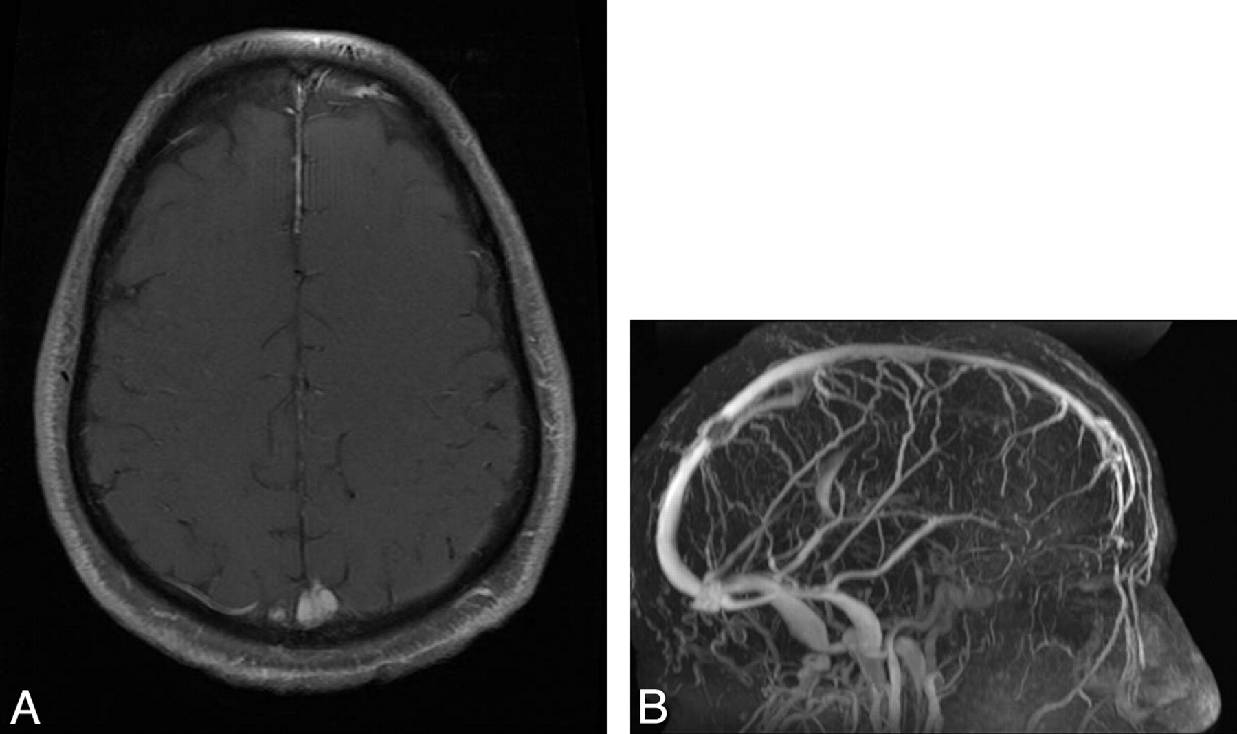
We report a case of a 57-year-old patient with a history of intracranial hypertension (visual impairment, headaches, and papillary edema) due to a meningioma exclusively located inside the superior sagittal sinus (SSS). The lesion was not detected on a nonenhanced brain CT. MR imaging revealed a centimetric round homogeneous lesion, hyperintense on T2-weighted images, intensely and homogeneously enhanced after gadolinium injection on T1-weighted sequences with fat suppression (Fig 1A), inside the distal part of the SSS. On venous MR angiography, the lesion appeared as a filling defect inside the SSS (Fig 1B). The tumor did not grow (1-year interval between the 2 MR imaging examinations), and its characteristics on MR imaging led to the diagnosis of stage I meningioma.

Fig 1.
  • Download figure
  • Open in new tab
  • Download powerpoint
Fig 1.

A, The T1-weighted sequence after gadolinium injection and fat suppression reveals an endovascular mass, intensely and homogeneously enhanced. The tumor's characteristics on all the sequences are consistent with meningioma. B, The MR venography shows a filling defect inside the SSS corresponding to an endoluminal tumor.

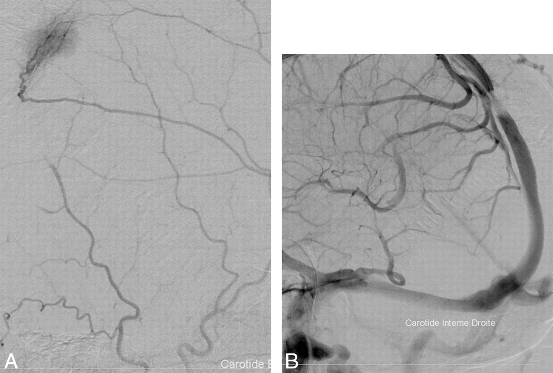
We performed an angiographic examination: The arterial phase revealed a tumor blush originating from the left middle cerebral artery, highly evocative of the hypervascularization of a meningioma (Fig 2A), and the venous phase showed an SSS stenosis correlating with the tumor blush (Fig 2B). The patient was treated with acetazolamide (Diamox), but after a year, headaches worsened and an angiographic treatment was required. A stent was inserted through a catheter directed into the SSS from a percutaneous jugular puncture. The patient was heparinized during the procedure and then given aspirin for 6 months. Clinical relief was achieved within 4 months. No histologic proof could be obtained, but the patient was referred to another hospital for stereotactic radiosurgery to treat the meningioma.

Fig 2.
  • Download figure
  • Open in new tab
  • Download powerpoint
Fig 2.

A, Angiographic exam: the arterial phase reveals a tumor blush typical of meningiomas' hypervascularization. The arterioles come from the left middle cerebral artery. B, The venous phase shows a SSS stenosis.

Meningiomas account for 13%–26% of all intracranial tumors and represent the most common nonglial primary tumors of the brain.1 The incidence is approximately 6 per 100,000 and is higher among the elderly. They are composed of neoplastic meningothelial cells originating from the arachnoid layer of the meninges. Atypical development sites are the ventricular system, the orbit, the scalp, the paranasal sinus, the nasopharynx, the neck, and the skin. In our patient, the lesion was exclusively located inside the SSS. To our knowledge, no such location has been reported so far, though intravenous extension of intracranial meningiomas occurs in approximately 20% of cases.2

There are several therapeutic options for treating meningiomas: Surgery can be used in 80% of cases.3 Radiation therapy is used in case of tumoral progression or incomplete resection, and radiosurgery has become the treatment of choice for lesions <3 cm and/or inaccessible to surgery.

The close correlation between CSF pressure and venous pressure in the SSS explains how a venous sinus obstruction causes intracranial hypertension. It was proved by simultaneous CSF and venous manometry4 that a venous pressure rise led to a CSF absorption dysfunction and, consequently, higher CSF volume and pressure. Benign intracranial hypertension can be classified into 2 categories depending on the underlying etiology4: the idiopathic type (also called pseudotumor cerebri) and the venogenic type when there is a primary pathologic condition of the venous sinus (thrombosis, vasculitis, extrinsic benign compression, or intraluminal tumor as in our patient).

Venous stent placement is a safe and effective procedure to relieve dural sinus obstructions.4 Full-dose heparin should be administered for 24–48 hours after the procedure; then, the heparin should be replaced by an antiplatelet agent until the stent is coated by the vascular epithelium (6 months).

References

  1. 1.↵
    1. O'Leary S,
    2. Adams WM,
    3. Parrish RW,
    4. et al
    . Atypical imaging appearances of intracranial meningiomas. Clin Radiol 2007;62:10–17
    CrossRefPubMed
  2. 2.↵
    1. Vachhrajani S,
    2. Jea A,
    3. Rutka JA,
    4. et al
    . Meningioma with dural venous sinus invasion and jugular vein extension. J Neurosurg Pediatr 2008;2:391–96
    CrossRefPubMed
  3. 3.↵
    1. Marosi C,
    2. Hassler M,
    3. Roessler K,
    4. et al
    . Meningioma. Crit Rev Oncol Hematol 2008;67:153–71
    CrossRefPubMed
  4. 4.↵
    1. Owler B,
    2. Parker G,
    3. Halmagyi GM,
    4. et al
    . Pseudotumor cerebri syndrome: venous sinus obstruction and its treatment with stent placement. J Neurosurg 2003;98:1045–55
    PubMed
  • Copyright © American Society of Neuroradiology
PreviousNext
Back to top

In this issue

American Journal of Neuroradiology: 31 (6)
American Journal of Neuroradiology
Vol. 31, Issue 6
1 Jun 2010
  • Table of Contents
  • Index by author
Advertisement
Print
Download PDF
Email Article

Thank you for your interest in spreading the word on American Journal of Neuroradiology.

NOTE: We only request your email address so that the person you are recommending the page to knows that you wanted them to see it, and that it is not junk mail. We do not capture any email address.

Enter multiple addresses on separate lines or separate them with commas.
A Meningioma Exclusively Located inside the Superior Sagittal Sinus Responsible for Intracranial Hypertension
(Your Name) has sent you a message from American Journal of Neuroradiology
(Your Name) thought you would like to see the American Journal of Neuroradiology web site.
CAPTCHA
This question is for testing whether or not you are a human visitor and to prevent automated spam submissions.
Cite this article
B. Szitkar
A Meningioma Exclusively Located inside the Superior Sagittal Sinus Responsible for Intracranial Hypertension
American Journal of Neuroradiology Jun 2010, 31 (6) E57-E58; DOI: 10.3174/ajnr.A2130

Citation Manager Formats

  • BibTeX
  • Bookends
  • EasyBib
  • EndNote (tagged)
  • EndNote 8 (xml)
  • Medlars
  • Mendeley
  • Papers
  • RefWorks Tagged
  • Ref Manager
  • RIS
  • Zotero
0 Responses
Respond to this article
Share
Bookmark this article
A Meningioma Exclusively Located inside the Superior Sagittal Sinus Responsible for Intracranial Hypertension
B. Szitkar
American Journal of Neuroradiology Jun 2010, 31 (6) E57-E58; DOI: 10.3174/ajnr.A2130
del.icio.us logo Twitter logo Facebook logo Mendeley logo
  • Tweet Widget
  • Facebook Like
  • Google Plus One
Purchase

Jump to section

  • Article
    • References
  • Figures & Data
  • Info & Metrics
  • Responses
  • References
  • PDF

Related Articles

  • No related articles found.
  • PubMed
  • Google Scholar

Cited By...

  • Pseudotumor Cerebri: Brief Review of Clinical Syndrome and Imaging Findings
  • Crossref (11)
  • Google Scholar

This article has been cited by the following articles in journals that are participating in Crossref Cited-by Linking.

  • Pseudotumor Cerebri: Brief Review of Clinical Syndrome and Imaging Findings
    A.J. Degnan, L.M. Levy
    American Journal of Neuroradiology 2011 32 11
  • Consensus recommendations for current treatments and accelerating clinical trials for patients with neurofibromatosis type 2
    Jaishri O. Blakeley, D. Gareth Evans, John Adler, Derald Brackmann, Ruihong Chen, Rosalie E. Ferner, C. Oliver Hanemann, Gordon Harris, Susan M. Huson, Abraham Jacob, Michel Kalamarides, Matthias A. Karajannis, Bruce R. Korf, Victor‐Felix Mautner, Andrea I. McClatchey, Harry Miao, Scott R. Plotkin, William Slattery, Anat O. Stemmer‐Rachamimov, D. Bradley Welling, Patrick Y. Wen, Brigitte Widemann, Kim Hunter‐Schaedle, Marco Giovannini
    American Journal of Medical Genetics Part A 2012 158A 1
  • Changing the paradigm of intracranial hypertension in brain tumor patients: a study based on non-invasive ICP measurements
    Jenny C. Kienzler, Rolandas Zakelis, Serge Marbacher, Sabrina Bäbler, Lucia Schwyzer, Elke Remonda, Javier Fandino
    BMC Neurology 2020 20 1
  • Acquired Sinus Pericranii Due To Parasagittal Meningiomatous Invasion of the Superior Sagittal Sinus and Bilateral Transverse Sinuses
    Krishna Amuluru, Fawaz Al-Mufti, Manan Shah, Charles J. Prestigiacomo, James K. Liu, Chirag D. Gandhi
    World Neurosurgery 2017 98
  • Cause or Consequence
    Sevim Türay, Nimet Kabakuş, Fatma Hanci, Aslihan Tunçlar, Mustafa Hizal
    The Neurologist 2019 24 5
  • Venous hypertension caused by a meningioma involving the sigmoid sinus: case report
    Koichiro Sumi, Naoki Otani, Fumi Mori, Shun Yamamuro, Hideki Oshima, Atsuo Yoshino
    BMC Neurology 2021 21 1
  • Stereotactic radiosurgery used to manage a meningioma filling the posterior two-thirds of the superior sagittal sinus
    Christopher P. Deibert, Douglas Kondziolka
    Journal of Neurosurgery 2013 119 5
  • Endovascular stenting for cerebral venous sinus stenosis secondary to meningioma invasion
    Gasim Ahmed, Mahmoud Abou-foul, William Sage, Norman McConachie
    Neuroradiology 2024 66 5
  • Intracranial Hypertension Secondary to Venous Sinus Stenosis by Meningioma: A Case Illustration With Literature Review, Tips for Diagnosis and Management
    Sadegh Bagherzadeh, Milad Shafizadeh, Leila Bahari, Hamidreza Golestaneh
    Clinical Case Reports 2025 13 4
  • Letter: Advances in the Understanding of Meningiomas Involving the Superior Sagittal Sinuses
    Tengkun Yin, Kai Lin, Jiyue Wang, Liyong Zhang, Shousen Wang
    Neurosurgery 2020 87 1

More in this TOC Section

  • Letter to the Editor regarding “Automated Volumetric Software in Dementia: Help or Hindrance to the Neuroradiologist?”
  • Reply:
  • Brain AVM’s Nidus: What if We Hadn’t Understood Anything?
Show more Letters

Similar Articles

Advertisement

Indexed Content

  • Current Issue
  • Accepted Manuscripts
  • Article Preview
  • Past Issues
  • Editorials
  • Editors Choice
  • Fellow Journal Club
  • Letters to the Editor

Cases

  • Case Collection
  • Archive - Case of the Week
  • Archive - Case of the Month
  • Archive - Classic Case

Special Collections

  • Special Collections

Resources

  • News and Updates
  • Turn around Times
  • Submit a Manuscript
  • Author Policies
  • Manuscript Submission Guidelines
  • Evidence-Based Medicine Level Guide
  • Publishing Checklists
  • Graphical Abstract Preparation
  • Imaging Protocol Submission
  • Submit a Case
  • Become a Reviewer/Academy of Reviewers
  • Get Peer Review Credit from Publons

Multimedia

  • AJNR Podcast
  • AJNR SCANtastic
  • Video Articles

About Us

  • About AJNR
  • Editorial Board
  • Not an AJNR Subscriber? Join Now
  • Alerts
  • Feedback
  • Advertise with us
  • Librarian Resources
  • Permissions
  • Terms and Conditions

American Society of Neuroradiology

  • Not an ASNR Member? Join Now

© 2025 by the American Society of Neuroradiology All rights, including for text and data mining, AI training, and similar technologies, are reserved.
Print ISSN: 0195-6108 Online ISSN: 1936-959X

Powered by HighWire