Advertisement

Main menu

  • Home
  • Content
    • Current Issue
    • Accepted Manuscripts
    • Article Preview
    • Past Issue Archive
    • AJNR Case Collection
    • Case of the Week Archive
    • Classic Case Archive
    • Case of the Month Archive
  • Special Collections
    • Spinal CSF Leak Articles (Jan 2020-June 2024)
    • 2024 AJNR Journal Awards
    • Most Impactful AJNR Articles
  • Multimedia
    • AJNR Podcast
    • AJNR Scantastics
    • Video Articles
  • For Authors
    • Submit a Manuscript
    • Author Policies
    • Fast publishing of Accepted Manuscripts
    • Graphical Abstract Preparation
    • Manuscript Submission Guidelines
    • Imaging Protocol Submission
    • Submit a Case for the Case Collection
  • About Us
    • About AJNR
    • Editorial Board
  • More
    • Become a Reviewer/Academy of Reviewers
    • Subscribers
    • Permissions
    • Alerts
    • Feedback
    • Advertisers
    • ASNR Home
  • Other Publications
    • ajnr

User menu

  • Alerts
  • Log in

Search

  • Advanced search
American Journal of Neuroradiology

American Journal of Neuroradiology

ASHNR American Society of Functional Neuroradiology ASHNR American Society of Pediatric Neuroradiology ASSR
  • Alerts
  • Log in

Advanced Search

  • Home
  • Content
    • Current Issue
    • Accepted Manuscripts
    • Article Preview
    • Past Issue Archive
    • AJNR Case Collection
    • Case of the Week Archive
    • Classic Case Archive
    • Case of the Month Archive
  • Special Collections
    • Spinal CSF Leak Articles (Jan 2020-June 2024)
    • 2024 AJNR Journal Awards
    • Most Impactful AJNR Articles
  • Multimedia
    • AJNR Podcast
    • AJNR Scantastics
    • Video Articles
  • For Authors
    • Submit a Manuscript
    • Author Policies
    • Fast publishing of Accepted Manuscripts
    • Graphical Abstract Preparation
    • Manuscript Submission Guidelines
    • Imaging Protocol Submission
    • Submit a Case for the Case Collection
  • About Us
    • About AJNR
    • Editorial Board
  • More
    • Become a Reviewer/Academy of Reviewers
    • Subscribers
    • Permissions
    • Alerts
    • Feedback
    • Advertisers
    • ASNR Home
  • Follow AJNR on Twitter
  • Visit AJNR on Facebook
  • Follow AJNR on Instagram
  • Join AJNR on LinkedIn
  • RSS Feeds

Welcome to the new AJNR, Updated Hall of Fame, and more. Read the full announcements.


AJNR is seeking candidates for the position of Associate Section Editor, AJNR Case Collection. Read the full announcement.


LetterLetter

MR Imaging Findings in Intracranial Dural Arteriovenous Fistula Shunt with Retrograde Cortical Venous Drainage Using Susceptibility-Weighted Angiography

J. Hodel, S. Gerber, M. Zins, M. Rodallec, X. Leclerc, R. Blanc and A. Rahmouni
American Journal of Neuroradiology November 2011, 32 (10) E196-E197; DOI: https://doi.org/10.3174/ajnr.A2875
J. Hodel
  • Find this author on Google Scholar
  • Find this author on PubMed
  • Search for this author on this site
S. Gerber
  • Find this author on Google Scholar
  • Find this author on PubMed
  • Search for this author on this site
M. Zins
  • Find this author on Google Scholar
  • Find this author on PubMed
  • Search for this author on this site
M. Rodallec
  • Find this author on Google Scholar
  • Find this author on PubMed
  • Search for this author on this site
X. Leclerc
  • Find this author on Google Scholar
  • Find this author on PubMed
  • Search for this author on this site
R. Blanc
  • Find this author on Google Scholar
  • Find this author on PubMed
  • Search for this author on this site
A. Rahmouni
  • Find this author on Google Scholar
  • Find this author on PubMed
  • Search for this author on this site
  • Article
  • Figures & Data
  • Info & Metrics
  • Responses
  • References
  • PDF
Loading

We read with special interest the article of Noguchi et al entitled “Intracranial Dural Arteriovenous Fistula with Retrograde Cortical Venous Drainage: Use of Susceptibility-Weighted Imaging in Combination with Dynamic Susceptibility Contrast Imaging.”1 The authors reported the use of SWI in combination with DSC for the imaging of related retrograde cortical venous drainage (RCVD) in patients with intracranial dural arteriovenous fistula (DAVF). They suggested that SWI in combination with DSC could be used to characterize the presence of RCVD in patients with DAVFs showing dilated cerebral veins on the surface of the brain. In this article, the RCVD was imaged by using minimal intensity projection (mIP) views.

Susceptibility-weighted angiography (SWAN) is a new 3D T2*-based gradient-echo sequence generating several echoes that are read out at different TE times.2 During each TR, the SWAN sequence captures multiple TE readouts: short TEs provide a time-of-flight (TOF) effect, whereas long TEs are responsible for the magnetic-susceptibility effect. Indeed, the SWAN sequence allows high-resolution visualization of both cerebral veins and arteries. By use of either mIP or maximum intensity projection (MIP) reconstructions, the hyperintense arteries can be distinguished from the hypointense veins.2 We report 2 cases of intracranial DAVFs with RCVD explored at 3T (Discovery MR 750; GE Healthcare, Milwaukee, Wisconsin) by using the SWAN sequence. The first case is a 32-year-old man with a right parietal DAVF supplied by the right middle meningeal artery (patient 1, Cognard classification type III). With the SWAN sequence, the RCVD is clearly identified with dilated parietal veins that appeared hyperintense (Fig 1). A 4D dynamic MR angiography (Time-Resolved Imaging of Contrast Kinetics [TRICKS]) showed the DAVF and the related RCVD with an abnormal early arterial filling of the parietal veins, which appeared bright on the SWAN images (Fig 1). The second case is a 25-year-old man with a right petrous DAVF supplied by the right middle meningeal artery with RCVD and venous ectasia (patient 2, Cognard classification type IV). With the SWAN sequence, both the RCVD and the venous ectasia appeared hyperintense (Fig 2). In this patient, a TOF sequence revealed a marked TOF effect through the venous ectasia related to an increased blood flow (Fig 2).

Fig 1.
  • Download figure
  • Open in new tab
  • Download powerpoint
Fig 1.

A, A 32-year-old man with a right parietal DAVF supplied by the right middle meningeal artery with venous drainage directly into the cortical veins (Cognard classification type III). On the native axial image of the SWAN sequence (A), brain arteries appeared bright (open arrows), whereas normal cerebral vein signal intensity was decreased (arrowheads). The SWAN sequence also revealed abnormal hyperintense parietal veins (arrows). B, A 32-year-old man with a right parietal DAVF supplied by the right middle meningeal artery with venous drainage directly into cortical veins (Cognard classification type III). With an oblique MIP reformation (B), this retrograde cortical venous drainage was easily detected. C, A 32-year-old man with a right parietal DAVF supplied by the right middle meningeal artery with venous drainage directly into the cortical veins (Cognard classification type III). The Sagittal MIP view of the 4D dynamic MR angiography (C) confirmed the early arterial phase filling of the abnormal parietal veins (arrows).

Fig 2.
  • Download figure
  • Open in new tab
  • Download powerpoint
Fig 2.

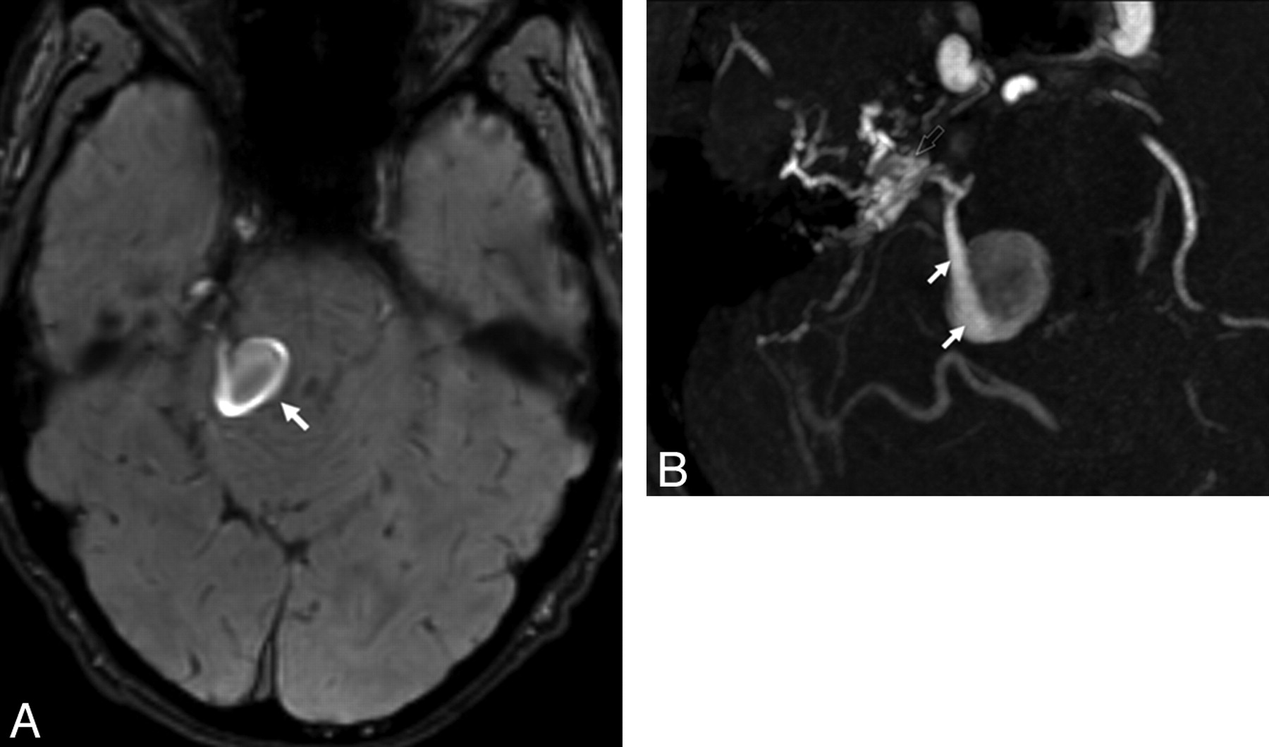
A, A 25-year-old man with a right petrous DAVF supplied by the right middle meningeal artery with retrograde cortical venous drainage and venous ectasia (Cognard classification type IV). On the native axial image of the SWAN sequence (A), the venous ectasia related to the right petrous DAVF appeared in hypersignal (arrow). B, A 25-year-old man with a right petrous DAVF supplied by the right middle meningeal artery with retrograde cortical venous drainage and venous ectasia (Cognard classification type IV). With the oblique MIP reformation of a TOF sequence (B), the petrous DAVF was visible (open arrow) as well as the venous ectasia (arrow). Note the TOF-related increased signal intensity due to the increased blood flow through the venous ectasia.

In the study of Noguchi et al,1 typical RCVD appeared in hyposignal on mIP reformations except for 1 patient with a type III DAVF. In this patient, mIP reformations of the SWI sequence failed to detect nondeoxygenated RCVD because in such cases, the arterial blood drained directly into the cortical veins. The typical hypointense pattern of RCVD described by Noguchi et al could be explained by the absence of TOF effect by using the SWI sequence. Indeed, the SWAN sequence differs from the SWI because of the short TEs, which provide a TOF effect within the images.

Our cases suggested that the venous drainage appeared hyperintense by using the SWAN sequence in either type III or IV DAVF and could be easily identified with MIP reconstructions. Moreover, considering the results obtained with SWI and SWAN sequences, we could speculate that the hyperintensity of the RCVD observed in our patients may be more related to a TOF effect than the presence of nondeoxygenated blood within the veins. Our findings indicate the potential value of the SWAN sequence for the diagnosis of intracranial DAVF. Indeed, the visualization of a venous hyperintensity on SWAN images may suggest a related intracranial arteriovenous shunt. Further studies are required to confirm this hypothesis, particularly concerning a DAVF with small and/or very slow-flowing venous drainage.

References

  1. 1.↵
    1. Noguchi K,
    2. Kuwayama N,
    3. Kubo M,
    4. et al
    . Intracranial dural arteriovenous fistula with retrograde cortical venous drainage: use of susceptibility-weighted imaging in combination with dynamic susceptibility contrast imaging. AJNR Am J Neuroradiol 2010;31:1903–10
    Abstract/FREE Full Text
  2. 2.↵
    1. Boeckh-Behrens T,
    2. Lutz J,
    3. Lummel N,
    4. et al
    . Susceptibility-weighted angiography (SWAN) of cerebral veins and arteries compared to TOF-MRA. Eur J Radiol 2011 Apr 3. [Epub ahead of print]
  • © 2011 by American Journal of Neuroradiology
PreviousNext
Back to top

In this issue

American Journal of Neuroradiology: 32 (10)
American Journal of Neuroradiology
Vol. 32, Issue 10
1 Nov 2011
  • Table of Contents
  • Index by author
Advertisement
Print
Download PDF
Email Article

Thank you for your interest in spreading the word on American Journal of Neuroradiology.

NOTE: We only request your email address so that the person you are recommending the page to knows that you wanted them to see it, and that it is not junk mail. We do not capture any email address.

Enter multiple addresses on separate lines or separate them with commas.
MR Imaging Findings in Intracranial Dural Arteriovenous Fistula Shunt with Retrograde Cortical Venous Drainage Using Susceptibility-Weighted Angiography
(Your Name) has sent you a message from American Journal of Neuroradiology
(Your Name) thought you would like to see the American Journal of Neuroradiology web site.
CAPTCHA
This question is for testing whether or not you are a human visitor and to prevent automated spam submissions.
Cite this article
J. Hodel, S. Gerber, M. Zins, M. Rodallec, X. Leclerc, R. Blanc, A. Rahmouni
MR Imaging Findings in Intracranial Dural Arteriovenous Fistula Shunt with Retrograde Cortical Venous Drainage Using Susceptibility-Weighted Angiography
American Journal of Neuroradiology Nov 2011, 32 (10) E196-E197; DOI: 10.3174/ajnr.A2875

Citation Manager Formats

  • BibTeX
  • Bookends
  • EasyBib
  • EndNote (tagged)
  • EndNote 8 (xml)
  • Medlars
  • Mendeley
  • Papers
  • RefWorks Tagged
  • Ref Manager
  • RIS
  • Zotero
0 Responses
Respond to this article
Share
Bookmark this article
MR Imaging Findings in Intracranial Dural Arteriovenous Fistula Shunt with Retrograde Cortical Venous Drainage Using Susceptibility-Weighted Angiography
J. Hodel, S. Gerber, M. Zins, M. Rodallec, X. Leclerc, R. Blanc, A. Rahmouni
American Journal of Neuroradiology Nov 2011, 32 (10) E196-E197; DOI: 10.3174/ajnr.A2875
del.icio.us logo Twitter logo Facebook logo Mendeley logo
  • Tweet Widget
  • Facebook Like
  • Google Plus One
Purchase

Jump to section

  • Article
    • References
  • Figures & Data
  • Info & Metrics
  • Responses
  • References
  • PDF

Related Articles

  • No related articles found.
  • PubMed
  • Google Scholar

Cited By...

  • The application of susceptibility-weighted MRI in pre-interventional evaluation of intracranial dural arteriovenous fistulas
  • Crossref (11)
  • Google Scholar

This article has been cited by the following articles in journals that are participating in Crossref Cited-by Linking.

  • Séquences IRM « SWAN, SWI et VenoBOLD » exploitant le phénomène de susceptibilité magnétique : principes techniques et applications cliniques
    J. Hodel, M. Rodallec, S. Gerber, R. Blanc, A. Maraval, S. Caron, L. Tyvaert, M. Zuber, M. Zins
    Journal of Neuroradiology 2012 39 2
  • MR Selective Flow-Tracking Cartography: A Postprocessing Procedure Applied to Four-dimensional Flow MR Imaging for Complete Characterization of Cranial Dural Arteriovenous Fistulas
    Myriam Edjlali, Pauline Roca, Cécile Rabrait, Denis Trystram, Christine Rodriguez-Régent, Kevin M. Johnson, Oliver Wieben, Patrick Turski, Jean-François Meder, Olivier Naggara, Catherine Oppenheim
    Radiology 2014 270 1
  • Susceptibility-weighted angiography for the detection of high-flow intracranial vascular lesions: preliminary study
    Jérôme Hodel, Raphaël Blanc, Mathieu Rodallec, Antoine Guillonnet, Sophie Gerber, Silvia Pistocchi, Rémi Sitta, Cécile Rabrait, Mathieu Zuber, Jean-Pierre Pruvo, Marc Zins, Xavier Leclerc
    European Radiology 2013 23 4
  • The application of susceptibility-weighted MRI in pre-interventional evaluation of intracranial dural arteriovenous fistulas
    Narendra Kumar Jain, Santhosh Kumar Kannath, Tirur Raman Kapilamoorthy, Bejoy Thomas
    Journal of NeuroInterventional Surgery 2017 9 5
  • Intracranial Dural Arteriovenous Fistula: Preliminary Report of Arterial Spin Labeling
    Kyo Noguchi, Hideto Kawabe, Hikaru Seto
    Open Journal of Medical Imaging 2013 03 01
  • ACCURACY OF SUSCEPTIBILITY WEIGHTED IMAGING AND 3D-TOF MRA IN DETECTION AND PRE-TREATMENT EVALUATION OF INTRACRANIAL DURAL ARTERIOVENOUS FISTULAS
    Minth Punpichet, Poonperm Sucharitpong, Norraseth Kittinorraseth, Sujima Tangjintanakan, Supakajee Saengruang-Orn
    Journal of Southeast Asian Medical Research 2024 8
  • Evaluation of bleeding risk in dural arteriovenous fistula using susceptibility-weighted imaging: A case report
    Satoshi Koizumi, Gakushi Yoshikawa, Masaaki Shojima, Yosuke Kitagawa, So Fujimoto, Atsushi Okano, Yuta Fukushima, Shikou Shimada, Kazuo Tsutsumi
    Interdisciplinary Neurosurgery 2014 1 4
  • Malformations artérioveineuses et fistules durales artérioveineuses intracrâniennes en réanimation
    S. Lenck, C. Damoisel, A. -L. Bernat, D. Bresson, M. -A. Labeyrie, D. Payen, S. Froelich, E. Houdart
    Réanimation 2015 24 5
  • The “Hypointense Focal Brain” on susceptibility-weighted imaging as a sign of venous congestion in cranial dural arteriovenous fistulas
    Zhao Hui Chen Zhou, Amaya Hilario, Elena Salvador Álvarez, Agustín María Cárdenas del Carre, Juan Romero Coronado, Carmen Lechuga Vázquez, Ana Martínez de Aragón, Ana Ramos González
    The Neuroradiology Journal 2025 38 1

More in this TOC Section

  • Brain AVM’s Nidus: What if We Hadn’t Understood Anything?
  • Letter to the Editor regarding “Automated Volumetric Software in Dementia: Help or Hindrance to the Neuroradiologist?”
  • Reply:
Show more Letters

Similar Articles

Advertisement

Indexed Content

  • Current Issue
  • Accepted Manuscripts
  • Article Preview
  • Past Issues
  • Editorials
  • Editors Choice
  • Fellow Journal Club
  • Letters to the Editor

Cases

  • Case Collection
  • Archive - Case of the Week
  • Archive - Case of the Month
  • Archive - Classic Case

Special Collections

  • Special Collections

Resources

  • News and Updates
  • Turn around Times
  • Submit a Manuscript
  • Author Policies
  • Manuscript Submission Guidelines
  • Evidence-Based Medicine Level Guide
  • Publishing Checklists
  • Graphical Abstract Preparation
  • Imaging Protocol Submission
  • Submit a Case
  • Become a Reviewer/Academy of Reviewers
  • Get Peer Review Credit from Publons

Multimedia

  • AJNR Podcast
  • AJNR SCANtastic
  • Video Articles

About Us

  • About AJNR
  • Editorial Board
  • Not an AJNR Subscriber? Join Now
  • Alerts
  • Feedback
  • Advertise with us
  • Librarian Resources
  • Permissions
  • Terms and Conditions

American Society of Neuroradiology

  • Not an ASNR Member? Join Now

© 2025 by the American Society of Neuroradiology All rights, including for text and data mining, AI training, and similar technologies, are reserved.
Print ISSN: 0195-6108 Online ISSN: 1936-959X

Powered by HighWire