Advertisement

Main menu

  • Home
  • Content
    • Current Issue
    • Accepted Manuscripts
    • Article Preview
    • Past Issue Archive
    • AJNR Case Collection
    • Case of the Week Archive
    • Classic Case Archive
    • Case of the Month Archive
  • Special Collections
    • Spinal CSF Leak Articles (Jan 2020-June 2024)
    • 2024 AJNR Journal Awards
    • Most Impactful AJNR Articles
  • Multimedia
    • AJNR Podcast
    • AJNR Scantastics
    • Video Articles
  • For Authors
    • Submit a Manuscript
    • Author Policies
    • Fast publishing of Accepted Manuscripts
    • Graphical Abstract Preparation
    • Manuscript Submission Guidelines
    • Imaging Protocol Submission
    • Submit a Case for the Case Collection
  • About Us
    • About AJNR
    • Editorial Board
  • More
    • Become a Reviewer/Academy of Reviewers
    • Subscribers
    • Permissions
    • Alerts
    • Feedback
    • Advertisers
    • ASNR Home
  • Other Publications
    • ajnr

User menu

  • Alerts
  • Log in

Search

  • Advanced search
American Journal of Neuroradiology

American Journal of Neuroradiology

ASHNR American Society of Functional Neuroradiology ASHNR American Society of Pediatric Neuroradiology ASSR
  • Alerts
  • Log in

Advanced Search

  • Home
  • Content
    • Current Issue
    • Accepted Manuscripts
    • Article Preview
    • Past Issue Archive
    • AJNR Case Collection
    • Case of the Week Archive
    • Classic Case Archive
    • Case of the Month Archive
  • Special Collections
    • Spinal CSF Leak Articles (Jan 2020-June 2024)
    • 2024 AJNR Journal Awards
    • Most Impactful AJNR Articles
  • Multimedia
    • AJNR Podcast
    • AJNR Scantastics
    • Video Articles
  • For Authors
    • Submit a Manuscript
    • Author Policies
    • Fast publishing of Accepted Manuscripts
    • Graphical Abstract Preparation
    • Manuscript Submission Guidelines
    • Imaging Protocol Submission
    • Submit a Case for the Case Collection
  • About Us
    • About AJNR
    • Editorial Board
  • More
    • Become a Reviewer/Academy of Reviewers
    • Subscribers
    • Permissions
    • Alerts
    • Feedback
    • Advertisers
    • ASNR Home
  • Follow AJNR on Twitter
  • Visit AJNR on Facebook
  • Follow AJNR on Instagram
  • Join AJNR on LinkedIn
  • RSS Feeds

Welcome to the new AJNR, Updated Hall of Fame, and more. Read the full announcements.


AJNR is seeking candidates for the position of Associate Section Editor, AJNR Case Collection. Read the full announcement.


Case ReportCase Report

Lingual Foregut Duplication in a Middle-Aged Adult

P. Patel, B.F. Branstetter and E.N. Myers
American Journal of Neuroradiology March 2011, 32 (3) E40-E41; DOI: https://doi.org/10.3174/ajnr.A1994
P. Patel
  • Find this author on Google Scholar
  • Find this author on PubMed
  • Search for this author on this site
B.F. Branstetter IV
  • Find this author on Google Scholar
  • Find this author on PubMed
  • Search for this author on this site
E.N. Myers
  • Find this author on Google Scholar
  • Find this author on PubMed
  • Search for this author on this site
  • Article
  • Figures & Data
  • Info & Metrics
  • Responses
  • References
  • PDF
Loading

Abstract

SUMMARY: Lingual duplication cysts are rare in the pediatric population and exceedingly rare in adults. Imaging is an important part of the evaluation of lingual lesions and is helpful in defining the location and extent for surgical planning. We present a lingual duplication cyst and discuss the imaging findings and radiologic differential diagnosis.

Migrational anomalies during fetal life can result in histologically normal tissue that arises in unexpected locations. When these anomalies result in mass lesions, they are called choristomas. Foregut duplication cysts are a form of choristoma in which gastrointestinal or respiratory epithelium is found in a mass arising in the neck or chest. Foregut duplications constitute approximately one-third of gastrointestinal duplication cysts, and they are usually divided into bronchogenic, esophageal, and neurenteric forms. However, foregut duplication cysts will occasionally arise in locations that do not correspond to this classification scheme. For example, 0.3% of enteric duplications occur in the oral cavity.1 One of the rarest locations for a foregut duplication cyst is within the anterior tongue. Although this type of duplication is occasionally seen in children, it almost never presents after the third decade of life.2–5 The differential diagnosis of an adult with an anterior tongue lesion includes benign and malignant neoplasms, venolymphatic malformation, dermoid cyst, and foregut duplication cyst. In these patients, cross-sectional imaging plays a critical role in narrowing the differential diagnosis and providing surgical guidance. We present a unique case of a 45-year-old patient with a lingual duplication cyst.

Case Report

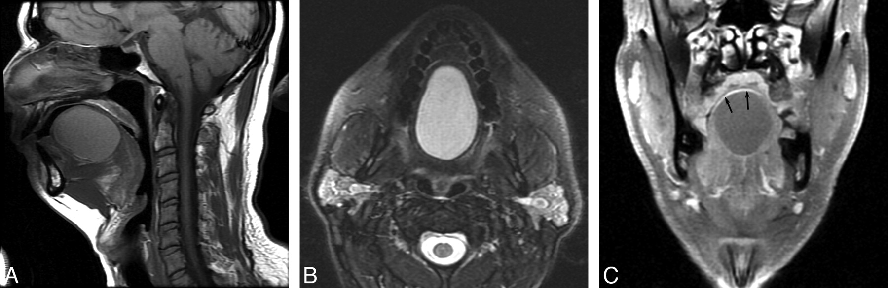
A 45-year-old man presented to the emergency department with a mass in his tongue that had acutely increased in size for 24 hours. The lesion had been present since childhood but had been about the size of a quarter until the previous day, when it enlarged to the size of a golf ball. He complained of difficulty in swallowing and in controlling secretions, as well as severe pain. His speech was impaired, but not his breathing. A contrast-enhanced CT of the neck failed to demonstrate the lesion due to artifacts from dental amalgam. MR imaging of the oral cavity showed a 5.1 × 3.7 cm well-circumscribed mass within the midline of the anterior tongue (Fig 1). The mass displayed intermediate signal intensity on T1, high signal intensity on T2, and partial rim enhancement on enhanced sequences. The differential diagnosis included lingual duplication cyst, dermoid cyst, and lymphatic malformation. The patient was treated with antibiotics and scheduled for surgery. Before surgery, the mass spontaneously discharged a small amount of brown fluid. At the time of surgery, the mass had decreased to 2.5 × 1.0 × 1.3 cm and was found to be a deep smoothly lined unilocular cyst in the submucosa. Histopathologic examination confirmed the mass as a foregut duplication cyst lined with squamous, ciliated columnar, and gastric mucosa. The patient was discharged on postoperative day 5 and recovered without further incident. There was no evidence of recurrence on clinical follow-up.

Fig 1.
  • Download figure
  • Open in new tab
  • Download powerpoint
Fig 1.

MR imaging of an adult lingual foregut duplication cyst. A, Sagittal T1-weighted image shows the location of the cyst within the intrinsic muscles of the tongue. The cyst is of intermediate T1 intensity due to its proteinaceous content. B, Axial T2-weighted image show the high T2 signal intensity. C, Fat-suppressed contrast-enhanced coronal T1-weighted image shows partial areas of rim enhancement (arrows) around the cyst.

Discussion

Embryologically, the primitive foregut gives rise to the pharynx, the respiratory tract, the esophagus, the stomach, and the duodenum to the level of the ampulla of Vater. There is no single theory that fully explains the embryogenesis of foregut duplications, but notochordal abnormalities appear to have a role.6 Lingual duplications may alternately reflect abnormal cellular migration because the primitive foregut and the pharyngeal arches (containing the developing tongue) are in close proximity.2 Foregut duplications are considered choristomas, with a wide range of ectopic tissues. In a prior review of 52 pediatric oral cavity choristomas, gastrointestinal epithelium lined most cysts, and the anterior two-thirds of the tongue was most frequently affected.2

The clinical presentation of any foregut duplication cyst is a reflection of its location, its mass effect, and complications of the ectopic mucosal lining.6 Oral cavity duplication cysts are usually asymptomatic, but there is a potential for respiratory and/or feeding problems in the infant. While mass effect is a common presentation in younger patients, degeneration into malignancy is a possible outcome in the adult, indicating that early surgical removal is preferred.7 The clinical differential diagnosis of a mass in the anterior tongue includes dermoid cyst, hamartoma, neurofibroma, teratoma, lymphoepithelial cyst, squamous cell carcinoma, and glandular neoplasms. Although radiologic findings usually cannot provide a definitive diagnosis, imaging has an important role in the evaluation of these patients, both for narrowing the differential diagnosis and for surgical planning. Complete excision of the cyst is the treatment of choice for foregut duplications, with no recurrences reported.8

MR imaging is the technique of choice for oral cavity lesions, providing excellent contrast resolution of soft tissues with multiple pulse sequences, but enhanced CT may also be useful in the absence of dental amalgam. Lingual duplications typically appear as nonenhancing or thinly rim-enhancing cystic lesions. On T1-weighted images, they may be of variable intensity, depending on the proteinaceous content. On T2-weighted images, they appear uniformly hyperintense.8 CT typically reveals a well-defined homogeneous cystic mass, but loculation is possible.9 Prenatally, sonography can also be of diagnostic use, followed by MR imaging for confirmation.1

The radiologic differential diagnosis of a cystic anterior tongue mass includes dermoid cyst, venolymphatic malformation, and foregut duplication cyst. Because dermoid cysts have various skin elements in their epithelial lining, they appear on CT as cystlike masses with variable (or even mixed) densities. On MR imaging, they appear as nonenhancing cystic lesions, which may have increased T1 signal intensity from fat content. The presence of proteinaceous fluid in duplication cysts produces similar signal intensities on T1 and T2 sequences, so these lesions may be difficult to distinguish.8 Mucocele of the tongue demonstrates the typical findings of a cystic lesion, with low signal intensity on T1 and high signal intensity on T2.10 Venolymphatic malformations display similar signal intensities on T1 and T2 sequences, with a heterogeneous and often infiltrative appearance.11,12 Thus, differentiating among these various lesions radiographically can be problematic, and excisional biopsy is needed for the final diagnosis.

In summary, lingual duplication cysts are uncommon in the pediatric population, and the diagnosis is exceedingly rare in adults. Nevertheless, this diagnosis must be considered even in middle-aged patients presenting with an anterior tongue mass.

References

  1. 1.↵
    1. Kong K,
    2. Walker P,
    3. Cassey J,
    4. et al
    . Foregut duplication cyst arising in the floor of mouth. Int J Pediatr Otorhinolaryngol 2004;68:827–30
    CrossRefPubMed
  2. 2.↵
    1. Manor Y,
    2. Buchner A,
    3. Peleg M,
    4. et al
    . Lingual cyst with respiratory epithelium: an entity of debatable histogenesis. J Oral Maxillofac Surg 1999;57:124–27
    CrossRefPubMed
  3. 3.↵
    1. Azanero WD,
    2. Mazzonetto R,
    3. Leon JE,
    4. et al
    . Lingual cyst with respiratory epithelium: a histopathological and immunohistochemical analysis of two cases. Int J Oral Maxillofac Surg 2009;38:388–92
    CrossRefPubMed
  4. 4.↵
    1. Martone CH,
    2. Wolf SM,
    3. Wesley RK
    . Heterotopic gastrointestinal cyst of the oral cavity. J Oral Maxillofac Surg 1992;50:1340–42
    CrossRefPubMed
  5. 5.↵
    1. Naidoo LC
    . Median lingual cyst: review of the literature and report of a case. J Oral Maxillofac Surg 1997;55:172–175
    CrossRefPubMed
  6. 6.↵
    1. Azzie G,
    2. Beasley S
    . Diagnosis and treatment of foregut duplications. Semin Pediatr Surg 2003;12:46–54
    CrossRefPubMed
  7. 7.↵
    1. Volchok J,
    2. Jaffer A,
    3. Cooper T,
    4. et al
    . Adenocarcinoma arising in a lingual foregut duplication cyst. Arch Otolaryngol Head Neck Surg 2007;133:717–19
    CrossRefPubMed
  8. 8.↵
    1. Eaton D,
    2. Billings K,
    3. Timmons C,
    4. et al
    . Congenital foregut duplication cysts of the anterior tongue. Arch Otolaryngol Head Neck Surg 2001;127:1484–87
    PubMed
  9. 9.↵
    1. Kim SH,
    2. Han MH,
    3. Park SW,
    4. et al
    . Radiologic-pathologic correlation of unusual lingual masses. Part II. Benign and malignant tumors. Korean J Radiol 2001;2:42–51
    CrossRefPubMed
  10. 10.↵
    1. Gray GG,
    2. Voigt E,
    3. Breuer F,
    4. et al
    . Imaging quiz case 2: intralingual mucocele. Arch Otolaryngol Head Neck Surg 1999;125:593–95
    PubMed
  11. 11.↵
    1. Huang TY,
    2. Hsin CH,
    3. Cheng PW,
    4. et al
    . Radiology quiz case 2: hypopharyngeal lymphangioma. Arch Otolaryngol Head Neck Surg 2009;135:613,615
    CrossRefPubMed
  12. 12.↵
    1. Kurabayashi T,
    2. Ida M,
    3. Yasumoto M,
    4. et al
    . MRI of ranulas. Neuroradiology 2000;42:917–22
    CrossRefPubMed
  • Received October 4, 2009.
  • Accepted after revision November 2, 2009.
  • Copyright © American Society of Neuroradiology
View Abstract
PreviousNext
Back to top

In this issue

American Journal of Neuroradiology: 32 (3)
American Journal of Neuroradiology
Vol. 32, Issue 3
1 Mar 2011
  • Table of Contents
  • Index by author
Advertisement
Print
Download PDF
Email Article

Thank you for your interest in spreading the word on American Journal of Neuroradiology.

NOTE: We only request your email address so that the person you are recommending the page to knows that you wanted them to see it, and that it is not junk mail. We do not capture any email address.

Enter multiple addresses on separate lines or separate them with commas.
Lingual Foregut Duplication in a Middle-Aged Adult
(Your Name) has sent you a message from American Journal of Neuroradiology
(Your Name) thought you would like to see the American Journal of Neuroradiology web site.
CAPTCHA
This question is for testing whether or not you are a human visitor and to prevent automated spam submissions.
Cite this article
P. Patel, B.F. Branstetter, E.N. Myers
Lingual Foregut Duplication in a Middle-Aged Adult
American Journal of Neuroradiology Mar 2011, 32 (3) E40-E41; DOI: 10.3174/ajnr.A1994

Citation Manager Formats

  • BibTeX
  • Bookends
  • EasyBib
  • EndNote (tagged)
  • EndNote 8 (xml)
  • Medlars
  • Mendeley
  • Papers
  • RefWorks Tagged
  • Ref Manager
  • RIS
  • Zotero
0 Responses
Respond to this article
Share
Bookmark this article
Lingual Foregut Duplication in a Middle-Aged Adult
P. Patel, B.F. Branstetter, E.N. Myers
American Journal of Neuroradiology Mar 2011, 32 (3) E40-E41; DOI: 10.3174/ajnr.A1994
del.icio.us logo Twitter logo Facebook logo Mendeley logo
  • Tweet Widget
  • Facebook Like
  • Google Plus One
Purchase

Jump to section

  • Article
    • Abstract
    • Case Report
    • Discussion
    • References
  • Figures & Data
  • Info & Metrics
  • Responses
  • References
  • PDF

Related Articles

  • No related articles found.
  • PubMed
  • Google Scholar

Cited By...

  • Prenatal Magnetic Resonance Imaging (MRI) Findings of a Foregut Duplication Cyst of the Tongue: Value of Real-time MRI Evaluation of the Fetal Swallowing Mechanism
  • Crossref (11)
  • Google Scholar

This article has been cited by the following articles in journals that are participating in Crossref Cited-by Linking.

  • Optimization of MR Parameters of 3D TOF-MRA for Various Intracranial Stents at 3.0T MRI
    Jin Woo Choi, Hong Gee Roh, Won-Jin Moon, Young Il Chun, Chung Hwan Kang
    Neurointervention 2011 6 2
  • Prenatal Magnetic Resonance Imaging (MRI) Findings of a Foregut Duplication Cyst of the Tongue
    Golbahar Houshmand, Keyanoosh Hosseinzadeh, John Ozolek
    Journal of Ultrasound in Medicine 2011 30 6
  • Histopathologic Finding of Both Gastric and Respiratory Epithelia in a Lingual Foregut Cyst
    Yangfan Luo, Nick Shillingford, Jeffrey A. Koempel
    Case Reports in Medicine 2015 2015
  • A Bronchogenic Cyst Masquerading as a Tongue Mass
    Zahra Aldawood, David J. Moyer, Sook-Bin Woo
    Head and Neck Pathology 2021 15 4
  • Heterotopic gastrointestinal cyst within the submandibular space in an adult: A case report and review of the literature
    Elliot Saleh, Mohammad Mansouri, Patrick J. Nolan, Robert Kelsch, Keivan Shifteh
    Radiology Case Reports 2022 17 9
  • Rapid Development of a Simple Ranula in a Child
    Dinesh Kumar Sathanantham, Gopi B. Shah, Seckin Ulüalp
    Annals of Otology, Rhinology & Laryngology 2015 124 4
  • Lingual Choristoma with Gastric Epithelium Revisited
    Christina M. Ombres, Isabelle Lestadi, Juan Putra
    Head and Neck Pathology 2022 16 2
  • Sperm protein antigen 17 and Sperm flagellar 1 cancer testis antigens are expressed in a rare case of ciliated foregut cyst of the common hepatic duct
    Fabio Grizzi, Maurizio Chiriva-Internati, Elena Miranda, Roxana Zaharie, Nadim Al Hajjar, Florin Zaharie, Cristina Díaz Del Arco, M.Jesús Fernández-Aceñero, Robert S. Bresalier, Emil Moiş
    Pathology - Research and Practice 2023 247
  • A case of lingual foregut cyst with the cyst wall lined by both respiratory and gastric epithelium
    Masaki KOBAYASHI, Maki MORITA, Natsuko YASUNAGA, Yoshinori HIRAI, Masaya AKASHI, Yasuyuki SHIBUYA
    Japanese Journal of Oral and Maxillofacial Surgery 2021 67 5
  • Heterotopic gastrointestinal cyst of the oral cavity: a rare clinical report and literature review
    Andrea Maldonado, Rubén Muñoz, Jesús Cabrera, José Alcides Almeida de Arruda, Bruno Augusto Benevenuto de Andrade, Mariana Villarroel-Dorrego, Isabella Bittencourt do Valle
    Journal of Stomatology Oral and Maxillofacial Surgery 2025

More in this TOC Section

  • Atypical Diffusion-Restricted Lesion in 5-Fluorouracil Encephalopathy
  • Multimodal Imaging of Spike Propagation: A Technical Case Report
  • Dural Infantile Hemangioma Masquerading as a Skull Vault Lesion
Show more Case Reports

Similar Articles

Advertisement

Indexed Content

  • Current Issue
  • Accepted Manuscripts
  • Article Preview
  • Past Issues
  • Editorials
  • Editors Choice
  • Fellow Journal Club
  • Letters to the Editor

Cases

  • Case Collection
  • Archive - Case of the Week
  • Archive - Case of the Month
  • Archive - Classic Case

Special Collections

  • Special Collections

Resources

  • News and Updates
  • Turn around Times
  • Submit a Manuscript
  • Author Policies
  • Manuscript Submission Guidelines
  • Evidence-Based Medicine Level Guide
  • Publishing Checklists
  • Graphical Abstract Preparation
  • Imaging Protocol Submission
  • Submit a Case
  • Become a Reviewer/Academy of Reviewers
  • Get Peer Review Credit from Publons

Multimedia

  • AJNR Podcast
  • AJNR SCANtastic
  • Video Articles

About Us

  • About AJNR
  • Editorial Board
  • Not an AJNR Subscriber? Join Now
  • Alerts
  • Feedback
  • Advertise with us
  • Librarian Resources
  • Permissions
  • Terms and Conditions

American Society of Neuroradiology

  • Not an ASNR Member? Join Now

© 2025 by the American Society of Neuroradiology All rights, including for text and data mining, AI training, and similar technologies, are reserved.
Print ISSN: 0195-6108 Online ISSN: 1936-959X

Powered by HighWire