Advertisement

Main menu

  • Home
  • Content
    • Current Issue
    • Accepted Manuscripts
    • Article Preview
    • Past Issue Archive
    • AJNR Case Collection
    • Case of the Week Archive
    • Classic Case Archive
    • Case of the Month Archive
  • Special Collections
    • Spinal CSF Leak Articles (Jan 2020-June 2024)
    • 2024 AJNR Journal Awards
    • Most Impactful AJNR Articles
  • Multimedia
    • AJNR Podcast
    • AJNR Scantastics
    • Video Articles
  • For Authors
    • Submit a Manuscript
    • Author Policies
    • Fast publishing of Accepted Manuscripts
    • Graphical Abstract Preparation
    • Manuscript Submission Guidelines
    • Imaging Protocol Submission
    • Submit a Case for the Case Collection
  • About Us
    • About AJNR
    • Editorial Board
  • More
    • Become a Reviewer/Academy of Reviewers
    • Subscribers
    • Permissions
    • Alerts
    • Feedback
    • Advertisers
    • ASNR Home
  • Other Publications
    • ajnr

User menu

  • Alerts
  • Log in

Search

  • Advanced search
American Journal of Neuroradiology

American Journal of Neuroradiology

ASHNR American Society of Functional Neuroradiology ASHNR American Society of Pediatric Neuroradiology ASSR
  • Alerts
  • Log in

Advanced Search

  • Home
  • Content
    • Current Issue
    • Accepted Manuscripts
    • Article Preview
    • Past Issue Archive
    • AJNR Case Collection
    • Case of the Week Archive
    • Classic Case Archive
    • Case of the Month Archive
  • Special Collections
    • Spinal CSF Leak Articles (Jan 2020-June 2024)
    • 2024 AJNR Journal Awards
    • Most Impactful AJNR Articles
  • Multimedia
    • AJNR Podcast
    • AJNR Scantastics
    • Video Articles
  • For Authors
    • Submit a Manuscript
    • Author Policies
    • Fast publishing of Accepted Manuscripts
    • Graphical Abstract Preparation
    • Manuscript Submission Guidelines
    • Imaging Protocol Submission
    • Submit a Case for the Case Collection
  • About Us
    • About AJNR
    • Editorial Board
  • More
    • Become a Reviewer/Academy of Reviewers
    • Subscribers
    • Permissions
    • Alerts
    • Feedback
    • Advertisers
    • ASNR Home
  • Follow AJNR on Twitter
  • Visit AJNR on Facebook
  • Follow AJNR on Instagram
  • Join AJNR on LinkedIn
  • RSS Feeds

Welcome to the new AJNR, Updated Hall of Fame, and more. Read the full announcements.


AJNR is seeking candidates for the position of Associate Section Editor, AJNR Case Collection. Read the full announcement.


Case ReportCase Report

The Emerging Echogenic Tract Sign of Pyriform Sinus Fistula: An Early Indicator in the Recovery Stage of Acute Suppurative Thyroiditis

N.H. Park, H.J. Park, C.S. Park, M.S. Kim and S.I. Park
American Journal of Neuroradiology March 2011, 32 (3) E44-E46; DOI: https://doi.org/10.3174/ajnr.A2015
N.H. Park
  • Find this author on Google Scholar
  • Find this author on PubMed
  • Search for this author on this site
H.J. Park
  • Find this author on Google Scholar
  • Find this author on PubMed
  • Search for this author on this site
C.S. Park
  • Find this author on Google Scholar
  • Find this author on PubMed
  • Search for this author on this site
M.S. Kim
  • Find this author on Google Scholar
  • Find this author on PubMed
  • Search for this author on this site
S.I. Park
  • Find this author on Google Scholar
  • Find this author on PubMed
  • Search for this author on this site
  • Article
  • Figures & Data
  • Info & Metrics
  • Responses
  • References
  • PDF
Loading

Abstract

SUMMARY: AST is commonly associated with pyriform sinus−thyroid fistula in children. Radiologic findings of AST are documented in a few reports. We report a new sign we term the “emerging echogenic tract sign,” which reflects a patent air-containing pyriform sinus−thyroid fistula on follow-up US. Recognition of this sign is an important finding suggesting an associated pyriform sinus−thyroid fistula in a patient with AST and also suggesting the adequate timing of barium esophagography to confirm the fistula.

Abbreviations

AST
acute suppurative thyroiditis
US
sonography

AST is a rare infectious disease affecting mainly children and young adults.1 That pyriform sinus fistula often causes suppurative thyroiditis has been well documented.2–4 However, when a pyriform sinus fistula is not readily identified in a patient with AST, the correct diagnosis can be difficult.

In the active phase of AST, demonstration of pyriform sinus fistula on US or esophagography is often difficult due to the inflammatory exudates within the fistula as well as a compression of the fistula by edema. With recovery of the thyroiditis, an echogenic tract suggesting the pyriform sinus fistula may emerge on US, and we termed this the “emerging echogenic tract sign.” This finding is suggestive of an associated pyriform sinus fistula and also suggests that a subsequent barium esophagography will be helpful in demonstrating it.

We report the emerging echogenic tract sign in the quiescence stage of AST with associated pyriform sinus fistula.

Case Reports

Case 1

A 7-year-old girl was referred for evaluation of high fever and painful swelling in the left side of the neck for 5 days.

US after admission (Fig 1A) showed swelling of the left lobe of the thyroid gland with heterogeneous hypoechoic change of perithyroidal soft tissue. On a color Doppler study, increased signals of blood flow within the lesion were detected. Definite abscess formation was not seen. CT of the neck and US performed 6 days later (Fig 1B, -C) showed marked improvement of the inflammatory change of the left lobe of thyroid gland and perithyroidal soft tissue. Subsequent barium esophagogram (Fig 1D) was performed for possible presence of associated pyriform sinus−thyroid fistula as the cause of AST, but it failed to demonstrate a fistula.

Fig 1.
  • Download figure
  • Open in new tab
  • Download powerpoint
Fig 1.

Images of a 7-year-old girl with fever and painful neck swelling for 5 days. A, Initial US of the thyroid gland shows a heterogeneous hypoechoic inflammatory change of perithyroid soft tissue with invasion into the anterior portion of the left lobe of the thyroid gland. B, CT scan at the level of the thyroid gland reveals a heterogeneous low-attenuated inflammatory mass anterior to the left lobe of the thyroid gland with invasion into the thyroid gland, displacing the ipsilateral carotid sheath laterally. C, Follow-up US 5 days after initial US shows improvement of the inflammatory change of the perithyroid soft tissue and the left lobe of the thyroid gland. D, Subsequent barium esophagography shows no demonstrable pyriform sinus fistula. E and F, Follow-up US obtained 3 months after the initial US shows marked improvement of the inflammatory change and a newly developed echogenic tractlike lesion (arrow) in the left lobe of the thyroid gland, suggestive of an associated pyriform sinus fistula. G, Subsequent barium esophagography shows the pyriform sinus fistula (arrows) originating from its apex.

Follow-up US performed 3 months after the initial US (Fig 1E, -F) showed a newly developed echogenic tract within the left lobe of the thyroid gland, suspicious for associated pyriform sinus-thyroid fistula. Repeat barium esophagography (Fig 1G) was performed and demonstrated this fistula.

Case 2

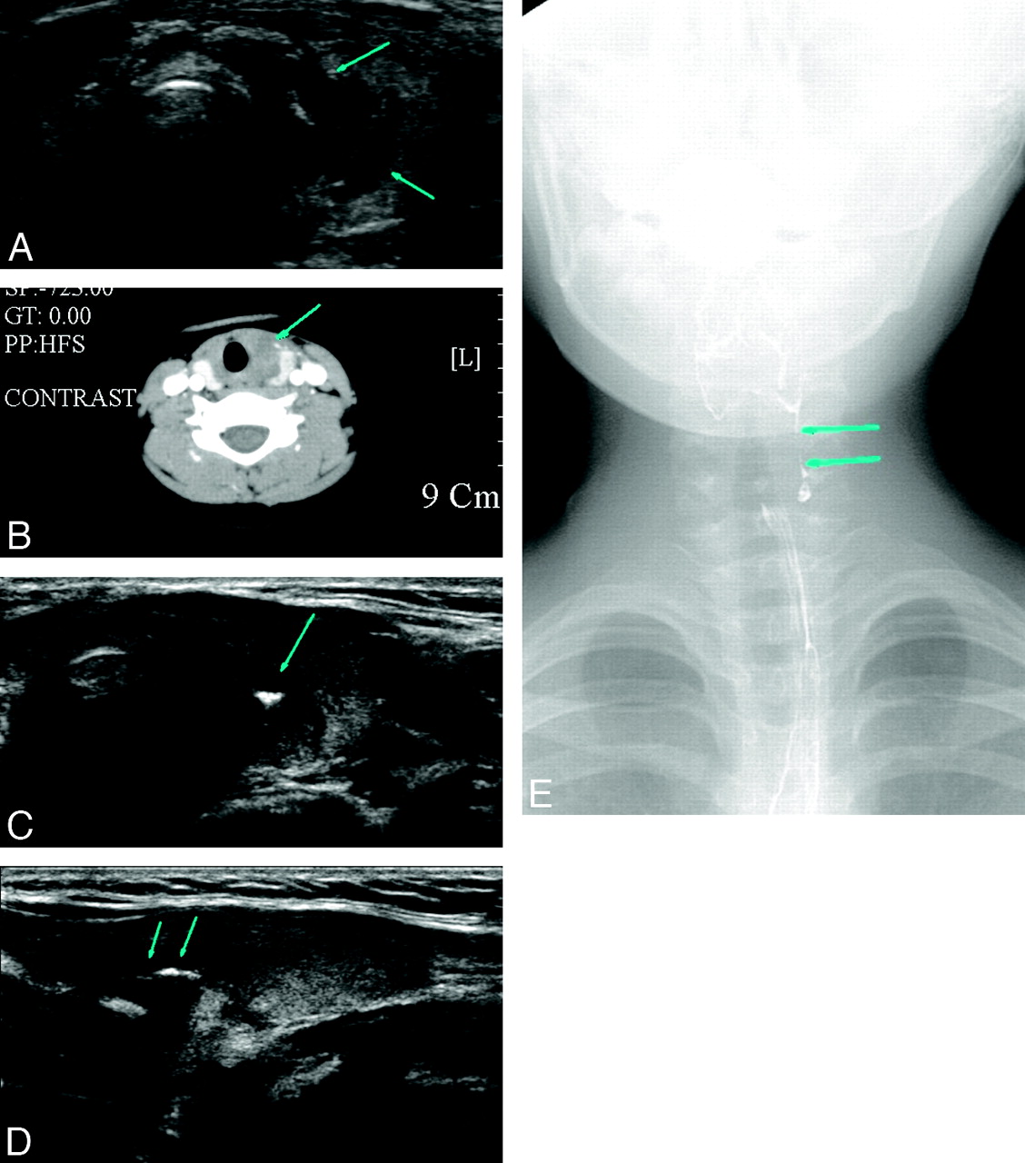
A 7-year-old girl presented with fever, poor oral intake, sore throat, and cough for 3 days and was referred for US. Initial US of the neck (Fig 2A) showed an ill-defined hypoechoic lesion with suspected microabscess in the left lobe of the thyroid gland and inflammatory change of overlying perithyroidal soft tissue. CT of the neck (Fig 2B) showed a heterogeneous low-attenuated lesion in the left lobe of the thyroid gland and inflammatory swelling of the overlying strap muscle.

Fig 2.
  • Download figure
  • Open in new tab
  • Download powerpoint
Fig 2.

Images of a 7-year-old girl with a sore throat and neck pain for 3 days. A, US shows an ill-defined heterogeneous hypoechoic lesion in the left lobe of the thyroid gland (arrows) and inflammatory change of the overlying strap muscle. B, CT scan shows a heterogeneous low-attenuated inflammatory change (arrow) in the medial aspect of the left lobe of the thyroid gland and overlying soft tissue. Tracheal deviation is noted by the inflammatory mass. C and D, Follow-up US performed 6 days after the initial US examination shows improvement of inflammatory change and a newly developed echogenic tractlike lesion (arrows) in the left lobe of the thyroid gland. E, Subsequent barium esophagography shows a pyriform sinus fistula (arrows) originating from the apex of the pyriform sinus.

Follow-up US (Fig 2C, -D) performed 6 days after initial US showed an improved state of intrathyroidal and perithyroidal inflammation and a newly developed echogenic tract in the left lobe of the thyroid gland. Barium esophagogram (Fig 2E) successfully demonstrated a pyriform sinus−thyroid fistula.

Discussion

AST and thyroid abscess are extremely rare disorders. A rich blood supply, a generous lymphatic drainage, a high iodine level that inhibits bacterial growth, and a complete protective fibrous capsule have been proposed as the contributing factors to the relatively high resistance of the thyroid gland to infections.1,5,6 Congenital pyriform sinus fistula is a remnant of the fourth branchial cleft and has recently been recognized as an underlying cause of AST or acute deep neck infection.2–4,6

Recurrent AST due to persistent pyriform sinus−thyroid fistula is likely more common than previously believed and usually becomes symptomatic before 10 years of age. Eighty percent of patients with recurrent AST due to persistent pyriform sinus−thyroid fistula present during the first decade of life (mean age, 7.6 years; age range, birth to 56 years) and 8%, during adulthood.3,7–10

In the acute phase, the echogenic tract sign was not demonstrated within the inflamed thyroid gland or perithyroid soft tissue, and it is presumed to be due to an obstruction of the fistula by inspissated inflammatory exudates.

After recovery from AST, follow-up US showed an echogenic tract, which we speculate represents an air-filled pyriform sinus fistula within the parenchyma of the left lobe of the thyroid gland. This emerging echogenic tract sign is highly suggestive of a pyriform sinus−thyroid fistula as the cause of AST and should lead to barium esophagography for confirmation.

In conclusion, the emerging echogenic tract sign, suggesting a pyriform sinus−thyroid fistula, may be appreciated only on follow-up US in the quiescence stage of AST. This finding appears helpful for the diagnosis of an associated pyriform sinus−thyroid fistula and should effect a confirmatory barium esophagography.

References

  1. 1.↵
    1. Berger SA,
    2. Zonszein J,
    3. Villamena P,
    4. et al
    . Infectious disease of the thyroid gland. Rev Infect Dis 1983;5:108–22
    PubMed
  2. 2.↵
    1. Park SW,
    2. Han MH,
    3. Sung MH,
    4. et al
    . Neck infection associated with pyriform sinus fistula: imaging findings. AJNR Am J Neuroradiol 2000;21:817–22
    Abstract/FREE Full Text
  3. 3.↵
    1. Bar-Ziv J,
    2. Slasky BS,
    3. Sichel JY,
    4. et al
    . Branchial pouch sinus tract from the piriform fossa causing acute suppurative thyroiditis, neck abscess, or both: CT appearance and the use of air as a contrast agent. AJR Am J Roentgenol 1996;167:1569–72
    CrossRefPubMed
  4. 4.↵
    1. Hatabu H,
    2. Kasagi K,
    3. Yamamoto K,
    4. et al
    . Acute suppurative thyroiditis associated with piriform sinus fistula: sonographic findings. AJR Am J Roentgenol 1990;155:845–47
    CrossRefPubMed
  5. 5.↵
    1. Womack NA,
    2. Cole WH
    . Thyroiditis. Surgery 1944;16:770–82
  6. 6.↵
    1. Miller D,
    2. Hill JL,
    3. Sun CC,
    4. et al
    . The diagnosis and management of pyriform sinus fistula in infants and young children. J Pediatr Surg 1983;18:377–81
    CrossRefPubMed
  7. 7.↵
    1. Himi T,
    2. Kataura A
    . Distribution of C cells in the thyroid gland with pyriform sinus fistula. Otolaryngol Head Neck Surg 1995;112:268–73
    CrossRefPubMed
  8. 8.↵
    1. Burge D,
    2. Middleton A
    . Persistent pharyngeal pouch derivatives in the neonate. J Pediatr Surg 1983;18:230–34
    CrossRefPubMed
  9. 9.↵
    1. Roediger WE,
    2. Kalk F,
    3. Spitz L,
    4. et al
    . Congenital thyroid cyst of ultimobranchial gland origin. J Pediatr Surg 1977;12:575–76
    CrossRefPubMed
  10. 10.↵
    1. Tucker HM,
    2. Skolnick ML
    . Fourth branchial cleft (pharyngeal pouch) remnant. Trans Am Acad Ophthalmol Otolaryngol 1973;77:ORL368–71
    PubMed
  • Received November 10, 2009.
  • Accepted after revision December 2, 2009.
  • Copyright © American Society of Neuroradiology
View Abstract
PreviousNext
Back to top

In this issue

American Journal of Neuroradiology: 32 (3)
American Journal of Neuroradiology
Vol. 32, Issue 3
1 Mar 2011
  • Table of Contents
  • Index by author
Advertisement
Print
Download PDF
Email Article

Thank you for your interest in spreading the word on American Journal of Neuroradiology.

NOTE: We only request your email address so that the person you are recommending the page to knows that you wanted them to see it, and that it is not junk mail. We do not capture any email address.

Enter multiple addresses on separate lines or separate them with commas.
The Emerging Echogenic Tract Sign of Pyriform Sinus Fistula: An Early Indicator in the Recovery Stage of Acute Suppurative Thyroiditis
(Your Name) has sent you a message from American Journal of Neuroradiology
(Your Name) thought you would like to see the American Journal of Neuroradiology web site.
CAPTCHA
This question is for testing whether or not you are a human visitor and to prevent automated spam submissions.
Cite this article
N.H. Park, H.J. Park, C.S. Park, M.S. Kim, S.I. Park
The Emerging Echogenic Tract Sign of Pyriform Sinus Fistula: An Early Indicator in the Recovery Stage of Acute Suppurative Thyroiditis
American Journal of Neuroradiology Mar 2011, 32 (3) E44-E46; DOI: 10.3174/ajnr.A2015

Citation Manager Formats

  • BibTeX
  • Bookends
  • EasyBib
  • EndNote (tagged)
  • EndNote 8 (xml)
  • Medlars
  • Mendeley
  • Papers
  • RefWorks Tagged
  • Ref Manager
  • RIS
  • Zotero
0 Responses
Respond to this article
Share
Bookmark this article
The Emerging Echogenic Tract Sign of Pyriform Sinus Fistula: An Early Indicator in the Recovery Stage of Acute Suppurative Thyroiditis
N.H. Park, H.J. Park, C.S. Park, M.S. Kim, S.I. Park
American Journal of Neuroradiology Mar 2011, 32 (3) E44-E46; DOI: 10.3174/ajnr.A2015
del.icio.us logo Twitter logo Facebook logo Mendeley logo
  • Tweet Widget
  • Facebook Like
  • Google Plus One
Purchase

Jump to section

  • Article
    • Abstract
    • Abbreviations
    • Case Reports
    • Discussion
    • References
  • Figures & Data
  • Info & Metrics
  • Responses
  • References
  • PDF

Related Articles

  • No related articles found.
  • PubMed
  • Google Scholar

Cited By...

  • No citing articles found.
  • Crossref (7)
  • Google Scholar

This article has been cited by the following articles in journals that are participating in Crossref Cited-by Linking.

  • The treatment of congenital pyriform sinus fistula: a single-center experience
    Liguang Xia, Zhengxiu Lin, Xiaokun Lin, Yongbiao Wang, Libin Zhu, Jinhan Lin, Yiming Zhao, Zhongrong Li
    Pediatric Surgery International 2020 36 7
  • Benign Conditions of the Thyroid Gland
    Amy Fan-Yee Juliano, Mary Beth Cunnane
    Seminars in Ultrasound, CT and MRI 2012 33 2
  • Empyema and mediastinal abscess in pyriform sinus fistula: A case report
    Hai-jiang Zhou, Wen-peng Yin, Tian-fei Lan, Shu-bin Guo
    World Journal of Emergency Medicine 2019 10 4
  • Thyroid Ultrasound and Ultrasound-Guided FNA
    Robert A. Sofferman
    2013
  • Pathology of the Head and Neck
    Catarina Eloy, Paula Soares, Valdemar Máximo, Manuel Sobrinho-Simões
    2016
  • Thyroid Disorders
    Henrique Vara Luiz, Isabel Manita, Jorge Portugal
    2016
  • Thyroid and Parathyroid Ultrasound and Ultrasound-Guided FNA
    Vinay T. Fernandes, Lisa A. Orloff
    2018

More in this TOC Section

  • Atypical Diffusion-Restricted Lesion in 5-Fluorouracil Encephalopathy
  • Multimodal Imaging of Spike Propagation: A Technical Case Report
  • Dural Infantile Hemangioma Masquerading as a Skull Vault Lesion
Show more Case Reports

Similar Articles

Advertisement

Indexed Content

  • Current Issue
  • Accepted Manuscripts
  • Article Preview
  • Past Issues
  • Editorials
  • Editors Choice
  • Fellow Journal Club
  • Letters to the Editor

Cases

  • Case Collection
  • Archive - Case of the Week
  • Archive - Case of the Month
  • Archive - Classic Case

Special Collections

  • Special Collections

Resources

  • News and Updates
  • Turn around Times
  • Submit a Manuscript
  • Author Policies
  • Manuscript Submission Guidelines
  • Evidence-Based Medicine Level Guide
  • Publishing Checklists
  • Graphical Abstract Preparation
  • Imaging Protocol Submission
  • Submit a Case
  • Become a Reviewer/Academy of Reviewers
  • Get Peer Review Credit from Publons

Multimedia

  • AJNR Podcast
  • AJNR SCANtastic
  • Video Articles

About Us

  • About AJNR
  • Editorial Board
  • Not an AJNR Subscriber? Join Now
  • Alerts
  • Feedback
  • Advertise with us
  • Librarian Resources
  • Permissions
  • Terms and Conditions

American Society of Neuroradiology

  • Not an ASNR Member? Join Now

© 2025 by the American Society of Neuroradiology All rights, including for text and data mining, AI training, and similar technologies, are reserved.
Print ISSN: 0195-6108 Online ISSN: 1936-959X

Powered by HighWire