
Article Figures & Data
Figures
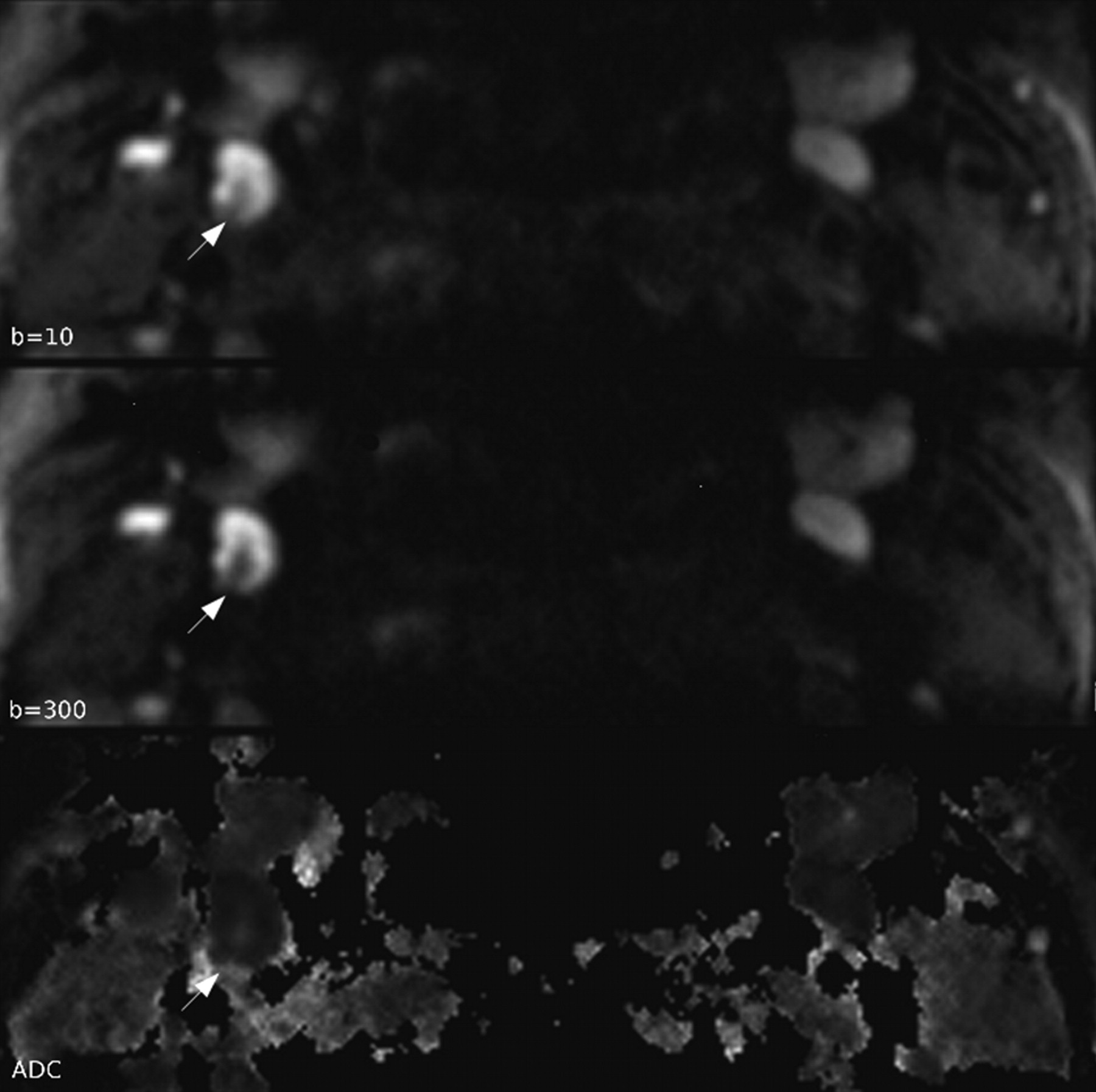
- Fig 1.
Typical DW images and ADC map. Normal LN hilum is seen as a central linear structure with low signal intensity on b = 10 and b = 300 images and high signal intensity on ADC map (white arrow) in the right side level 2 LN with a 6.8-mm diameter. ADC of hilum is 2.152 × 10⁻³ mm2/s, which is close to the diffusivity of bulk water.
- Fig 2.
There is significant ADC difference in the 15 LNs in that hilum is identified by DWI and ADC map (A). ADC from ROIs excluding hilum is significantly lower than ADC from ROIs including hilum. The variation of ADC is smaller when the hilum was excluded within ROIs. Also, there is significant difference in ADC values measured on a total of 58 LNs according to the hilar inclusion (B). ADC is significantly lower in LNs with ROIs in which the hilum is excluded.


Tables
Results of ADC values according to hilum inclusion
LNs with Visible Hilum (n = 15) Total LNs (n = 58) Hilum(−) Hilum(+) P Hilum(−) Hilum(+) P ADC 0.983 ± 0.169 1.206 ± 0.244 <.0001 1.034 ± 0.183 1.095 ± 0.213 .0002 -
Note:—Hilum(−) means that the hilum was excluded within the ROIs. Hilum(+) means that the hilum was included within the ROIs.
-
In this issue


Jump to section
Related Articles
Cited By...
- No citing articles found.