Advertisement

Main menu

  • Home
  • Content
    • Current Issue
    • Accepted Manuscripts
    • Article Preview
    • Past Issue Archive
    • AJNR Case Collection
    • Case of the Week Archive
    • Classic Case Archive
    • Case of the Month Archive
  • Special Collections
    • Spinal CSF Leak Articles (Jan 2020-June 2024)
    • 2024 AJNR Journal Awards
    • Most Impactful AJNR Articles
  • Multimedia
    • AJNR Podcast
    • AJNR Scantastics
    • Video Articles
  • For Authors
    • Submit a Manuscript
    • Author Policies
    • Fast publishing of Accepted Manuscripts
    • Graphical Abstract Preparation
    • Manuscript Submission Guidelines
    • Imaging Protocol Submission
    • Submit a Case for the Case Collection
  • About Us
    • About AJNR
    • Editorial Board
  • More
    • Become a Reviewer/Academy of Reviewers
    • Subscribers
    • Permissions
    • Alerts
    • Feedback
    • Advertisers
    • ASNR Home
  • Other Publications
    • ajnr

User menu

  • Alerts
  • Log in

Search

  • Advanced search
American Journal of Neuroradiology

American Journal of Neuroradiology

ASHNR American Society of Functional Neuroradiology ASHNR American Society of Pediatric Neuroradiology ASSR
  • Alerts
  • Log in

Advanced Search

  • Home
  • Content
    • Current Issue
    • Accepted Manuscripts
    • Article Preview
    • Past Issue Archive
    • AJNR Case Collection
    • Case of the Week Archive
    • Classic Case Archive
    • Case of the Month Archive
  • Special Collections
    • Spinal CSF Leak Articles (Jan 2020-June 2024)
    • 2024 AJNR Journal Awards
    • Most Impactful AJNR Articles
  • Multimedia
    • AJNR Podcast
    • AJNR Scantastics
    • Video Articles
  • For Authors
    • Submit a Manuscript
    • Author Policies
    • Fast publishing of Accepted Manuscripts
    • Graphical Abstract Preparation
    • Manuscript Submission Guidelines
    • Imaging Protocol Submission
    • Submit a Case for the Case Collection
  • About Us
    • About AJNR
    • Editorial Board
  • More
    • Become a Reviewer/Academy of Reviewers
    • Subscribers
    • Permissions
    • Alerts
    • Feedback
    • Advertisers
    • ASNR Home
  • Follow AJNR on Twitter
  • Visit AJNR on Facebook
  • Follow AJNR on Instagram
  • Join AJNR on LinkedIn
  • RSS Feeds

Welcome to the new AJNR, Updated Hall of Fame, and more. Read the full announcements.


AJNR is seeking candidates for the position of Associate Section Editor, AJNR Case Collection. Read the full announcement.


Research ArticleHead and NeckF

The Relation of Carotid Calcium Volume with Carotid Artery Stenosis in Symptomatic Patients

H.A. Marquering, C.B.L.M. Majoie, L. Smagge, A.G. Kurvers, H.A. Gratama van Andel, R. van den Berg and P.J. Nederkoorn
American Journal of Neuroradiology August 2011, 32 (7) 1182-1187; DOI: https://doi.org/10.3174/ajnr.A2519
H.A. Marquering
  • Find this author on Google Scholar
  • Find this author on PubMed
  • Search for this author on this site
C.B.L.M. Majoie
  • Find this author on Google Scholar
  • Find this author on PubMed
  • Search for this author on this site
L. Smagge
  • Find this author on Google Scholar
  • Find this author on PubMed
  • Search for this author on this site
A.G. Kurvers
  • Find this author on Google Scholar
  • Find this author on PubMed
  • Search for this author on this site
H.A. Gratama van Andel
  • Find this author on Google Scholar
  • Find this author on PubMed
  • Search for this author on this site
R. van den Berg
  • Find this author on Google Scholar
  • Find this author on PubMed
  • Search for this author on this site
P.J. Nederkoorn
  • Find this author on Google Scholar
  • Find this author on PubMed
  • Search for this author on this site
  • Article
  • Figures & Data
  • Info & Metrics
  • Responses
  • References
  • PDF
Loading

Article Figures & Data

Figures

  • Tables
  • Fig 1.
    • Download figure
    • Open in new tab
    • Download powerpoint
    Fig 1.

    Illustration of the calcium volume measurement according to the method of McKinney et al.8 First, the carotid bifurcation is sculpted (left). After further adjustment of the window level and width, only the calcium is displayed (right). With the volume calculation tool, the calcium volume is displayed.

  • Fig 2.
    • Download figure
    • Open in new tab
    • Download powerpoint
    Fig 2.

    Scatterplot showing all measurements for the symptomatic and asymptomatic ICAs. The stenosis categories are illustrated with the gray vertical lines.

  • Fig 3.
    • Download figure
    • Open in new tab
    • Download powerpoint
    Fig 3.

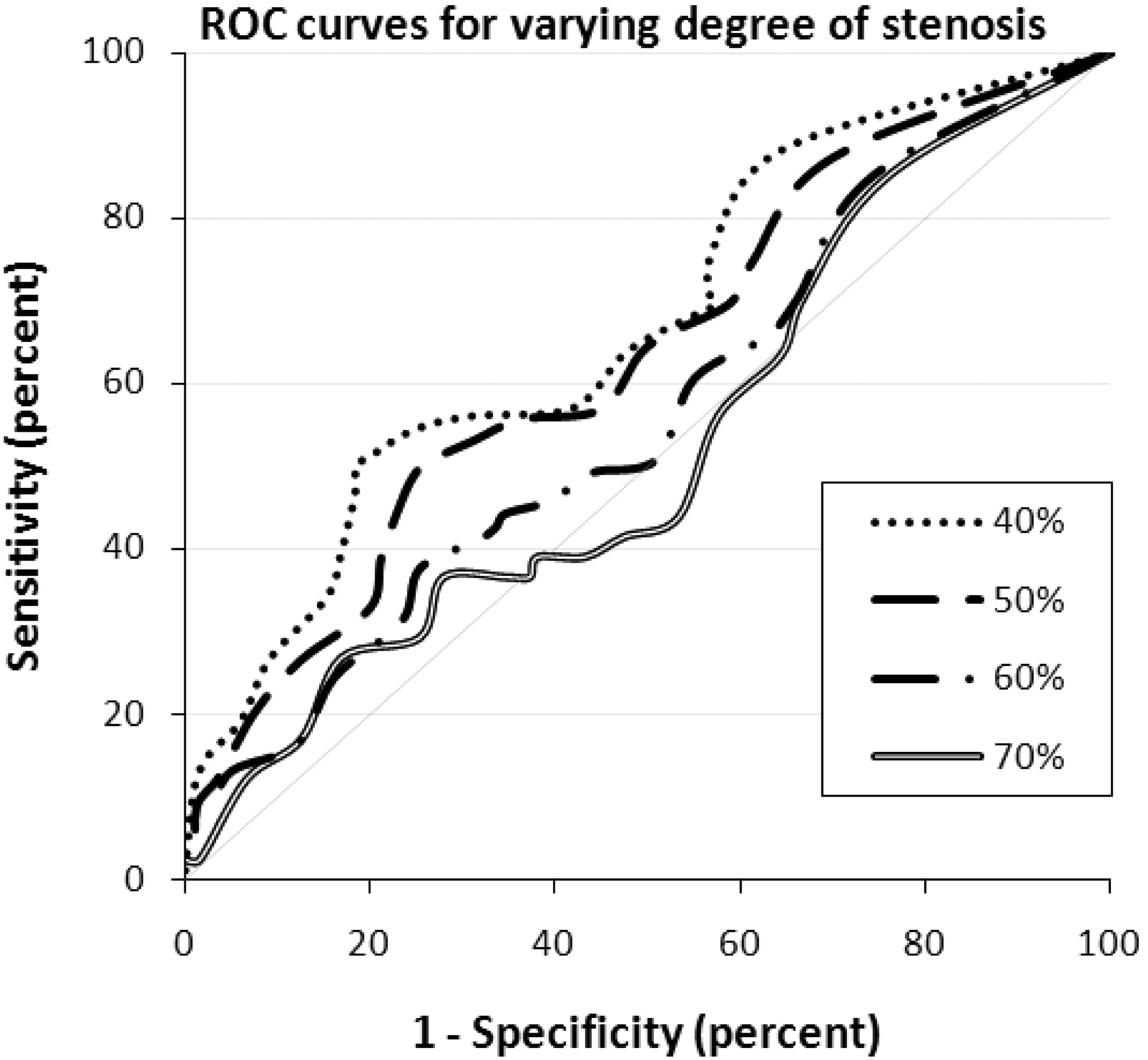
    ROC curves for various calcium volume−based degrees of stenosis determination, illustrating the sensitivity and false-positive rate (1-specificity) for the tests. The best possible test yields a point in the upper left corner; a random guess gives a point along the diagonal line. The higher the curve is from the diagonal, the better the prediction test. This figure illustrates that the performance of the calcium volume−based stenosis determination decreases with increasing stenosis degree.
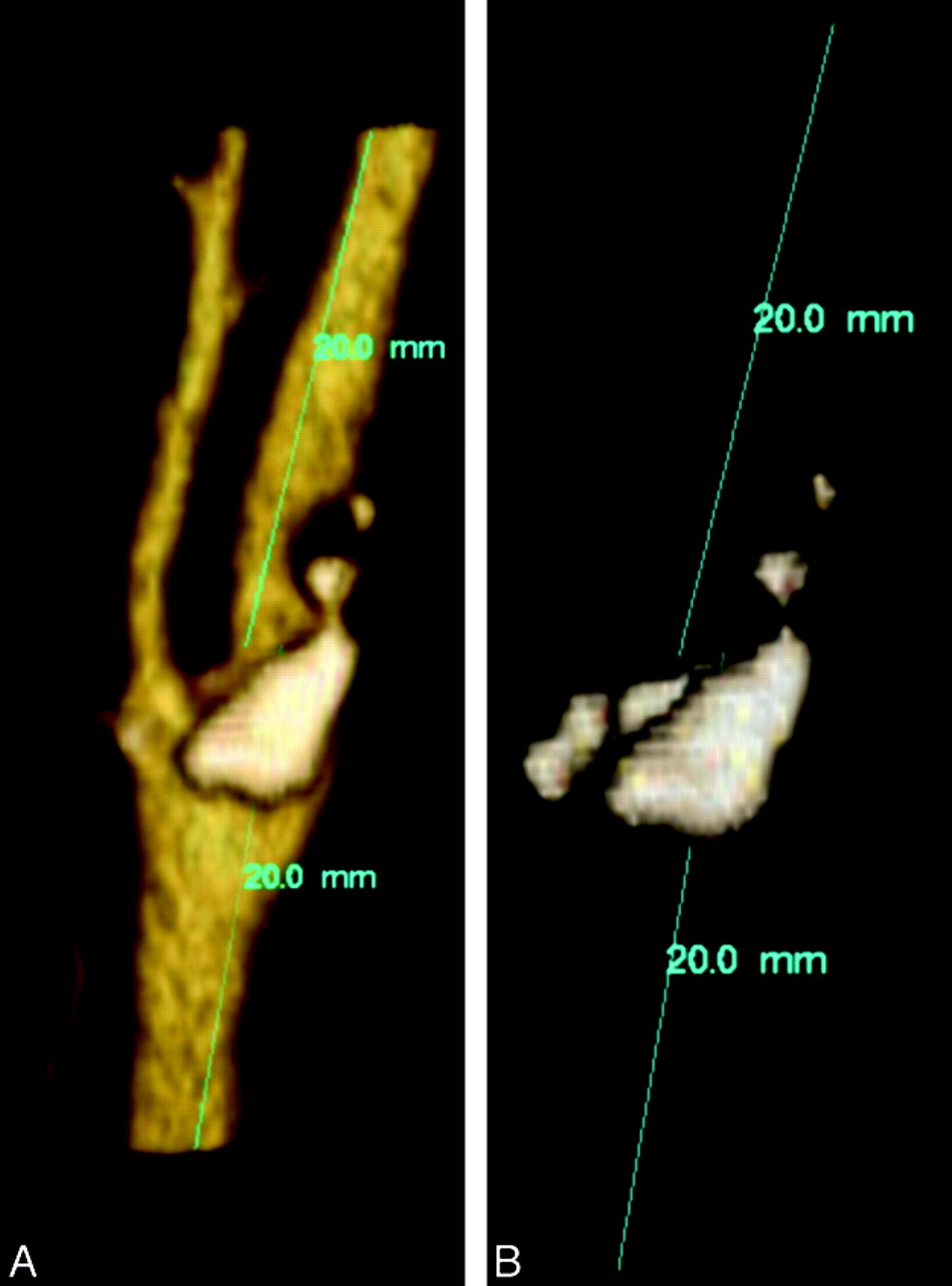
  • Fig 4.
    • Download figure
    • Open in new tab
    • Download powerpoint
    Fig 4.

    VR and MPR reconstructions showing a severe stenosis of 75% according to the NASCET criteria. B shows a display of calcium of the same bifurcation as in A. Total calcium volume is 0.01 mL.
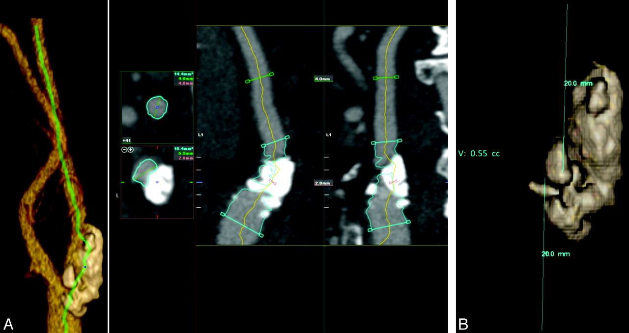
  • Fig 5.
    • Download figure
    • Open in new tab
    • Download powerpoint
    Fig 5.

    VR and MPR reconstructions showing a low-level stenosis of 29% according to the NASCET criteria. B is a display of calcium of the same bifurcation as Fig 4A. Total calcium volume is 0.55 mL.

Tables

  • Figures
    • View popup
    Table 1:

    Calcium volume for stenosis degree groupsa

    Stenosis CategoriesGroup Size (Symptomatic)Average Calcium Volume (mL)
    SymptomaticAsymptomaticAll
    Minimal (0%–29%)49 (8)0.05 (± 0.06)0.05 (± 0.07)0.05 (± 0.06)
    Mild (30%–49%)29 (10)0.11 (± 0.17)0.08 (± 0.10)0.09 (± 0.12)
    Moderate (50%–69%)40 (22)0.12 (± 0.13)0.17 (± 0.16)0.14 (± 0.14)
    Severe (70%–99%)28 (20)0.15 (± 0.19)0.13 (± 0.17)0.15 (± 0.19)
    Occlusion13 (7)0.02 (± 0.03)0.07 (± 0.12)0.04 (± 0.09)
    All159 (67)0.11 (± 0.15)0.09 (± 0.12)0.10 (± 0.13)
    • a In the second column, the number of arteries per category is given. The number of arteries that are labelled “symptomatic side” is given in parentheses. Columns 3–5 give the average calcium volume per artery. The SD is given in parentheses.

    • View popup
    Table 2:

    Average calcium volume, stenosis degree, correlation for symptomatic and asymptomatic arteries, and the combination of botha

    Pearson Correlation (Significance)Average Calcium Volume (mL)Average Degree of Stenosis (%)Group Size
    Symptomatic0.04 (0.7)0.11 (± 0.15)61 (± 26)67
    Asymptomatic0.29 (0.005)0.09 (± 0.12)37 (± 30)92
    All0.20 (0.012)0.10 (± 0.13)47 (± 31)159
    • a The second column displays the Pearson correlation coefficient relating the stenosis degree with the calcium volume. The significance is given in parentheses. The average calcium volume is displayed in the third column with the SD in parentheses. The fourth column shows the average degree of stenosis of the arteries with its SD shown in parentheses.

    • View popup
    Table 3:

    Performance of the calcium volume test determining a stenosis degree of ≥40%a

    Calcium Volume Threshold = 0.03 mLCalcium Volume Threshold = 0.09 mL
    SymptomaticAsymptomaticAllSymptomaticAsymptomaticAll
    Sensitivity63% (49%–74%)66% (50%–79%)64% (54%–73%)48% (36%–61%)45% (30%–60%)47% (37%–57%)
    Specificity73% (43%–90%)48% (35%–61%)52% (40%–64%)73% (43%–90%)83% (71%–91%)82% (70%–89%)
    PPV92% (79%–97%)47% (34%–60%)66% (56%–75%)90% (74%–97%)65% (46%–81%)79% (66%–87%)
    NPV28% (15%–46%)67% (51%–79%)50% (38%–62%)22% (11%–37%)68% (56%–78%)51% (42%–61%)
    • a The test was performed for a calcium-volume threshold of 0.03 mL (left) and 0.09 mL The 95% confidence interval is given in parentheses.

    • View popup
    Table 4:

    Performance of the calcium volume test determining a stenosis degree of ≥50%a

    Calcium Volume Threshold = 0.03 mLCalcium Volume Threshold = 0.09 mL
    SymptomaticAsymptomaticAllSymptomaticAsymptomaticAll
    Sensitivity65% (50%–77%)65% (47%–79%)65% (54%–74%)48% (34%–62%)45% (29%–62%)47% (36%–58%)
    Specificity63% (41%–81%)46% (34%–58%)50% (39%–61%)63% (41%–81%)80% (69%–88%)76% (66%–84%)
    PPV82% (67%–91%)38% (26%–51%)56% (46%–66%)77% (59%–88%)54% (35%–71%)66% (53%–77%)
    NPV41% (26%–59%)72% (56%–83%)59% (47%–70%)32% (20%–49%)74% (63%–83%)59% (50%–68%)
    • a The test has been performed for a calcium volume threshold of 0.03 mL (left) and 0.09 mL (right). The 95% confidence interval is given in parentheses.

    • View popup
    Table 5:

    Performance of the calcium volume test determining a stenosis ≥70%a

    Calcium Volume Threshold = 0.09 mL
    SymptomaticAsymptomaticAll
    Sensitivity41% (25%–59%)29% (12%–55%)37% (24%–52%)
    Specificity53% (37%–67%)72% (61%–81%)65% (56%–73%)
    PPV37% (22%–54%)15% (6%–34%)27% (17%–40%)
    NPV57% (41%–71%)85% (74%–92%)7%5 (66%–82%)
    • a All values of a calcium volume threshold of 0.03 are worse and, therefore, are not presented here.

PreviousNext
Back to top

In this issue

American Journal of Neuroradiology: 32 (7)
American Journal of Neuroradiology
Vol. 32, Issue 7
1 Aug 2011
  • Table of Contents
  • Index by author
Advertisement
Print
Download PDF
Email Article

Thank you for your interest in spreading the word on American Journal of Neuroradiology.

NOTE: We only request your email address so that the person you are recommending the page to knows that you wanted them to see it, and that it is not junk mail. We do not capture any email address.

Enter multiple addresses on separate lines or separate them with commas.
The Relation of Carotid Calcium Volume with Carotid Artery Stenosis in Symptomatic Patients
(Your Name) has sent you a message from American Journal of Neuroradiology
(Your Name) thought you would like to see the American Journal of Neuroradiology web site.
CAPTCHA
This question is for testing whether or not you are a human visitor and to prevent automated spam submissions.
Cite this article
H.A. Marquering, C.B.L.M. Majoie, L. Smagge, A.G. Kurvers, H.A. Gratama van Andel, R. van den Berg, P.J. Nederkoorn
The Relation of Carotid Calcium Volume with Carotid Artery Stenosis in Symptomatic Patients
American Journal of Neuroradiology Aug 2011, 32 (7) 1182-1187; DOI: 10.3174/ajnr.A2519

Citation Manager Formats

  • BibTeX
  • Bookends
  • EasyBib
  • EndNote (tagged)
  • EndNote 8 (xml)
  • Medlars
  • Mendeley
  • Papers
  • RefWorks Tagged
  • Ref Manager
  • RIS
  • Zotero
0 Responses
Respond to this article
Share
Bookmark this article
The Relation of Carotid Calcium Volume with Carotid Artery Stenosis in Symptomatic Patients
H.A. Marquering, C.B.L.M. Majoie, L. Smagge, A.G. Kurvers, H.A. Gratama van Andel, R. van den Berg, P.J. Nederkoorn
American Journal of Neuroradiology Aug 2011, 32 (7) 1182-1187; DOI: 10.3174/ajnr.A2519
del.icio.us logo Twitter logo Facebook logo Mendeley logo
  • Tweet Widget
  • Facebook Like
  • Google Plus One
Purchase

Jump to section

  • Article
    • Abstract
    • Abbreviations
    • Materials and Methods
    • Results
    • Discussion
    • Conclusions
    • Footnotes
    • References
  • Figures & Data
  • Info & Metrics
  • Responses
  • References
  • PDF

Related Articles

  • No related articles found.
  • PubMed
  • Google Scholar

Cited By...

  • Does Vascular Calcification Accelerate Inflammation?: A Substudy of the dal-PLAQUE Trial
  • Diagnostic Accuracy of 4 Commercially Available Semiautomatic Packages for Carotid Artery Stenosis Measurement on CTA
  • Association Between Arterial Calcifications and Nonlacunar and Lacunar Ischemic Strokes
  • Spectrum Bias: Patient Selection May Affect Performance of Calcium Volume in Detecting Carotid Stenosis
  • Reply:
  • Crossref (22)
  • Google Scholar

This article has been cited by the following articles in journals that are participating in Crossref Cited-by Linking.

  • Multimodality Imaging of Vertebrobasilar Dolichoectasia: Clinical Presentations and Imaging Spectrum
    Mohammad Samim, Alan Goldstein, Joseph Schindler, Michele H. Johnson
    RadioGraphics 2016 36 4
  • Does Vascular Calcification Accelerate Inflammation?
    Francis R. Joshi, Nikil K. Rajani, Markus Abt, Mark Woodward, Jan Bucerius, Venkatesh Mani, Ahmed Tawakol, David Kallend, Zahi A. Fayad, James H.F. Rudd
    Journal of the American College of Cardiology 2016 67 1
  • Coronary and carotid atherosclerosis: How useful is the imaging?
    Pranvera Ibrahimi, Fisnik Jashari, Rachel Nicoll, Gani Bajraktari, Per Wester, Michael Y. Henein
    Atherosclerosis 2013 231 2
  • Association Between Arterial Calcifications and Nonlacunar and Lacunar Ischemic Strokes
    Anouk C. van Dijk, Susanne Fonville, Taihra Zadi, Antonius M.G. van Hattem, Ghesrouw Saiedie, Peter J. Koudstaal, Aad van der Lugt
    Stroke 2014 45 3
  • Calcium detection, its quantification, and grayscale morphology-based risk stratification using machine learning in multimodality big data coronary and carotid scans: A review
    Sumit K. Banchhor, Narendra D. Londhe, Tadashi Araki, Luca Saba, Petia Radeva, Narendra N. Khanna, Jasjit S. Suri
    Computers in Biology and Medicine 2018 101
  • Carotid calcification in panoramic radiographs: radiographic appearance and the degree of carotid stenosis
    Maria Garoff, Jan Ahlqvist, Eva Levring Jäghagen, Elias Johansson, Per Wester
    Dentomaxillofacial Radiology 2016 45 6
  • Performance of Semiautomatic Assessment of Carotid Artery Stenosis on CT Angiography: Clarification of Differences with Manual Assessment
    H.A. Marquering, P.J. Nederkoorn, L. Smagge, H.A. Gratama van Andel, R. van den Berg, C.B. Majoie
    American Journal of Neuroradiology 2012 33 4
  • Calcium quantity in carotid plaques: detection in panoramic radiographs and association with degree of stenosis
    Maria Garoff, Elias Johansson, Jan Ahlqvist, Conny Arnerlöv, Eva Levring Jäghagen, Per Wester
    Oral Surgery, Oral Medicine, Oral Pathology and Oral Radiology 2015 120 2
  • Validation of a method for quantifying carotid artery calcification from panoramic radiographs
    Amy C. Alman, Lonnie R. Johnson, David C. Calverley, Gary K. Grunwald, Dennis C. Lezotte, John E. Hokanson
    Oral Surgery, Oral Medicine, Oral Pathology and Oral Radiology 2013 116 4
  • Diagnostic Accuracy of 4 Commercially Available Semiautomatic Packages for Carotid Artery Stenosis Measurement on CTA
    J. Borst, H.A. Marquering, M. Kappelhof, T. Zadi, A.C. van Dijk, P.J. Nederkoorn, R. van den Berg, A. van der Lugt, C.B.L.M. Majoie
    American Journal of Neuroradiology 2015 36 10

More in this TOC Section

  • Correlation of Apparent Diffusion Coefficient at 3T with Prognostic Parameters of Retinoblastoma
  • Parathyroid Lesions: Characterization with Dual-Phase Arterial and Venous Enhanced CT of the Neck
  • MR Diagnosis of Facial Neuritis: Diagnostic Performance of Contrast-Enhanced 3D-FLAIR Technique Compared with Contrast-Enhanced 3D-T1-Fast-Field Echo with Fat Suppression
Show more Head and Neck

Similar Articles

Advertisement

Indexed Content

  • Current Issue
  • Accepted Manuscripts
  • Article Preview
  • Past Issues
  • Editorials
  • Editors Choice
  • Fellow Journal Club
  • Letters to the Editor

Cases

  • Case Collection
  • Archive - Case of the Week
  • Archive - Case of the Month
  • Archive - Classic Case

Special Collections

  • Special Collections

Resources

  • News and Updates
  • Turn around Times
  • Submit a Manuscript
  • Author Policies
  • Manuscript Submission Guidelines
  • Evidence-Based Medicine Level Guide
  • Publishing Checklists
  • Graphical Abstract Preparation
  • Imaging Protocol Submission
  • Submit a Case
  • Become a Reviewer/Academy of Reviewers
  • Get Peer Review Credit from Publons

Multimedia

  • AJNR Podcast
  • AJNR SCANtastic
  • Video Articles

About Us

  • About AJNR
  • Editorial Board
  • Not an AJNR Subscriber? Join Now
  • Alerts
  • Feedback
  • Advertise with us
  • Librarian Resources
  • Permissions
  • Terms and Conditions

American Society of Neuroradiology

  • Not an ASNR Member? Join Now

© 2025 by the American Society of Neuroradiology All rights, including for text and data mining, AI training, and similar technologies, are reserved.
Print ISSN: 0195-6108 Online ISSN: 1936-959X

Powered by HighWire