Advertisement

Main menu

  • Home
  • Content
    • Current Issue
    • Accepted Manuscripts
    • Article Preview
    • Past Issue Archive
    • AJNR Case Collection
    • Case of the Week Archive
    • Classic Case Archive
    • Case of the Month Archive
  • Special Collections
    • Spinal CSF Leak Articles (Jan 2020-June 2024)
    • 2024 AJNR Journal Awards
    • Most Impactful AJNR Articles
  • Multimedia
    • AJNR Podcast
    • AJNR Scantastics
    • Video Articles
  • For Authors
    • Submit a Manuscript
    • Author Policies
    • Fast publishing of Accepted Manuscripts
    • Graphical Abstract Preparation
    • Manuscript Submission Guidelines
    • Imaging Protocol Submission
    • Submit a Case for the Case Collection
  • About Us
    • About AJNR
    • Editorial Board
  • More
    • Become a Reviewer/Academy of Reviewers
    • Subscribers
    • Permissions
    • Alerts
    • Feedback
    • Advertisers
    • ASNR Home
  • Other Publications
    • ajnr

User menu

  • Alerts
  • Log in

Search

  • Advanced search
American Journal of Neuroradiology

American Journal of Neuroradiology

ASHNR American Society of Functional Neuroradiology ASHNR American Society of Pediatric Neuroradiology ASSR
  • Alerts
  • Log in

Advanced Search

  • Home
  • Content
    • Current Issue
    • Accepted Manuscripts
    • Article Preview
    • Past Issue Archive
    • AJNR Case Collection
    • Case of the Week Archive
    • Classic Case Archive
    • Case of the Month Archive
  • Special Collections
    • Spinal CSF Leak Articles (Jan 2020-June 2024)
    • 2024 AJNR Journal Awards
    • Most Impactful AJNR Articles
  • Multimedia
    • AJNR Podcast
    • AJNR Scantastics
    • Video Articles
  • For Authors
    • Submit a Manuscript
    • Author Policies
    • Fast publishing of Accepted Manuscripts
    • Graphical Abstract Preparation
    • Manuscript Submission Guidelines
    • Imaging Protocol Submission
    • Submit a Case for the Case Collection
  • About Us
    • About AJNR
    • Editorial Board
  • More
    • Become a Reviewer/Academy of Reviewers
    • Subscribers
    • Permissions
    • Alerts
    • Feedback
    • Advertisers
    • ASNR Home
  • Follow AJNR on Twitter
  • Visit AJNR on Facebook
  • Follow AJNR on Instagram
  • Join AJNR on LinkedIn
  • RSS Feeds

Welcome to the new AJNR, Updated Hall of Fame, and more. Read the full announcements.


AJNR is seeking candidates for the position of Associate Section Editor, AJNR Case Collection. Read the full announcement.


Research ArticleHead and Neck
Open Access

Diffusion Tensor Imaging of the Optic Nerve in Subacute Anterior Ischemic Optic Neuropathy at 3T

M.-Y. Wang, P.-H. Qi and D.-P. Shi
American Journal of Neuroradiology August 2011, 32 (7) 1188-1194; DOI: https://doi.org/10.3174/ajnr.A2487
M.-Y. Wang
  • Find this author on Google Scholar
  • Find this author on PubMed
  • Search for this author on this site
P.-H. Qi
  • Find this author on Google Scholar
  • Find this author on PubMed
  • Search for this author on this site
D.-P. Shi
  • Find this author on Google Scholar
  • Find this author on PubMed
  • Search for this author on this site
  • Article
  • Figures & Data
  • Info & Metrics
  • Responses
  • References
  • PDF
Loading

Article Figures & Data

Figures

  • Tables
  • Fig 1.
    • Download figure
    • Open in new tab
    • Download powerpoint
    Fig 1.

    A−C, DTI maps of the central section of the ON. The regions of interest are placed on the b=0 averaged images, which are defined in the coronal map (A) and adjusted in the sagittal (B) and axial (C) images obtained by using multiplanar reconstruction. D−G, Then the regions of interest are transferred onto the ADC (D), FA (E), λ‖ (F), and λ⊥ (G) maps.
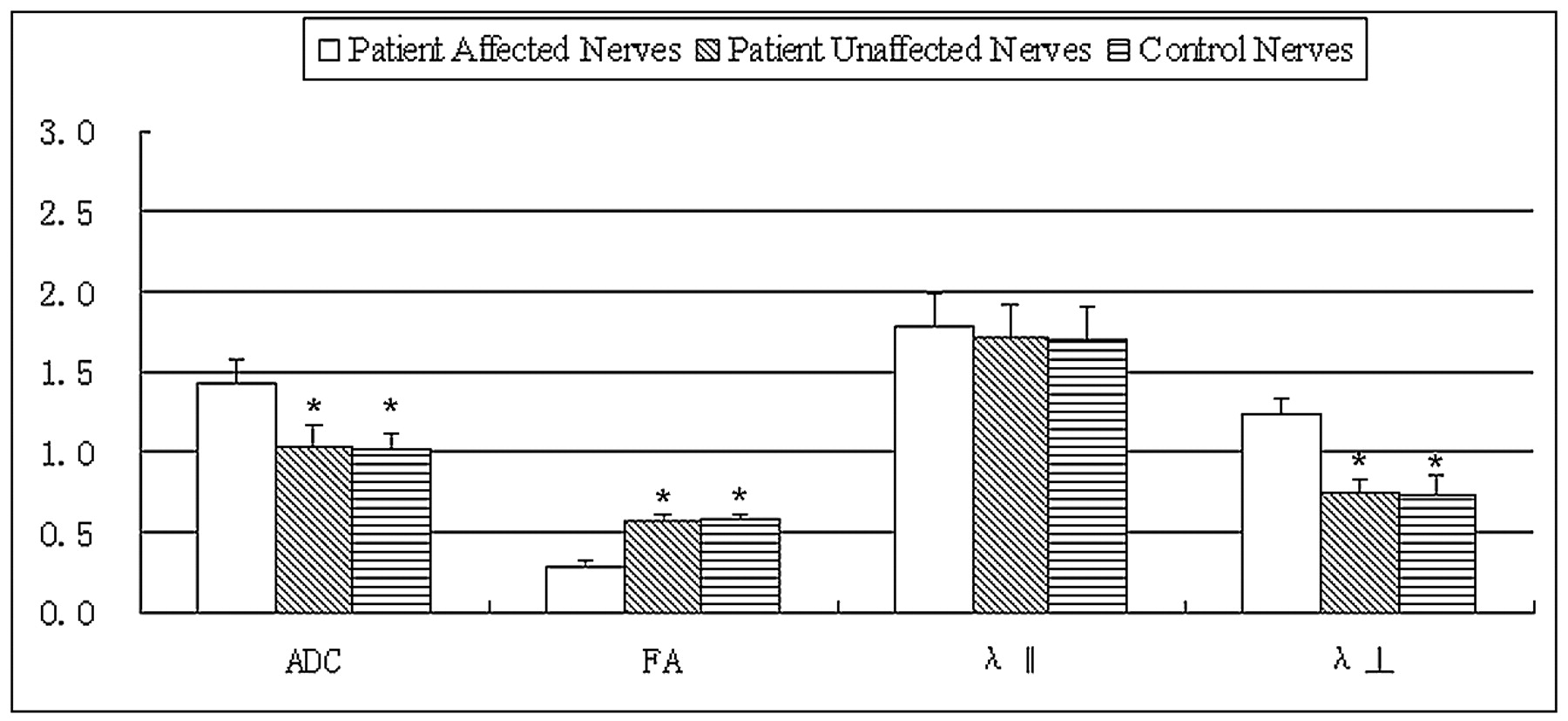
  • Fig 2.
    • Download figure
    • Open in new tab
    • Download powerpoint
    Fig 2.

    Bar graphs of the ADC, FA, principal eigenvalue λ‖, and the orthogonal eigenvalues λ⊥ averaged across the affected and unaffected contralateral ONs of 26 patients with ION compared with normal ONs of 15 controls. The error bars denote the SDs across subjects. The units of the ADC, λ‖, and λ⊥ measures are in ×10−3 square millimeters per second −1. The asterisk indicates a significant difference compared with the affected nerves of patients. There is no significant difference between unaffected ONs of patients and ONs of controls in all DTI measures.

  • Fig 3.
    • Download figure
    • Open in new tab
    • Download powerpoint
    Fig 3.

    Plot of ADC (A) and the orthogonal eigenvalue λ⊥ (B) versus whole-field VEP amplitude averaged across the pixels inside the regions of interest along the affected ONs of 26 patients with ischemic ION, respectively.

Tables

  • Figures
    • View popup
    Table 1:

    Demographic data and visual function of patientsa

    MeanSDRangeRatio
    Male/female–––14:12
    Age (yr)53.239.5327–69–
    Timeb20.658.846–30–
    Standard logarithmic visual acuity
        Affected0.220.210.01–0.80–
        Not affected0.970.160.6–1.2–
    Visual field mean deviation (dB)
        Affected22.985.9510.93–32–
        Not affected1.721.870.10–7.70–
    • a —indicates patients with AION.

    • b Since onset of AION (days).

    • View popup
    Table 2:

    Demographic data and visual function of controls

    MeanSDRangeRatio
    Male/female8:7
    Age (yr)508.2330–58
    Standard logarithmic visual acuity, selected1.120.110.8–1.4
    Visual field mean deviation (dB), selected1.561.220.1–6.2
    • View popup
    Table 3:

    DTI parameter and electrophysiologic group dataa

    FAb,cADC (×10−6 mm2s−1)b,cλ‖(×10−6 mm2s−1)λ⊥(×10−6 mm2s−1)b,cWhole-Field VEP Amplitude (μV)b,cWhole-Field VEP Latency (ms)b,c
    Patients' affected nerves (n = 26)0.283 ± 0.0731429 ± 1981786 ± 1591245 ± 1724.53 ± 1.17132.7 ± 17.48
    Patients' unaffected nerves (n = 26)0.569 ± 0.0461028 ± 1461720 ± 199746 ± 789.67 ± 2.5294.86 ± 9.07
    Control nerves (n = 15)0.587 ± 0.0231008 ± 1111702 ± 199729 ± 1309.40 ± 3.0397.77 ± 4.62
    • a The values are mean ± SD.

    • b Significant difference between nerves of controls and affected nerves of patients.

    • c Significant difference between unaffected contralateral nerves and affected nerves in patients. There is no significant difference between unaffected ONs of patients and ONs of controls in all DTI measures.

PreviousNext
Back to top

In this issue

American Journal of Neuroradiology: 32 (7)
American Journal of Neuroradiology
Vol. 32, Issue 7
1 Aug 2011
  • Table of Contents
  • Index by author
Advertisement
Print
Download PDF
Email Article

Thank you for your interest in spreading the word on American Journal of Neuroradiology.

NOTE: We only request your email address so that the person you are recommending the page to knows that you wanted them to see it, and that it is not junk mail. We do not capture any email address.

Enter multiple addresses on separate lines or separate them with commas.
Diffusion Tensor Imaging of the Optic Nerve in Subacute Anterior Ischemic Optic Neuropathy at 3T
(Your Name) has sent you a message from American Journal of Neuroradiology
(Your Name) thought you would like to see the American Journal of Neuroradiology web site.
CAPTCHA
This question is for testing whether or not you are a human visitor and to prevent automated spam submissions.
Cite this article
M.-Y. Wang, P.-H. Qi, D.-P. Shi
Diffusion Tensor Imaging of the Optic Nerve in Subacute Anterior Ischemic Optic Neuropathy at 3T
American Journal of Neuroradiology Aug 2011, 32 (7) 1188-1194; DOI: 10.3174/ajnr.A2487

Citation Manager Formats

  • BibTeX
  • Bookends
  • EasyBib
  • EndNote (tagged)
  • EndNote 8 (xml)
  • Medlars
  • Mendeley
  • Papers
  • RefWorks Tagged
  • Ref Manager
  • RIS
  • Zotero
0 Responses
Respond to this article
Share
Bookmark this article
Diffusion Tensor Imaging of the Optic Nerve in Subacute Anterior Ischemic Optic Neuropathy at 3T
M.-Y. Wang, P.-H. Qi, D.-P. Shi
American Journal of Neuroradiology Aug 2011, 32 (7) 1188-1194; DOI: 10.3174/ajnr.A2487
del.icio.us logo Twitter logo Facebook logo Mendeley logo
  • Tweet Widget
  • Facebook Like
  • Google Plus One
Purchase

Jump to section

  • Article
    • Abstract
    • Abbreviations
    • Materials and Methods
    • Results
    • Discussion
    • Conclusions
    • References
  • Figures & Data
  • Info & Metrics
  • Responses
  • References
  • PDF

Related Articles

  • No related articles found.
  • PubMed
  • Google Scholar

Cited By...

  • The Central Bright Spot Sign: A Potential New MR Imaging Sign for the Early Diagnosis of Anterior Ischemic Optic Neuropathy due to Giant Cell Arteritis
  • Reduced Field-of-View Diffusion Tensor Imaging of the Optic Nerve in Retinitis Pigmentosa at 3T
  • Crossref (24)
  • Google Scholar

This article has been cited by the following articles in journals that are participating in Crossref Cited-by Linking.

  • Effects of white matter microstructure on phase and susceptibility maps
    Samuel Wharton, Richard Bowtell
    Magnetic Resonance in Medicine 2015 73 3
  • Reduced-FOV excitation decreases susceptibility artifact in diffusion-weighted MRI with endorectal coil for prostate cancer detection
    Natalie Korn, John Kurhanewicz, Suchandrima Banerjee, Olga Starobinets, Emine Saritas, Susan Noworolski
    Magnetic Resonance Imaging 2015 33 1
  • Diffusion restriction of the optic nerve in patients with acute visual deficit
    Benjamin Bender, Christian Heine, Sören Danz, Felix Bischof, Katrin Reimann, Miriam Bender, Thomas Nägele, Ulrike Ernemann, Andreas Korn
    Journal of Magnetic Resonance Imaging 2014 40 2
  • Quantitative 3-T diffusion tensor imaging in detecting optic nerve degeneration in patients with glaucoma: association with retinal nerve fiber layer thickness and clinical severity
    Mei-Yun Wang, Ke Wu, Jun-Min Xu, Jianping Dai, Wei Qin, Jiangang Liu, Jie Tian, Dapeng Shi
    Neuroradiology 2013 55 4
  • The Central Bright Spot Sign: A Potential New MR Imaging Sign for the Early Diagnosis of Anterior Ischemic Optic Neuropathy due to Giant Cell Arteritis
    P. Remond, A. Attyé, A. Lecler, L. Lamalle, N. Boudiaf, F. Aptel, A. Krainik, C. Chiquet
    American Journal of Neuroradiology 2017 38 7
  • Diffusion-Weighted Imaging and Post-contrast Enhancement in Differentiating Optic Neuritis and Non-arteritic Anterior Optic Neuropathy
    Ore-ofe O. Adesina, J. Scott McNally, Karen L. Salzman, Bradley J. Katz, Judith E. A. Warner, Molly McFadden, Kathleen B. Digre
    Neuro-Ophthalmology 2018 42 2
  • Thyroid-Associated Orbitopathy: Evaluating Microstructural Changes of Extraocular Muscles and Optic Nerves Using Readout-Segmented Echo-Planar Imaging-Based Diffusion Tensor Imaging
    Huan-Huan Chen, Hao Hu, Wen Chen, Dai Cui, Xiao-Quan Xu, Fei-Yun Wu, Tao Yang
    Korean Journal of Radiology 2020 21 3
  • Quantitative assessment of optic nerve in patients with Leber’s hereditary optic neuropathy using reduced field-of-view diffusion tensor imaging
    Ling Wang, Ke Fan, Yanqiu Zhang, Yanzi Chen, Qin Tian, Dapeng Shi
    European Journal of Radiology 2017 93
  • Reduced Field-of-View Diffusion Tensor Imaging of the Optic Nerve in Retinitis Pigmentosa at 3T
    Y. Zhang, X. Guo, M. Wang, L. Wang, Q. Tian, D. Zheng, D. Shi
    American Journal of Neuroradiology 2016 37 8
  • Diffusion-weighted imaging in posterior ischemic optic neuropathy
    Nagham Al-Zubidi, Scott Stevens, Steve H. Fung, Andrew G. Lee
    Canadian Journal of Ophthalmology 2014 49 1

More in this TOC Section

  • Correlation of Apparent Diffusion Coefficient at 3T with Prognostic Parameters of Retinoblastoma
  • Parathyroid Lesions: Characterization with Dual-Phase Arterial and Venous Enhanced CT of the Neck
  • Comparison of Contrast Effect on the Cochlear Perilymph after Intratympanic and Intravenous Gadolinium Injection
Show more Head and Neck

Similar Articles

Advertisement

Indexed Content

  • Current Issue
  • Accepted Manuscripts
  • Article Preview
  • Past Issues
  • Editorials
  • Editors Choice
  • Fellow Journal Club
  • Letters to the Editor

Cases

  • Case Collection
  • Archive - Case of the Week
  • Archive - Case of the Month
  • Archive - Classic Case

Special Collections

  • Special Collections

Resources

  • News and Updates
  • Turn around Times
  • Submit a Manuscript
  • Author Policies
  • Manuscript Submission Guidelines
  • Evidence-Based Medicine Level Guide
  • Publishing Checklists
  • Graphical Abstract Preparation
  • Imaging Protocol Submission
  • Submit a Case
  • Become a Reviewer/Academy of Reviewers
  • Get Peer Review Credit from Publons

Multimedia

  • AJNR Podcast
  • AJNR SCANtastic
  • Video Articles

About Us

  • About AJNR
  • Editorial Board
  • Not an AJNR Subscriber? Join Now
  • Alerts
  • Feedback
  • Advertise with us
  • Librarian Resources
  • Permissions
  • Terms and Conditions

American Society of Neuroradiology

  • Not an ASNR Member? Join Now

© 2025 by the American Society of Neuroradiology All rights, including for text and data mining, AI training, and similar technologies, are reserved.
Print ISSN: 0195-6108 Online ISSN: 1936-959X

Powered by HighWire