Advertisement

Main menu

  • Home
  • Content
    • Current Issue
    • Accepted Manuscripts
    • Article Preview
    • Past Issue Archive
    • AJNR Case Collection
    • Case of the Week Archive
    • Classic Case Archive
    • Case of the Month Archive
  • Special Collections
    • Spinal CSF Leak Articles (Jan 2020-June 2024)
    • 2024 AJNR Journal Awards
    • Most Impactful AJNR Articles
  • Multimedia
    • AJNR Podcast
    • AJNR Scantastics
    • Video Articles
  • For Authors
    • Submit a Manuscript
    • Author Policies
    • Fast publishing of Accepted Manuscripts
    • Graphical Abstract Preparation
    • Manuscript Submission Guidelines
    • Imaging Protocol Submission
    • Submit a Case for the Case Collection
  • About Us
    • About AJNR
    • Editorial Board
  • More
    • Become a Reviewer/Academy of Reviewers
    • Subscribers
    • Permissions
    • Alerts
    • Feedback
    • Advertisers
    • ASNR Home
  • Other Publications
    • ajnr

User menu

  • Alerts
  • Log in

Search

  • Advanced search
American Journal of Neuroradiology

American Journal of Neuroradiology

ASHNR American Society of Functional Neuroradiology ASHNR American Society of Pediatric Neuroradiology ASSR
  • Alerts
  • Log in

Advanced Search

  • Home
  • Content
    • Current Issue
    • Accepted Manuscripts
    • Article Preview
    • Past Issue Archive
    • AJNR Case Collection
    • Case of the Week Archive
    • Classic Case Archive
    • Case of the Month Archive
  • Special Collections
    • Spinal CSF Leak Articles (Jan 2020-June 2024)
    • 2024 AJNR Journal Awards
    • Most Impactful AJNR Articles
  • Multimedia
    • AJNR Podcast
    • AJNR Scantastics
    • Video Articles
  • For Authors
    • Submit a Manuscript
    • Author Policies
    • Fast publishing of Accepted Manuscripts
    • Graphical Abstract Preparation
    • Manuscript Submission Guidelines
    • Imaging Protocol Submission
    • Submit a Case for the Case Collection
  • About Us
    • About AJNR
    • Editorial Board
  • More
    • Become a Reviewer/Academy of Reviewers
    • Subscribers
    • Permissions
    • Alerts
    • Feedback
    • Advertisers
    • ASNR Home
  • Follow AJNR on Twitter
  • Visit AJNR on Facebook
  • Follow AJNR on Instagram
  • Join AJNR on LinkedIn
  • RSS Feeds

Welcome to the new AJNR, Updated Hall of Fame, and more. Read the full announcements.


AJNR is seeking candidates for the position of Associate Section Editor, AJNR Case Collection. Read the full announcement.


Research ArticleBrain

Assessment of Lateral Geniculate Nucleus Atrophy with 3T MR Imaging and Correlation with Clinical Stage of Glaucoma

H. Dai, K.T. Mu, J.P. Qi, C.Y. Wang, W.Z. Zhu, L.M. Xia, Z.Q. Chen, H. Zhang, F. Ai and J.N. Morelli
American Journal of Neuroradiology August 2011, 32 (7) 1347-1353; DOI: https://doi.org/10.3174/ajnr.A2486
H. Dai
  • Find this author on Google Scholar
  • Find this author on PubMed
  • Search for this author on this site
K.T. Mu
  • Find this author on Google Scholar
  • Find this author on PubMed
  • Search for this author on this site
J.P. Qi
  • Find this author on Google Scholar
  • Find this author on PubMed
  • Search for this author on this site
C.Y. Wang
  • Find this author on Google Scholar
  • Find this author on PubMed
  • Search for this author on this site
W.Z. Zhu
  • Find this author on Google Scholar
  • Find this author on PubMed
  • Search for this author on this site
L.M. Xia
  • Find this author on Google Scholar
  • Find this author on PubMed
  • Search for this author on this site
Z.Q. Chen
  • Find this author on Google Scholar
  • Find this author on PubMed
  • Search for this author on this site
H. Zhang
  • Find this author on Google Scholar
  • Find this author on PubMed
  • Search for this author on this site
F. Ai
  • Find this author on Google Scholar
  • Find this author on PubMed
  • Search for this author on this site
J.N. Morelli
  • Find this author on Google Scholar
  • Find this author on PubMed
  • Search for this author on this site
  • Article
  • Figures & Data
  • Info & Metrics
  • Responses
  • References
  • PDF
Loading

Article Figures & Data

Figures

  • Tables
  • Fig 1.
    • Download figure
    • Open in new tab
    • Download powerpoint
    Fig 1.

    The pie chart shows the distribution of 52 eyes in 26 patients into different clinical stages.

  • Fig 2.
    • Download figure
    • Open in new tab
    • Download powerpoint
    Fig 2.

    Oblique coronal localizer sequence demonstrates the orientation of acquired images for both the PD and GM sequences, perpendicular to the long axis of the temporal lobe or medial hippocampus.

  • Fig 3.
    • Download figure
    • Open in new tab
    • Download powerpoint
    Fig 3.

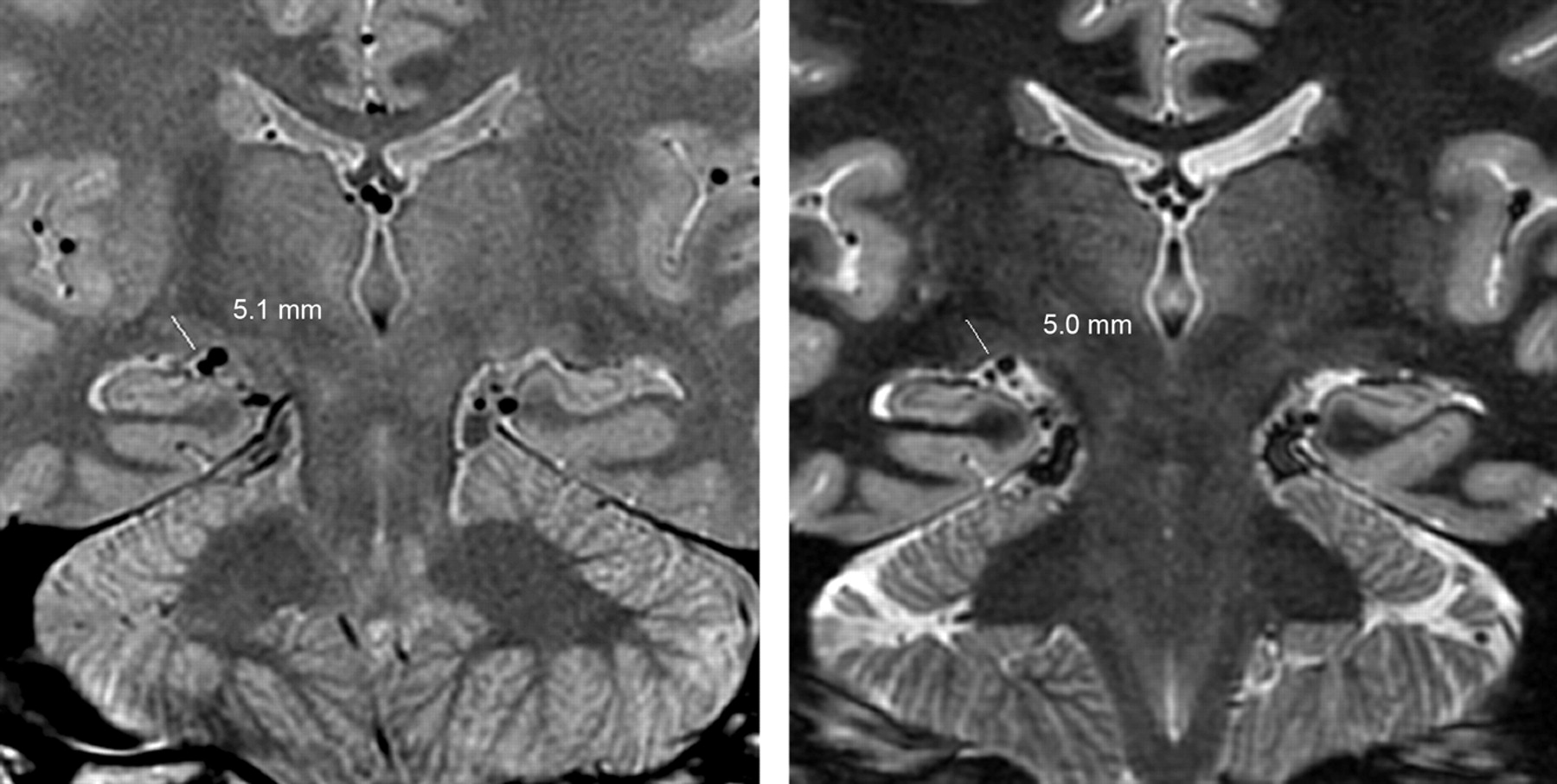
    PD (left) and GM (right) images obtained in the identical oblique coronal plane demonstrate LGN height measurements in a 43-year-old healthy male volunteer. Specifically, LGN height is determined by drawing a line from the apex of the convexity perpendicular to the base of the LGN. The left image is from the PD sequence, and the right one, from GM sequence.

  • Fig 4.
    • Download figure
    • Open in new tab
    • Download powerpoint
    Fig 4.

    A 36-year-old man with bilateral stage 4 open-angle glaucoma demonstrates bilateral LGN atrophy as shown on both PD (left) and GM (right) sequences.

  • Fig 5.
    • Download figure
    • Open in new tab
    • Download powerpoint
    Fig 5.

    GM images demonstrate visualization of the LGN on 2 different oblique coronal sections in a healthy volunteer and show outlining of the LGN as performed for volume analysis.

  • Fig 6.
    • Download figure
    • Open in new tab
    • Download powerpoint
    Fig 6.

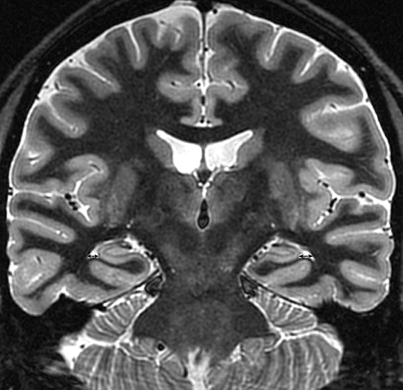
    Temporal horns are measured on the GM sequence as a control brain region used to correct all the measurements of LGN size obtained from the PD and GM sequences.

  • Fig 7.
    • Download figure
    • Open in new tab
    • Download powerpoint
    Fig 7.

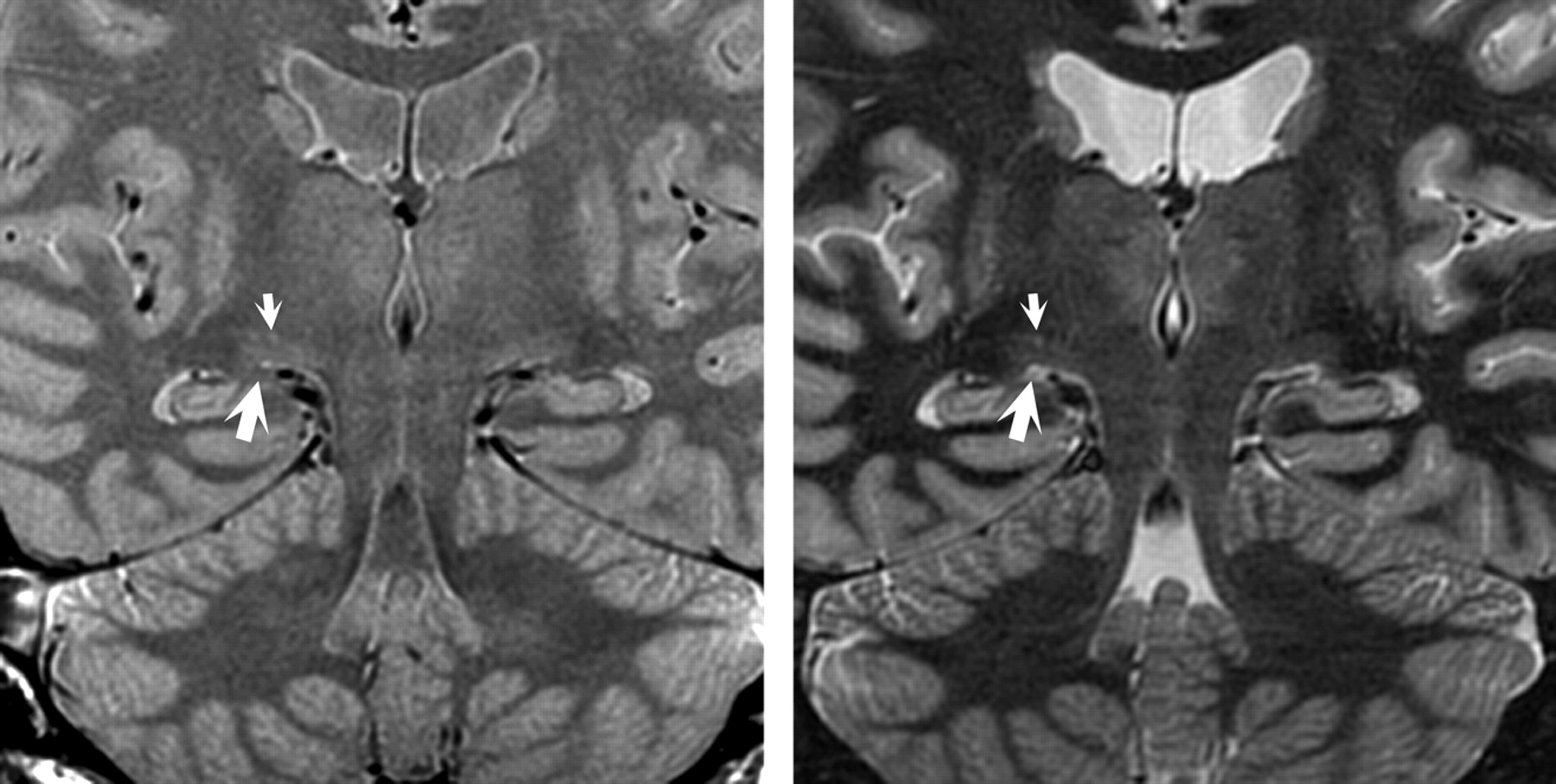
    Clearer demarcation of gray and white matter is demonstrated in the GM sequence image (right), compared with the PD sequence image (left). Higher signal intensity of the Virchow-Robin space (small white arrows) and the CSF (large white arrows) adjacent to the LGN is notable on the GM sequences.

Tables

  • Figures
    • View popup
    Table 1:

    Comparison of LGN size in patients with glaucoma and volunteers as assessed by various parametersa,b

    PDhRPDhLPDvRPDvLGMhRGMhLGMvRGMvL
    Patients4.36 ± 0.614.31 ± 0.6198.0 ± 27.293.7 ± 25.84.20 ± 0.714.00 ± 0.8585.2 ± 27.180.5 ± 23.6
    Controls5.05 ± 0.414.99 ± 0.41143.5 ± 22.3143.1 ± 19.74.88 ± 0.514.77 ± 0.47131.7 ± 18.5129.6 ± 21.0
    • a Covariates appearing in the model are evaluated at the following value: temporal horn width (cumulative) = 4.3885.

    • b All P values < 10−3.

    • View popup
    Table 2:

    Correlation between LGN size and glaucoma stage

    StagePDhRPDhLSumPDvRPDvLSumGMhRGMhLSumGMvRGMvLSum
    L−.353a,b−.428−.405−.468−.525−.523−.400−.512−.473−.499−.478−.512
    .077b,c.029.040.016.006.006.043.008.015.010.014.008
    R−.374b−.514−.461−.460−.401−.455−.330b−.395−.375b−.350b−.386b−.379b
    .060c.007.018.018.042.020.099c.046.059c.080c.093c.057c
    A−.455−.589−.541−.581−.582−.613−.458−.570−.533−.547−.520−.560
    .019.002.004.002.002.001.019.002.005.004.006.003
    • a r value.

    • b Parameters without significant difference (P> .05).

    • c P value.

PreviousNext
Back to top

In this issue

American Journal of Neuroradiology: 32 (7)
American Journal of Neuroradiology
Vol. 32, Issue 7
1 Aug 2011
  • Table of Contents
  • Index by author
Advertisement
Print
Download PDF
Email Article

Thank you for your interest in spreading the word on American Journal of Neuroradiology.

NOTE: We only request your email address so that the person you are recommending the page to knows that you wanted them to see it, and that it is not junk mail. We do not capture any email address.

Enter multiple addresses on separate lines or separate them with commas.
Assessment of Lateral Geniculate Nucleus Atrophy with 3T MR Imaging and Correlation with Clinical Stage of Glaucoma
(Your Name) has sent you a message from American Journal of Neuroradiology
(Your Name) thought you would like to see the American Journal of Neuroradiology web site.
CAPTCHA
This question is for testing whether or not you are a human visitor and to prevent automated spam submissions.
Cite this article
H. Dai, K.T. Mu, J.P. Qi, C.Y. Wang, W.Z. Zhu, L.M. Xia, Z.Q. Chen, H. Zhang, F. Ai, J.N. Morelli
Assessment of Lateral Geniculate Nucleus Atrophy with 3T MR Imaging and Correlation with Clinical Stage of Glaucoma
American Journal of Neuroradiology Aug 2011, 32 (7) 1347-1353; DOI: 10.3174/ajnr.A2486

Citation Manager Formats

  • BibTeX
  • Bookends
  • EasyBib
  • EndNote (tagged)
  • EndNote 8 (xml)
  • Medlars
  • Mendeley
  • Papers
  • RefWorks Tagged
  • Ref Manager
  • RIS
  • Zotero
0 Responses
Respond to this article
Share
Bookmark this article
Assessment of Lateral Geniculate Nucleus Atrophy with 3T MR Imaging and Correlation with Clinical Stage of Glaucoma
H. Dai, K.T. Mu, J.P. Qi, C.Y. Wang, W.Z. Zhu, L.M. Xia, Z.Q. Chen, H. Zhang, F. Ai, J.N. Morelli
American Journal of Neuroradiology Aug 2011, 32 (7) 1347-1353; DOI: 10.3174/ajnr.A2486
del.icio.us logo Twitter logo Facebook logo Mendeley logo
  • Tweet Widget
  • Facebook Like
  • Google Plus One
Purchase

Jump to section

  • Article
    • Abstract
    • Abbreviations
    • Materials and Methods
    • Results
    • Discussion
    • Conclusions
    • References
  • Figures & Data
  • Info & Metrics
  • Responses
  • References
  • PDF

Related Articles

  • No related articles found.
  • PubMed
  • Google Scholar

Cited By...

  • Retinotopic remapping of the visual system in deaf adults
  • Damage of the lateral geniculate nucleus in MS: Assessing the missing node of the visual pathway
  • Visualization of the Medial and Lateral Geniculate Nucleus on Phase Difference Enhanced Imaging
  • An Investigation of Lateral Geniculate Nucleus Volume in Patients With Primary Open-Angle Glaucoma Using 7 Tesla Magnetic Resonance Imaging
  • Structural Brain Abnormalities in Patients with Primary Open-Angle Glaucoma: A Study with 3T MR Imaging
  • Crossref (52)
  • Google Scholar

This article has been cited by the following articles in journals that are participating in Crossref Cited-by Linking.

  • Glaucoma and the brain: Trans-synaptic degeneration, structural change, and implications for neuroprotection
    Mitchell Lawlor, Helen Danesh-Meyer, Leonard A. Levin, Indran Davagnanam, Enrico De Vita, Gordon T. Plant
    Survey of Ophthalmology 2018 63 3
  • Neuroprotective agents in the management of glaucoma
    C. Nucci, A. Martucci, C. Giannini, L. A. Morrone, G. Bagetta, R. Mancino
    Eye 2018 32 5
  • Glaucoma as a Neurodegenerative Disease
    Helen V. Danesh-Meyer, Leonard A. Levin
    Journal of Neuro-Ophthalmology 2015 35 Supplement 1
  • Glaucoma: A Degenerative Optic Neuropathy Related to Neuroinflammation?
    Stéphane Mélik Parsadaniantz, Annabelle Réaux-le Goazigo, Anaïs Sapienza, Christophe Habas, Christophe Baudouin
    Cells 2020 9 3
  • Brain involvement in glaucoma: advanced neuroimaging for understanding and monitoring a new target for therapy
    Carlo Nucci, Alessio Martucci, Massimo Cesareo, Raffaele Mancino, Rossella Russo, Giacinto Bagetta, Luciano Cerulli, Francesco G Garaci
    Current Opinion in Pharmacology 2013 13 1
  • Altered Spontaneous Brain Activity in Primary Open Angle Glaucoma: A Resting-State Functional Magnetic Resonance Imaging Study
    Yinwei Song, Ketao Mu, Junming Wang, Fuchun Lin, Zhiqi Chen, Xiaoqin Yan, Yonghong Hao, Wenzhen Zhu, Hong Zhang, Sanjoy Bhattacharya
    PLoS ONE 2014 9 2
  • Selective reduction of fMRI responses to transient achromatic stimuli in the magnocellular layers of the LGN and the superficial layer of the SC of early glaucoma patients
    Peng Zhang, Wen Wen, Xinghuai Sun, Sheng He
    Human Brain Mapping 2016 37 2
  • Whole-brain voxel-based analysis of diffusion tensor MRI parameters in patients with primary open angle glaucoma and correlation with clinical glaucoma stage
    Hui Dai, Dazhi Yin, Chunhong Hu, John N. Morelli, Su Hu, Xu Yan, Dongrong Xu
    Neuroradiology 2013 55 2
  • In vivo Probabilistic Structural Atlas of the Inferior and Superior Colliculi, Medial and Lateral Geniculate Nuclei and Superior Olivary Complex in Humans Based on 7 Tesla MRI
    María G. García-Gomar, Christian Strong, Nicola Toschi, Kavita Singh, Bruce R. Rosen, Lawrence L. Wald, Marta Bianciardi
    Frontiers in Neuroscience 2019 13
  • Congenital blindness affects diencephalic but not mesencephalic structures in the human brain
    Luca Cecchetti, Emiliano Ricciardi, Giacomo Handjaras, Ron Kupers, Maurice Ptito, Pietro Pietrini
    Brain Structure and Function 2016 221 3

More in this TOC Section

  • Fast Contrast-Enhanced 4D MRA and 4D Flow MRI Using Constrained Reconstruction (HYPRFlow): Potential Applications for Brain Arteriovenous Malformations
  • Quiet PROPELLER MRI Techniques Match the Quality of Conventional PROPELLER Brain Imaging Techniques
  • Predictors of Reperfusion in Patients with Acute Ischemic Stroke
Show more Brain

Similar Articles

Advertisement

Indexed Content

  • Current Issue
  • Accepted Manuscripts
  • Article Preview
  • Past Issues
  • Editorials
  • Editors Choice
  • Fellow Journal Club
  • Letters to the Editor

Cases

  • Case Collection
  • Archive - Case of the Week
  • Archive - Case of the Month
  • Archive - Classic Case

Special Collections

  • Special Collections

Resources

  • News and Updates
  • Turn around Times
  • Submit a Manuscript
  • Author Policies
  • Manuscript Submission Guidelines
  • Evidence-Based Medicine Level Guide
  • Publishing Checklists
  • Graphical Abstract Preparation
  • Imaging Protocol Submission
  • Submit a Case
  • Become a Reviewer/Academy of Reviewers
  • Get Peer Review Credit from Publons

Multimedia

  • AJNR Podcast
  • AJNR SCANtastic
  • Video Articles

About Us

  • About AJNR
  • Editorial Board
  • Not an AJNR Subscriber? Join Now
  • Alerts
  • Feedback
  • Advertise with us
  • Librarian Resources
  • Permissions
  • Terms and Conditions

American Society of Neuroradiology

  • Not an ASNR Member? Join Now

© 2025 by the American Society of Neuroradiology All rights, including for text and data mining, AI training, and similar technologies, are reserved.
Print ISSN: 0195-6108 Online ISSN: 1936-959X

Powered by HighWire