
Article Figures & Data
Figures
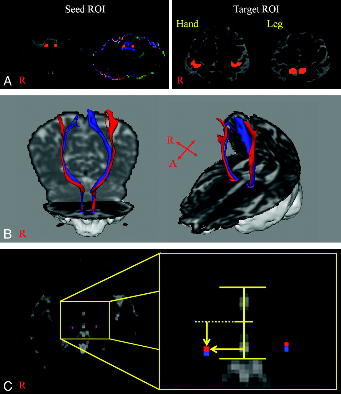
- Fig 1.
A, A seed region of interest is placed on the posterolateral medulla. Target regions of interest are shown at the postcentral gyrus posterior to the precentral knob for the hand and the postcentral gyrus posterior to the leg somatotopy of the precentral gyrus for the leg. B, The STP of a healthy subject (a 65-year-old woman) (red is the STP for the hand; blue is the STP for the leg). C, The highest probabilistic locations are measured laterally from the midline of the ACPC line in the mediolateral direction and posteriorly from the midpoint of the ACPC line in the anteroposterior direction (red is the highest probabilistic location of the STP for the hand; blue is the highest probabilistic location of the STP for the leg).
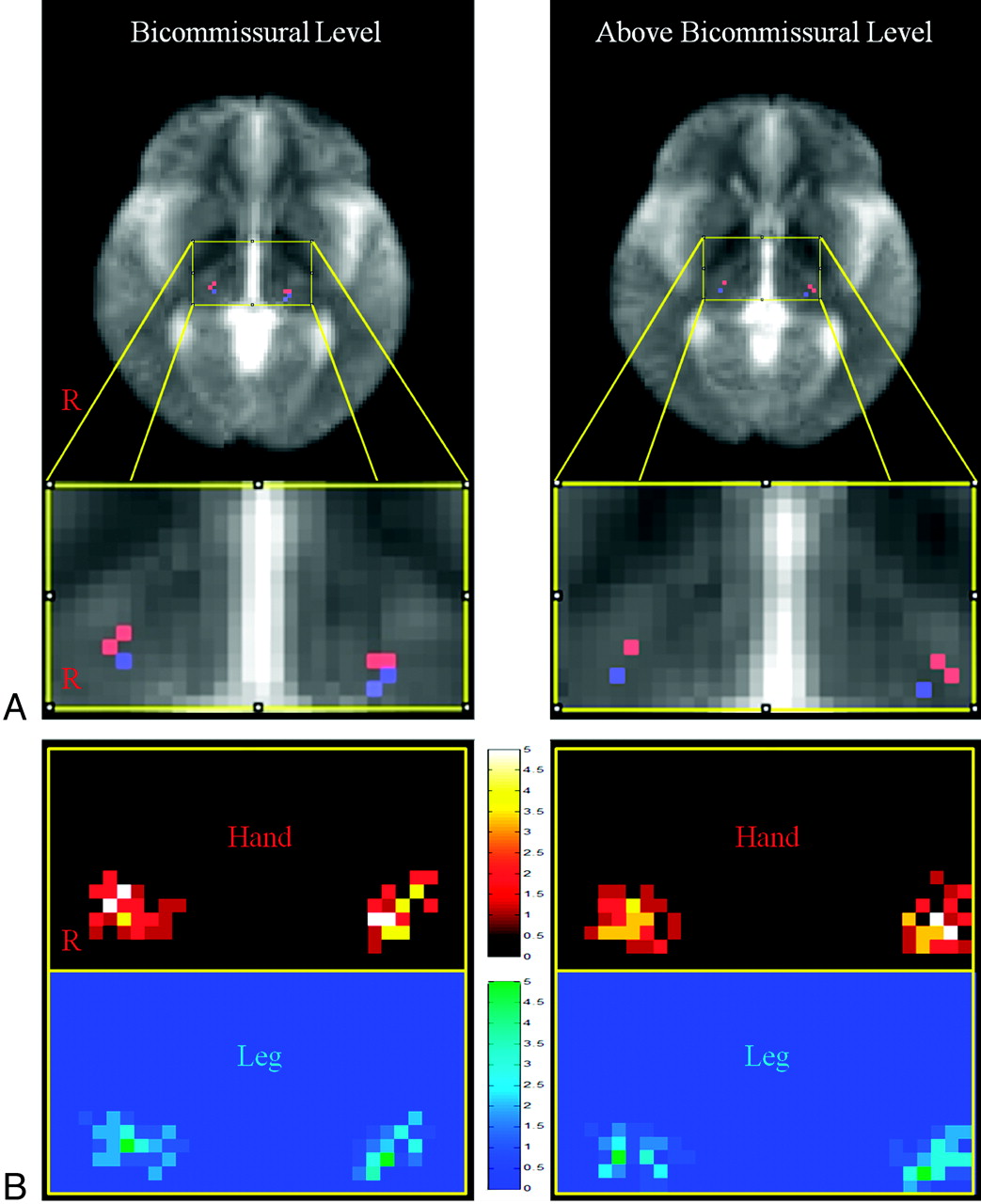
- Fig 2.
A, The most overlapping voxel of the probabilistic map for the STP superimposed on the mean of the non-diffusion-weighted image (red is the STP for the hand; blue is the STP for the leg). B, Probabilistic maps at the bicommissural level and above the bicommissural level. The probabilistic map shows overlapping voxels that are color-coded from 0 (black, blue) to 5 (white, green).


Tables
Somatotopic locations of the highest probability point of the STP at the VPL nucleus of the thalamusa
Level Mediolateral Direction (mm) Anteroposterior Direction (mm) Hand Leg P Value Hand Leg P Value Bicommissural Right 17.07 16.69 7.34 8.40 (±2.63) (±3.02) (±1.96) (±1.80) Left 16.65 16.06 7.72 9.03 (±2.21) (±2.42) (±2.29) (±1.92) Total 16.86 16.37 .231 7.53 8.71 .000 (±2.42) (±2.74) (±2.13) (±1.87) Above-bicommissural Right 19.24 18.90 7.17 8.31 (±2.48) (±2.90) (±1.96) (±1.68) Left 19.11 18.80 7.72 8.73 (±2.12) (±2.42) (±2.26) (±1.97) Total 19.18 18.85 .400 7.45 8.52 .001 (±2.29) (±2.66) (±2.12) (±1.92) -
a Values are means. Each location of the point was calculated as a pixel unit and converted to millimeters. Voxel size is 1.73 × 1.73 × 2.3 mm.
-


