
Article Figures & Data
Figures
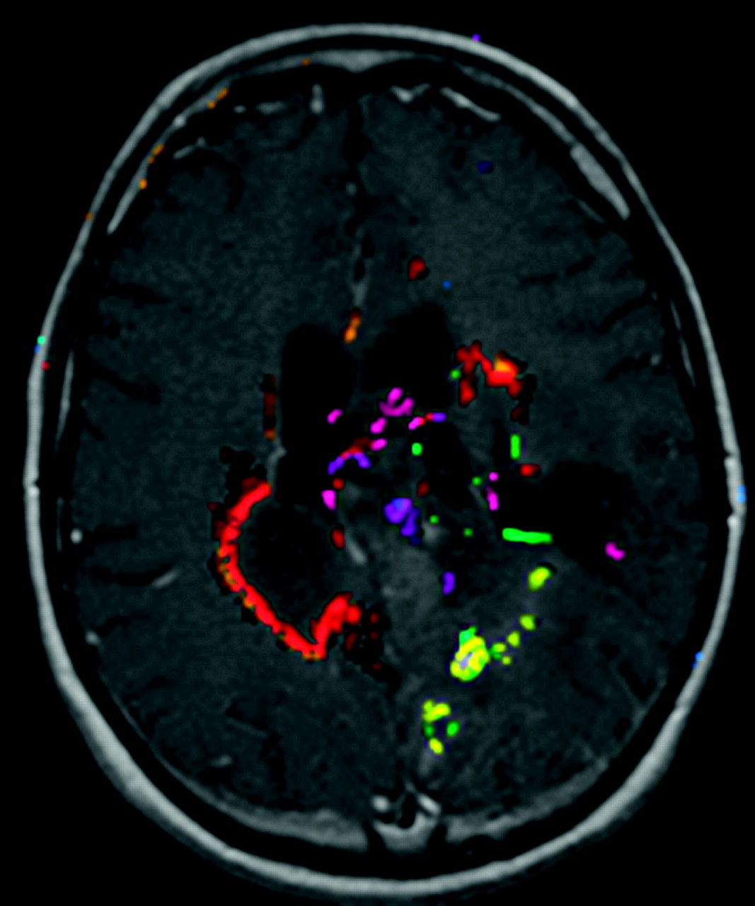
- Fig 1.
Example output of the automated change detection algorithm. Compared with simple image subtraction, change detection combines information from all multiple MR pulse sequences and uses knowledge about how progression or regression appears on the sequences, as well as standardized ways to set thresholds for true changes. Different colors represent different types of change, eg, red means new enhancement and T2 signal intensity abnormality, yellow means new nonenhancing T2 signal intensity abnormality, green means reduced T2 signal intensity abnormality, and purple means less enhancement and less T2 signal intensity abnormality.
- Fig 2.
A, Display application showing old (B = baseline) and new (F = follow-up) examinations in an above/below format. Note that this case allows the user to view the automated change detection overlay (the “Show CD Overlay” checkbox is enabled) but not image subtraction (“Subtract Images” is disabled). This also allows flicker mode, because the “Base Examination on Top” checkbox is enabled. Checking or unchecking that box changes whether the top row shows the baseline or follow-up examination. B, Display application with subtraction image, showing slight enlargement of the tumor nodule in the right frontal region (white arrow). C, Display application showing color change detection overlay on the images, as well as radiologist marking indicating progression, with confidence level of 3 (third image, top row).
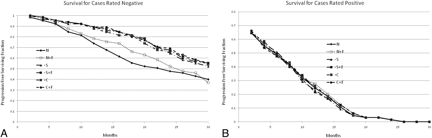
- Fig 3.
A, Survival curves for each display method, when all of the raters determined there was no progression. This graph demonstrates that methods by using flicker display help to correctly identify the cases that are negative (that will have long times until progression). The N method was significantly different from the others at the P < .05 level, but there was no difference between the other methods. B, Survival curves for each display method, when all raters determined that there was tumor progression. This graph suggests that the “normal” display mode (with or with flicker) identified some cases as progressers that actually will not progress in the near term. The differences were not statistically significant.


In this issue


Jump to section
Related Articles
Cited By...
- No citing articles found.