
Article Figures & Data
Figures
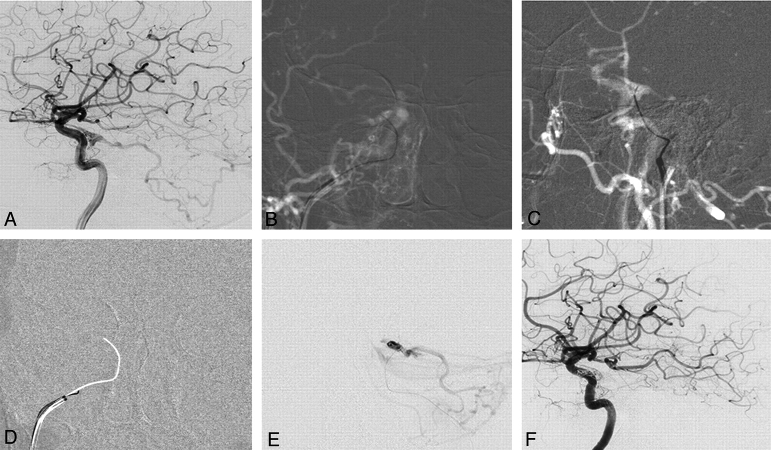
- Fig 1.
DAVF of the cavernous sinus with reflux into the posterior fossa. A 76-year-old woman presented with right-sided chemosis and diplopia. A, Right internal carotid angiogram shows a DAVF at the cavernous sinus fed by cavernous branches and the meningeohypophyseal artery of the ICA with marked posterior fossa reflux. There is no flow visible into the IPS. B and C, Following an arterial roadmap, AP view (B) and lateral view (C), the guiding catheter is oriented superiorly, medially, and anteriority toward the presumed origin of the IPS. A 00.35-inch guidewire is used to reopen the occluded IPS by gentle rotation and is advanced through the occluded sinus under roadmap guidance. D, Once access is gained with the guidewire as demonstrated by the arterial roadmap, a blank roadmap is initiated, and the guidewire is removed, and a track is left for the microcatheter to enter the cavernous sinus. E, The location of the microcatheter is checked by a careful contrast injection before coil embolization to demonstrate the exact origin of the cortical venous reflux and to verify that the right compartment is coiled. F, Right internal carotid angiogram following embolization demonstrates complete obliteration of the DAVF.
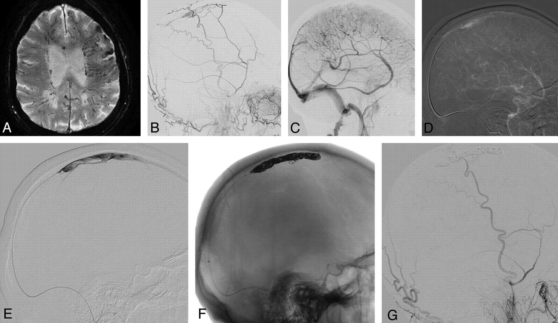
- Fig 2.
DAVF into a left “isolated” transverse sinus with cortical venous reflux. This 59-year-old female patient presented with alteration of consciousness and vertigo. A, Axial T2-weighted image shows dilation of cortical veins and hypersignal of the brain parenchyma as a sign of venous congestion in the left temporal and occipital lobes. B and C, Vertebral artery injections in an AP plane (B) and lateral occipital artery injections (C) demonstrate a DAVF at the left transverse sinus with an isolated enlarged venous pouch and cortical venous reflux with supply from multiple small branches of the occipital arteries and a dural branch of the left vertebral artery. D, With the guiding catheter in the left jugular bulb, a 0.035-inch guidewire is advanced through the occluded sigmoid sinus into the venous pouch. E, The track is used by a microcatheter, and the position of the microcatheter is checked with a careful injection once in the venous pouch. F and G, Following packing of the sinus with coils, control angiograms of the left vertebral artery (F) and the left common carotid artery (G) demonstrate complete obliterations of the DAVF.
- Fig 3.
Dural AVF of an isolated SSS with cortical venous reflux. An 87-year-old man presented with seizure, alteration of consciousness, and progressive weakness of his right hand. A, A T2 gradient-echo sequence demonstrates multiple dilated transmedullary veins and cortical microhemorrhages as a sign of chronic venous congestion. B and C, ECA (B) and ICA (C) runs demonstrate a DAVF into the SSS with occlusion at the distal third of the SSS with venous congestion. A 6F Shuttle-SL guide sheath (Cook, Bloomington, Indiana) is placed into the right jugular bulb, and a 6F Neuron (Penumbra, Alameda, California) catheter is advanced into the proximal SSS. D, From here, a 0.035-inch guidewire is advanced into the venous pouch. E–G, Via the thus-opened track, a microcatheter is advanced (E) with confirmation of the tip of the catheter in the SSS, and complete occlusion of the isolated sinus with coils is performed (F), leading to complete obliteration of the DAVF (G).
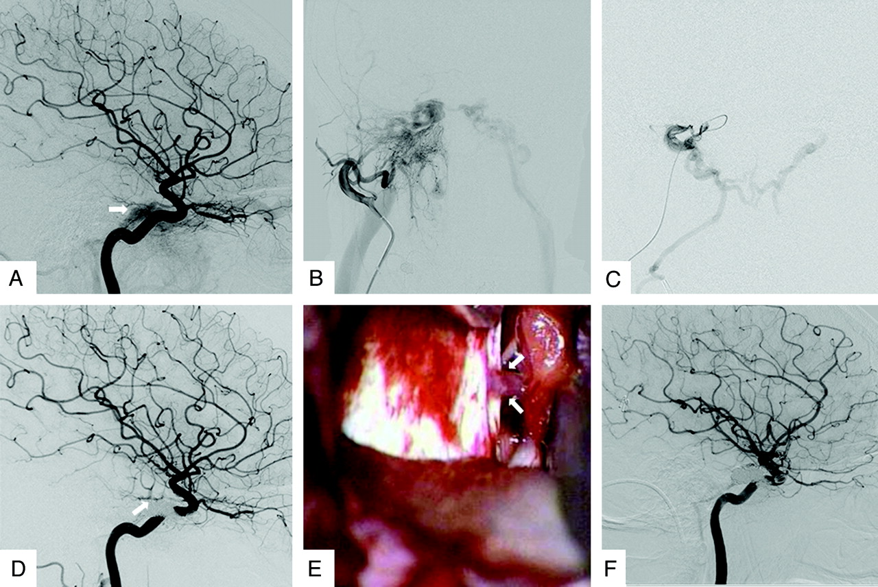
- Fig 4.
Dural AVF with incomplete occlusion following transvenous therapy. A 66-year-old woman presented with proptosis and chemosis of the right eye with mild diplopia. A and B, Cerebral angiogram reveals a caroticocavernous DAVF at the right cavernous sinus with arterial supply from the meningohypophyseal trunk (A) and meningeal branches from the ECA (B), with cortical venous reflux toward the posterior fossa and nonvisualization of the right IPV. With the previously described transvenous reopening technique, a microcatheter is advanced into the cavernous sinus. C, Microcatheter injections reveal the cortical venous reflux. The position of the microcatheter is deemed sufficient for subsequent coil embolization with the aim of first occluding the potential cortical venous reflux toward the posterior fossa. During deployment of the first coil, the microcatheter was pushed back, and access to the posteriorly directed venous outlet toward the posterior fossa was lost. Because access to this outlet could not be regained immediately, it was erroneously deemed sufficient to perform a dense packing of the cavernous sinus to obliterate the shunt. D, While ECA control injections (used during the coiling procedure to control for obliteration of venous reflux) showed obliteration of the DAVF, the final control injection into the ICA, however, still demonstrated reflux to the posterior fossa; and despite multiple attempts, it was not possible to regain access to the vein responsible for the cortical venous reflux, given the coil mass deposited into the sinus. Therefore, surgical disconnection of the remaining cortical venous reflux was deemed necessary. E, During surgical exposure of the cavernous sinus region, the posteriorly directed vein from the cavernous sinus that was responsible for the residual shunt is detected and the vein is coagulated. F, Follow-up angiogram after surgery demonstrates obliteration of the cortical venous reflux. This case demonstrates that it is always necessary to disconnect the cortical venous reflux before obliterating the venous sinus because packing of the sinus may preclude subsequent catheterization of cortical venous outflow tracks. This case also demonstrates that if cortical venous reflux is demonstrated to arise from multiple vessels (in this case both the ICA and the ECA), arterial control injections should be performed in both vessels to verify obliteration of the cortical venous reflux.


In this issue


Jump to section
Related Articles
Cited By...
- Correspondence on 'Embolization strategies for intracranial dural arteriovenous fistulas with an isolated sinus: a single-center experience in 20 patients by Hendriks et al
- Embolization strategies for intracranial dural arteriovenous fistulas with an isolated sinus: a single-center experience in 20 patients
- Ethmoidal dural arteriovenous fistulas: endovascular transvenous embolization technique
- Toward a Better Understanding of Dural Arteriovenous Fistula Angioarchitecture: Superselective Transvenous Embolization of a Sigmoid Common Arterial Collector
- Transvenous embolization of dural carotid cavernous fistulas: the role of liquid embolic agents in association with coils on patient outcomes
- Quantifying the Cerebral Hemodynamics of Dural Arteriovenous Fistula in Transverse Sigmoid Sinus Complicated by Sinus Stenosis: A Retrospective Cohort Study