Advertisement

Main menu

  • Home
  • Content
    • Current Issue
    • Accepted Manuscripts
    • Article Preview
    • Past Issue Archive
    • AJNR Case Collection
    • Case of the Week Archive
    • Classic Case Archive
    • Case of the Month Archive
  • Special Collections
    • Spinal CSF Leak Articles (Jan 2020-June 2024)
    • 2024 AJNR Journal Awards
    • Most Impactful AJNR Articles
  • Multimedia
    • AJNR Podcast
    • AJNR Scantastics
    • Video Articles
  • For Authors
    • Submit a Manuscript
    • Author Policies
    • Fast publishing of Accepted Manuscripts
    • Graphical Abstract Preparation
    • Manuscript Submission Guidelines
    • Imaging Protocol Submission
    • Submit a Case for the Case Collection
  • About Us
    • About AJNR
    • Editorial Board
  • More
    • Become a Reviewer/Academy of Reviewers
    • Subscribers
    • Permissions
    • Alerts
    • Feedback
    • Advertisers
    • ASNR Home
  • Other Publications
    • ajnr

User menu

  • Alerts
  • Log in

Search

  • Advanced search
American Journal of Neuroradiology

American Journal of Neuroradiology

ASHNR American Society of Functional Neuroradiology ASHNR American Society of Pediatric Neuroradiology ASSR
  • Alerts
  • Log in

Advanced Search

  • Home
  • Content
    • Current Issue
    • Accepted Manuscripts
    • Article Preview
    • Past Issue Archive
    • AJNR Case Collection
    • Case of the Week Archive
    • Classic Case Archive
    • Case of the Month Archive
  • Special Collections
    • Spinal CSF Leak Articles (Jan 2020-June 2024)
    • 2024 AJNR Journal Awards
    • Most Impactful AJNR Articles
  • Multimedia
    • AJNR Podcast
    • AJNR Scantastics
    • Video Articles
  • For Authors
    • Submit a Manuscript
    • Author Policies
    • Fast publishing of Accepted Manuscripts
    • Graphical Abstract Preparation
    • Manuscript Submission Guidelines
    • Imaging Protocol Submission
    • Submit a Case for the Case Collection
  • About Us
    • About AJNR
    • Editorial Board
  • More
    • Become a Reviewer/Academy of Reviewers
    • Subscribers
    • Permissions
    • Alerts
    • Feedback
    • Advertisers
    • ASNR Home
  • Follow AJNR on Twitter
  • Visit AJNR on Facebook
  • Follow AJNR on Instagram
  • Join AJNR on LinkedIn
  • RSS Feeds

Welcome to the new AJNR, Updated Hall of Fame, and more. Read the full announcements.


AJNR is seeking candidates for the position of Associate Section Editor, AJNR Case Collection. Read the full announcement.


Research ArticleTechnical Note

Safety of 1000 CT-Guided Steroid Injections with Air Used to Localize the Epidural Space

A. Chang, S. Pochert, C. Romano, A. Brook and T. Miller
American Journal of Neuroradiology October 2011, 32 (9) E175-E177; DOI: https://doi.org/10.3174/ajnr.A2316
A. Chang
  • Find this author on Google Scholar
  • Find this author on PubMed
  • Search for this author on this site
S. Pochert
  • Find this author on Google Scholar
  • Find this author on PubMed
  • Search for this author on this site
C. Romano
  • Find this author on Google Scholar
  • Find this author on PubMed
  • Search for this author on this site
A. Brook
  • Find this author on Google Scholar
  • Find this author on PubMed
  • Search for this author on this site
T. Miller
  • Find this author on Google Scholar
  • Find this author on PubMed
  • Search for this author on this site
  • Article
  • Figures & Data
  • Info & Metrics
  • Responses
  • References
  • PDF
Loading

Abstract

SUMMARY: Historically, ESIs were performed without any imaging guidance, resulting in erroneous placement in up to 30% of injections. Fluoroscopic imaging is now used to guide most procedures. Recently, several reports have described the use of CT to guide ESIs instead of fluoroscopy. CT provides the ability to use air as contrast to localize the epidural space. This retrospective review will discuss findings in 1000 CT-guided ESIs with air localization.

Abbreviations

ADR
adverse drug reaction
ESI
epidural spinal injection

ESIs have a low complication rate1 and are minimally invasive. While the efficacy of ESIs is still unclear, observational studies have largely supported their effectiveness. However, there are few well-designed randomized double-blinded studies.2

Historically, ESIs were performed without any imaging guidance, resulting in erroneous placement in up to 30% of injections.3 Because of this and the potential for intrathecal and intravascular injections, imaging is now used to guide most procedures. Although fluoroscopy is currently the standard for ESIs, it has its limitations. Among these is the poor demonstration of soft-tissue structures, which necessitates contrast injection to localize the epidural space or nerve roots. It has been reported that ESIs performed without contrast are at increased risk of being intravascular injections (transforaminal),4 which can, in turn, cause untoward side effects. Furthermore, contrast use runs the risk of inducing allergic reactions in contrast-sensitive patients.

More recently, several reports have described the use of CT to guide ESIs instead of fluoroscopy. CT has several advantages, including increased anatomic precision and visualization of needle placement with millimeter accuracy. The superior tissue contrast offered by CT provides the ability to use air as contrast to localize the epidural space. Air is used in much the same way as iodinated contrast—it is injected to verify that the needle is indeed within the epidural space and not intrathecal. Air has several advantages, including lack of risk of allergic reactions and no cost for materials. There is profound ability to discriminate between air and soft-tissue by using CT, which makes air an excellent contrast agent. Because incorrect needle placement and incorrect steroid injection are the most frequent complications of ESIs, it is important to find a safe and reliable way to localize the epidural space.

Because this method, to our knowledge, has not been previously described in the literature, this retrospective review will discuss findings in 1000 CT-guided ESIs with air localization.

Materials and Methods

This institutional review board−approved protocol is a retrospective review composed of a record search in the computerized radiology information system from 2006 to 2009. The search included all CT-guided ESIs in the lumbar region for which air was used for localization. Cases in which the additional use of contrast was used for needle localization were included as well. The data collected included age, sex, level treated, needle size, amount of air, type and amount of steroid used, contrast use and amount, and any reported complications.

Technique

The CT-guided epidural access was typically performed with the patient prone; adjustments were made for patient comfort. A standard sterile prep and drape were used. A scout lateral image was obtained. Five axial images through the affected level (3-mm helical, 120 kVp, 50 mA) were obtained for planning. The trajectory was planned on the CT console. The skin entry site and depth were recorded. Local anesthesia was administered with 2% lidocaine both superficially and deep at the planned entry location. The needle was inserted 1 cm short of the planned depth. A 3-mL luer-slip syringe with 1 mL of air was attached to the spinal needle, and the loss of resistance technique was used to cross the ligamentum flavum. Confirmatory images were obtained once loss of resistance was felt by the operator.

Results

We identified 751 patients who underwent 1000 procedures. The average patient age was 59.2 years, 439 (58.5%) were women and 312 (41.5%) were men. Seventy-five percent of the treated levels were L5/S1, with L4/5 and L3/4 much less frequently treated (15.5% and 4.9%). Rarely, procedures targeted L2/3 (1.3%), L1/2 (0.8%), and T12/L1 (0.1%). A spinal needle was used for all procedures, with 90% using a 22-ga spinal needle. Of the total number of cases, 575 reported a specific amount of air injected; the typical dose was 0.5 mL, with a range of 0.05–2 mL (Fig 1). A total of 132 cases required further administration of iodinated contrast, predominantly iohexal (Omnipaque 180; GE Healthcare, Princeton, New Jersey), to confirm needle position. Of those cases, 103 reported a specific amount of contrast; the usual dose was 1.0 mL, with a range of 0.5–5.0 mL

Fig 1.
  • Download figure
  • Open in new tab
  • Download powerpoint
Fig 1.

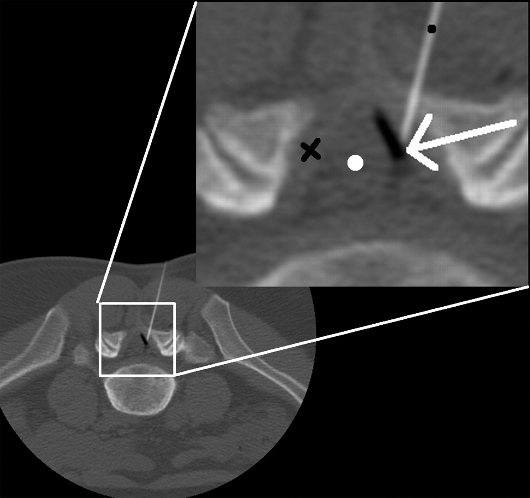
Axial CT image through L5/S1 (3-mm section, 100 KVp, 50 mA). Image demonstrates 22-g spinal needle (black dot) traversing the ligamentum flavum (black X) tip in the epidural space. The white arrow demonstrates the interface of injected air (black) on top of epidural fat (gray). The air in the epidural space pushes the thecal sac medially (white dot).

The most common steroid administered was betamethasone acetate (Celestone; Schering, Kenilworth, New Jersey) (in 91.5% of cases), with methylprednisolone acetate (Depo-Medrol; Schering) administered 4.7% of the time. Eighteen millligrams of Celestone and 3 mL of Sensorcaine (bupivacaine HCL 0.25%; APP Pharmaceuticals, Schaumburg, Illinois) were given in most cases (65.2%). Of the 1000 procedures identified, no immediate or delayed clinically significant complications were reported during a standard 24-hour and 1-week follow-up (99% of patients had 24-hour follow-up and 93% had 2-week follow-up via phone or office consultation).

Procedure time averaged 15 minutes (range, 10–32 minutes). Average radiation dose measured as an effective dose calculated from the dose-length product and abdomen and pelvis conversion was 1.5 mSv (range, 0.9–3 mSv).5

Discussion

Air localization is a technique used in conjunction with CT guidance to identify the epidural space and verify correct needle placement. This technique provides an alternative to iodine-based contrasts.

In 87% of cases, the epidural space was visualized by using air alone. The remainder required contrast to verify needle localization to the epidural space. When we compared the groups with air and contrast, the distribution of ages and levels treated was the same. Further demographic analysis did not demonstrate a clear difference between the groups. More severe spondylosis was suspected as a cause for increased contrast use; this information was not collected. Further analysis and prospective evaluation may be required to determine in which patients contrast should be used as the primary agent for localizing the epidural space.

In general, ESIs have a very low rate of complications, though they are not completely free from risk. Reported complications include spinal headache after dural puncture, infection, intravascular injections, and nerve trauma.6 There have also been reports of rarer but usually more serious complications, including pneumocephalus,7 epidural hematomas,8 paraplegia,9 vision loss,10 epidural abscess,11 and meningitis.12

While the relative risks and complications of ESI have been described in the literature, the safety of the air localization technique for use in ESI has not been previously discussed, to our knowledge. Our study detected no clinically significant complications with the use of air localization or with the procedure overall. Other studies have reported overall complication rates from 0.04%.13

The reported incidence of complications is very low, and this study reproduced that finding. In theory, air should reduce the risk of anaphylactic reactions to the contrast medium, and it may also reduce the rate of other adverse drug reactions from contrast. However, given the retrospective nature of this study, it is uncertain whether the lower rate of complications is attributable to the use of air in place of contrast or whether some other factor is influencing the results.

Several studies have evaluated the safety of contrast. Currently, nonionic contrast is preferred over ionic contrast due to a lower rate of ADRs.14 A large-scale study of 337,647 cases of intravenous contrast use found the ionic contrast ADR rate to be 12.66% and the nonionic ADR rate to be 3.13%; severe ADRs were found in 0.22% of the ionic and 0.04% of the nonionic groups.15 The overall incidence of ADRs from contrast is low, though ADRs were found to be more likely in patients with a history of ADRs to contrast and in those with a history of allergy.15 One advantage of air localization is that it reduces the potential of anaphylactic reactions due to contrast allergies, and this may play a role in the reduced rates of complications we observed.

Although contrast such as gadolinium is available for use in patients with contrast allergies,15 gadolinium has the disadvantage of being significantly more expensive than regular contrast. Even a commonly used nonionic contrast such as Omnipaque-180 costs $431 for ten 10 mL vials, and the cumulative cost can be significant for an institution that performs a large volume of ESIs. Another benefit of air as a localizing agent is its cost-saving potential.

The use of CT guidance for ESIs offers many potential advantages. The ability to use air as a contrast agent is one. Another is precise needle guidance to actual soft-tissue structures, instead of inference from bony landmarks or contrast location. Room time was similar to that of a CT scanner in the hospital radiology department. Radiation dose demonstrated in this large series was reasonable compared with that found by Kim et al 16 and should not be a deterrent to CT use. Our results of 1.5 mSv are similar to the typical effective dose from continuous fluoroscopic guidance for ESI reported by Schmid et al (0.43–1.25 mSv for 1–3 minutes of guidance).17

Conclusions

The use of air to localize the epidural space in CT-guided ESIs has a high success rate and a very low rate of complications.

References

  1. 1.↵
    1. Koes BW,
    2. Scholten RJ,
    3. Mens JM,
    4. et al
    . Efficacy of epidural steroid injections for low-back pain and sciatica: a systematic review of randomized clinical trials. Pain 1995; 63: 279–88
    CrossRefPubMed
  2. 2.↵
    1. Abram S
    . Treatment of lumbosacral radiculopathy with epidural steroids. Anesthesiology 1999; 91: 1937–41
    CrossRefPubMed
  3. 3.↵
    1. White AH,
    2. Derby R,
    3. Wynne G
    . Epidural injections for the diagnosis and treatment of low back pain. Spine 1980; 5: 67–86
  4. 4.↵
    1. Furman M,
    2. O'Brien E,
    3. Zgleszewski T
    . Incidence of intravascular penetration in transforaminal lumbosacral epidural steroid injections. Spine (Phila Pa 1976) 2000; 25: 2628–32
    CrossRef
  5. 5.↵
    1. Mettler FA Jr.,
    2. Huda W,
    3. Yoshizumi TT,
    4. et al
    . Effective doses in radiology and diagnostic nuclear medicine: a catalog. Radiology 2008; 248: 254–63
    CrossRefPubMed
  6. 6.↵
    1. Goodman BS,
    2. Posecion LW,
    3. Mallempati S,
    4. et al
    . Complications and pitfalls of lumbar interlaminar and transforaminal epidural injections. Curr Rev Musculoskelet Med 2008; 1: 212–22. Epub 2008 Aug 15
    CrossRefPubMed
  7. 7.↵
    1. Simopoulos T,
    2. Peeters-Asdourian C
    . Pneumocephalus after cervical epidural steroid injection. Anesth Analg 2001; 92: 1576–77
    PubMed
  8. 8.↵
    1. Williams K,
    2. Jackowski A,
    3. Evans P
    . Epidural haematoma requiring surgical decompression following repeated cervical epidural steroid injections for chronic pain. Pain 1990; 42: 197–99
    CrossRefPubMed
  9. 9.↵
    1. Tripathi M,
    2. Nath S,
    3. Gupta R
    . Paraplegia after intracord injection during attempted epidural steroid injection in an awake-patient. Anesth Analg 2005; 101: 1209–11, table of contents
    CrossRefPubMed
  10. 10.↵
    1. Young W
    . Transient blindness after lumbar epidural steroid injection: a case report and literature review. Spine (Phila Pa 1976) 2002; 27: E476–77
    CrossRef
  11. 11.↵
    1. Hooten W,
    2. Kinney M,
    3. Huntoon M
    . Epidural abscess and meningitis after epidural corticosteroid injection. Mayo Clin Proc 2004; 79: 682–86
    CrossRefPubMed
  12. 12.↵
    1. White A,
    2. Derby R,
    3. Wynne G
    . Epidural injections for the diagnosis and treatment of low-back pain. Spine (Phila Pa 1976); 5: 78–86
  13. 13.↵
    1. el-Khoury G,
    2. Ehara S,
    3. Weinstein J,
    4. et al
    . Epidural steroid injection: a procedure ideally performed with fluoroscopic control. Radiology 1988; 168: 554–57
    PubMed
  14. 14.↵
    1. Wolf G,
    2. Mishkin M,
    3. Roux S,
    4. et al
    . Comparison of the rates of adverse drug reactions: ionic contrast agents, ionic agents combined with steroids, and nonionic agents. Invest Radiol 1991; 26: 404–10
    CrossRefPubMed
  15. 15.↵
    1. Safriel Y,
    2. Ali M,
    3. Hayt M
    . Gadolinium use in spine procedures for patients with allergy to iodinated contrast: experience of 127 procedures. AJNR Am J Neuroradiol 2006; 27: 1194–97
    Abstract/FREE Full Text
  16. 16.↵
    1. Kim S,
    2. Toncheva G,
    3. Anderson-Evans C,
    4. et al
    . Kerma area product method for effective dose estimation during lumbar epidural steroid injection procedures: phantom study. AJR Am J Roentgenol 2009; 192: 1726–30
    CrossRefPubMed
  17. 17.↵
    1. Schmid G,
    2. Schmitz A,
    3. Borchardt D,
    4. et al
    . Effective dose of CT- and fluoroscopy-guided perineural/epidural injections of the lumbar spine: a comparative study. Cardiovasc Intervent Radiol 2006: 29: 84–91
    CrossRefPubMed
  • Received June 29, 2010.
  • Accepted after revision August 9, 2010.
  • © 2011 by American Journal of Neuroradiology
View Abstract
PreviousNext
Back to top

In this issue

American Journal of Neuroradiology: 32 (9)
American Journal of Neuroradiology
Vol. 32, Issue 9
1 Oct 2011
  • Table of Contents
  • Index by author
Advertisement
Print
Download PDF
Email Article

Thank you for your interest in spreading the word on American Journal of Neuroradiology.

NOTE: We only request your email address so that the person you are recommending the page to knows that you wanted them to see it, and that it is not junk mail. We do not capture any email address.

Enter multiple addresses on separate lines or separate them with commas.
Safety of 1000 CT-Guided Steroid Injections with Air Used to Localize the Epidural Space
(Your Name) has sent you a message from American Journal of Neuroradiology
(Your Name) thought you would like to see the American Journal of Neuroradiology web site.
CAPTCHA
This question is for testing whether or not you are a human visitor and to prevent automated spam submissions.
Cite this article
A. Chang, S. Pochert, C. Romano, A. Brook, T. Miller
Safety of 1000 CT-Guided Steroid Injections with Air Used to Localize the Epidural Space
American Journal of Neuroradiology Oct 2011, 32 (9) E175-E177; DOI: 10.3174/ajnr.A2316

Citation Manager Formats

  • BibTeX
  • Bookends
  • EasyBib
  • EndNote (tagged)
  • EndNote 8 (xml)
  • Medlars
  • Mendeley
  • Papers
  • RefWorks Tagged
  • Ref Manager
  • RIS
  • Zotero
0 Responses
Respond to this article
Share
Bookmark this article
Safety of 1000 CT-Guided Steroid Injections with Air Used to Localize the Epidural Space
A. Chang, S. Pochert, C. Romano, A. Brook, T. Miller
American Journal of Neuroradiology Oct 2011, 32 (9) E175-E177; DOI: 10.3174/ajnr.A2316
del.icio.us logo Twitter logo Facebook logo Mendeley logo
  • Tweet Widget
  • Facebook Like
  • Google Plus One
Purchase

Jump to section

  • Article
    • Abstract
    • Abbreviations
    • Materials and Methods
    • Technique
    • Results
    • Discussion
    • Conclusions
    • References
  • Figures & Data
  • Info & Metrics
  • Responses
  • References
  • PDF

Related Articles

  • No related articles found.
  • PubMed
  • Google Scholar

Cited By...

  • Comparison of CT and fluoroscopic guidance for lumbar puncture in an obese population with prior failed unguided attempt
  • Optimal Contrast Concentration for CT-Guided Epidural Steroid Injections
  • Radiation Dose for 345 CT-Guided Interlaminar Lumbar Epidural Steroid Injections
  • Ultrasound Guided Versus CT-Controlled Pararadicular Injections in the Lumbar Spine: A Prospective Randomized Clinical Trial
  • Lateral Decubitus Positioning for Cervical Nerve Root Block Using CT Image Guidance Minimizes Effective Radiation Dose and Procedural Time
  • Crossref (19)
  • Google Scholar

This article has been cited by the following articles in journals that are participating in Crossref Cited-by Linking.

  • The Lumbar Neural Foramen and Transforaminal Epidural Steroid Injections: An Anatomic Review With Key Safety Considerations in Planning the Percutaneous Approach
    Jacob C. Mandell, Gregory J. Czuczman, Glenn C. Gaviola, Varand Ghazikhanian, Charles H. Cho
    American Journal of Roentgenology 2017 209 1
  • Emerging Applications of Abdominal 4D Flow MRI
    Alejandro Roldán-Alzate, Christopher J. Francois, Oliver Wieben, Scott B. Reeder
    American Journal of Roentgenology 2016 207 1
  • Ultrasound Guided Versus CT-Controlled Pararadicular Injections in the Lumbar Spine: A Prospective Randomized Clinical Trial
    A. Loizides, H. Gruber, S. Peer, K. Galiano, R. Bale, J. Obernauer
    American Journal of Neuroradiology 2013 34 2
  • Comparison of CT and fluoroscopic guidance for lumbar puncture in an obese population with prior failed unguided attempt
    Andrew D Brook, Judah Burns, Edward Dauer, Alan H Schoendfeld, Todd S Miller
    Journal of NeuroInterventional Surgery 2014 6 4
  • Ultrasound-guided Versus Fluoroscopy-controlled Lumbar Transforaminal Epidural Injections
    Ge Yang, Jinfeng Liu, Liangjuan Ma, Zhenhua Cai, Chao Meng, Sihua Qi, Huacheng Zhou
    The Clinical Journal of Pain 2016 32 2
  • Lateral Decubitus Positioning for Cervical Nerve Root Block Using CT Image Guidance Minimizes Effective Radiation Dose and Procedural Time
    T.S. Miller, K. Fruauff, J. Farinhas, D. Pasquale, C. Romano, A.H. Schoenfeld, A. Brook
    American Journal of Neuroradiology 2013 34 1
  • Radiation Dose for 345 CT-Guided Interlaminar Lumbar Epidural Steroid Injections
    A.L. Chang, A.H. Schoenfeld, A.L. Brook, T.S. Miller
    American Journal of Neuroradiology 2013 34 10
  • In vitrovalidation of endovascular Doppler-derived flow rates in models of the cerebral circulation
    P M McGah, J D Nerva, R P Morton, M C Barbour, M R Levitt, P D Mourad, L J Kim, A Aliseda
    Physiological Measurement 2015 36 11
  • Radiation dose of fluoroscopy-guided versus ultralow-dose CT-fluoroscopy-guided lumbar spine epidural steroid injections
    Ged G. Wieschhoff, Nityanand P. Miskin, Jeom Soon Kim, Leena M. Hamberg, Jacob C. Mandell
    Skeletal Radiology 2022 51 5
  • Back to the scanner: expected and unexpected imaging findings following spinal puncture and access
    Judah Burns, Meir H. Scheinfeld
    Emergency Radiology 2013 20 4

More in this TOC Section

  • 3D Fast Spin-Echo T1 Black-Blood Imaging for the Diagnosis of Cervical Artery Dissection
  • The Kety-Schmidt Technique for Quantitative Perfusion and Oxygen Metabolism Measurements in the MR Imaging Environment
  • Feasibility of Placement of an Anterior Cervical Epidural Blood Patch for Spontaneous Intracranial Hypotension
Show more Technical Note

Similar Articles

Advertisement

Indexed Content

  • Current Issue
  • Accepted Manuscripts
  • Article Preview
  • Past Issues
  • Editorials
  • Editors Choice
  • Fellow Journal Club
  • Letters to the Editor

Cases

  • Case Collection
  • Archive - Case of the Week
  • Archive - Case of the Month
  • Archive - Classic Case

Special Collections

  • Special Collections

Resources

  • News and Updates
  • Turn around Times
  • Submit a Manuscript
  • Author Policies
  • Manuscript Submission Guidelines
  • Evidence-Based Medicine Level Guide
  • Publishing Checklists
  • Graphical Abstract Preparation
  • Imaging Protocol Submission
  • Submit a Case
  • Become a Reviewer/Academy of Reviewers
  • Get Peer Review Credit from Publons

Multimedia

  • AJNR Podcast
  • AJNR SCANtastic
  • Video Articles

About Us

  • About AJNR
  • Editorial Board
  • Not an AJNR Subscriber? Join Now
  • Alerts
  • Feedback
  • Advertise with us
  • Librarian Resources
  • Permissions
  • Terms and Conditions

American Society of Neuroradiology

  • Not an ASNR Member? Join Now

© 2025 by the American Society of Neuroradiology All rights, including for text and data mining, AI training, and similar technologies, are reserved.
Print ISSN: 0195-6108 Online ISSN: 1936-959X

Powered by HighWire