Advertisement

Main menu

  • Home
  • Content
    • Current Issue
    • Accepted Manuscripts
    • Article Preview
    • Past Issue Archive
    • AJNR Case Collection
    • Case of the Week Archive
    • Classic Case Archive
    • Case of the Month Archive
  • Special Collections
    • Spinal CSF Leak Articles (Jan 2020-June 2024)
    • 2024 AJNR Journal Awards
    • Most Impactful AJNR Articles
  • Multimedia
    • AJNR Podcast
    • AJNR Scantastics
    • Video Articles
  • For Authors
    • Submit a Manuscript
    • Author Policies
    • Fast publishing of Accepted Manuscripts
    • Graphical Abstract Preparation
    • Manuscript Submission Guidelines
    • Imaging Protocol Submission
    • Submit a Case for the Case Collection
  • About Us
    • About AJNR
    • Editorial Board
  • More
    • Become a Reviewer/Academy of Reviewers
    • Subscribers
    • Permissions
    • Alerts
    • Feedback
    • Advertisers
    • ASNR Home
  • Other Publications
    • ajnr

User menu

  • Alerts
  • Log in

Search

  • Advanced search
American Journal of Neuroradiology

American Journal of Neuroradiology

ASHNR American Society of Functional Neuroradiology ASHNR American Society of Pediatric Neuroradiology ASSR
  • Alerts
  • Log in

Advanced Search

  • Home
  • Content
    • Current Issue
    • Accepted Manuscripts
    • Article Preview
    • Past Issue Archive
    • AJNR Case Collection
    • Case of the Week Archive
    • Classic Case Archive
    • Case of the Month Archive
  • Special Collections
    • Spinal CSF Leak Articles (Jan 2020-June 2024)
    • 2024 AJNR Journal Awards
    • Most Impactful AJNR Articles
  • Multimedia
    • AJNR Podcast
    • AJNR Scantastics
    • Video Articles
  • For Authors
    • Submit a Manuscript
    • Author Policies
    • Fast publishing of Accepted Manuscripts
    • Graphical Abstract Preparation
    • Manuscript Submission Guidelines
    • Imaging Protocol Submission
    • Submit a Case for the Case Collection
  • About Us
    • About AJNR
    • Editorial Board
  • More
    • Become a Reviewer/Academy of Reviewers
    • Subscribers
    • Permissions
    • Alerts
    • Feedback
    • Advertisers
    • ASNR Home
  • Follow AJNR on Twitter
  • Visit AJNR on Facebook
  • Follow AJNR on Instagram
  • Join AJNR on LinkedIn
  • RSS Feeds

Welcome to the new AJNR, Updated Hall of Fame, and more. Read the full announcements.


AJNR is seeking candidates for the position of Associate Section Editor, AJNR Case Collection. Read the full announcement.


Case ReportCase Report

Contrast Agent Induced Obliteration of a Spinal Arteriovenous Malformation

R.J. Nijenhuis, M. Sluzewski and W.J. van Rooij
American Journal of Neuroradiology January 2012, 33 (1) E1-E3; DOI: https://doi.org/10.3174/ajnr.A2436
R.J. Nijenhuis
  • Find this author on Google Scholar
  • Find this author on PubMed
  • Search for this author on this site
M. Sluzewski
  • Find this author on Google Scholar
  • Find this author on PubMed
  • Search for this author on this site
W.J. van Rooij
  • Find this author on Google Scholar
  • Find this author on PubMed
  • Search for this author on this site
  • Article
  • Figures & Data
  • Info & Metrics
  • Responses
  • References
  • PDF
Loading

Abstract

SUMMARY: We present a patient undergoing spinal angiography and suspected of having an AVM. During the first injection of nonionic contrast, a nidal AVM supplied by the great anterior radiculomedullary artery at T9 left was found. The second injection at T9 left no longer showed the contrast filling of the AVM. Follow-up angiography showed persistent AVM obliteration with an intact arterial spinal axis. The patient's symptoms resolved. Interaction between nonionic contrast, blood, and vessel wall most likely induced the obliteration.

Abbreviations

AVM
arteriovenous malformation

AVMs of the spinal cord are rare congenital vascular disorders characterized by a perimedullary fistula or an intramedullary nidus. The feeding vessels arise from the anterior or posterior spinal arteries with drainage into the perimedullary venous plexus. Spinal AVMs most often present in young adulthood with symptoms of myelopathy or acute neurologic deterioration secondary to intramedullary or subarachnoid hemorrhage. The presence of a spinal AVM is readily diagnosed with MR imaging. Spinal angiography is needed to evaluate the angioarchitecture for treatment planning. Treatment can be endovascular or surgical; both methods carry a serious risk of occluding normal vessels to the cord with consequent ischemic damage and neurologic deficit.1 Spontaneous closure of spinal AVMs has occasionally been described.1–3 We present a case of a spinal AVM that was obliterated during angiography.

Case Report

A 35-year-old man presented with painful sensations along his right buttock and thigh. Spinal MR imaging demonstrated a small AVM posteriorly located in the cone with dilated perimedullary draining veins. There were no signs of myelopathy or previous hemorrhages (Fig 1A, -B). Spinal angiography with use of a nonionic contrast agent (Iohexol, Omnipaque 300; GE Healthcare, Eindhoven, the Netherlands) confirmed the presence of the AVM (Fig 2A, -B), with the anterior spinal artery arising from the intercostal artery at T9 left as a single feeder to the nidus; the adjacent intercostal and lumbar arteries did not supply the AVM. The anterior spinal artery coursed under the distal part of the conus to supply the posteriorly located AVM nidus. Most surprising, during the second selective injection at the same pedicle, slow flow in the anterior spinal artery was noticed without visualization of the nidus (Fig 2C, -D). Also injections 10–15 minutes later and additional injections in the adjacent segmental arteries failed to allow visualization of the AVM. The procedure was terminated. In the following days, the patient's symptoms gradually improved, and he was discharged home. Three months later all symptoms had resolved. Follow-up MR imaging and MR angiography4,5 at 3 and 6 months and spinal angiography at 3 months confirmed the lasting complete obliteration of the spinal AVM with an intact anterior spinal axis (Fig 3A−D).

Fig 1.
  • Download figure
  • Open in new tab
  • Download powerpoint
Fig 1.

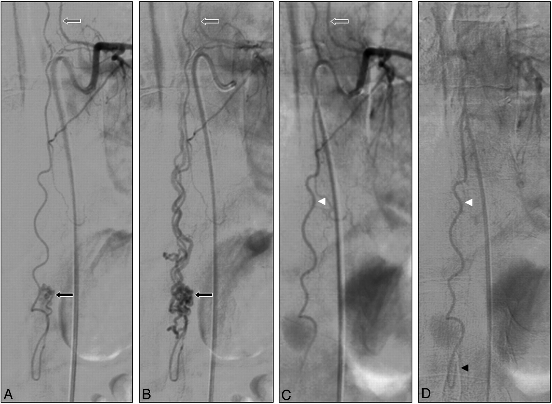
A and B, Sagittal T2-weighted MR images showing tortuous enlarged perimedullary and radicular veins (white arrows) and the posteriorly located AVM nidus (black arrow, B).

Fig 2.
  • Download figure
  • Open in new tab
  • Download powerpoint
Fig 2.

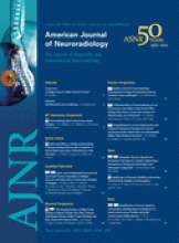
A and B, Spinal angiograms showing the great anterior radiculomedullary artery at T9 left (gray arrow) supplying the AVM in the arterial (A) and venous (B) phase. Note that the anterior spinal artery runs all the way down and around the conus to supply the posteriorly located AVM (black arrow). C and D, Later injections at the same pedicle show no contrast filling of the AVM in the arterial (C) and venous phase (D) and a patent anterior spinal artery (white arrowhead), which runs around the conus (black arrowhead, D).

Fig 3.
  • Download figure
  • Open in new tab
  • Download powerpoint
Fig 3.

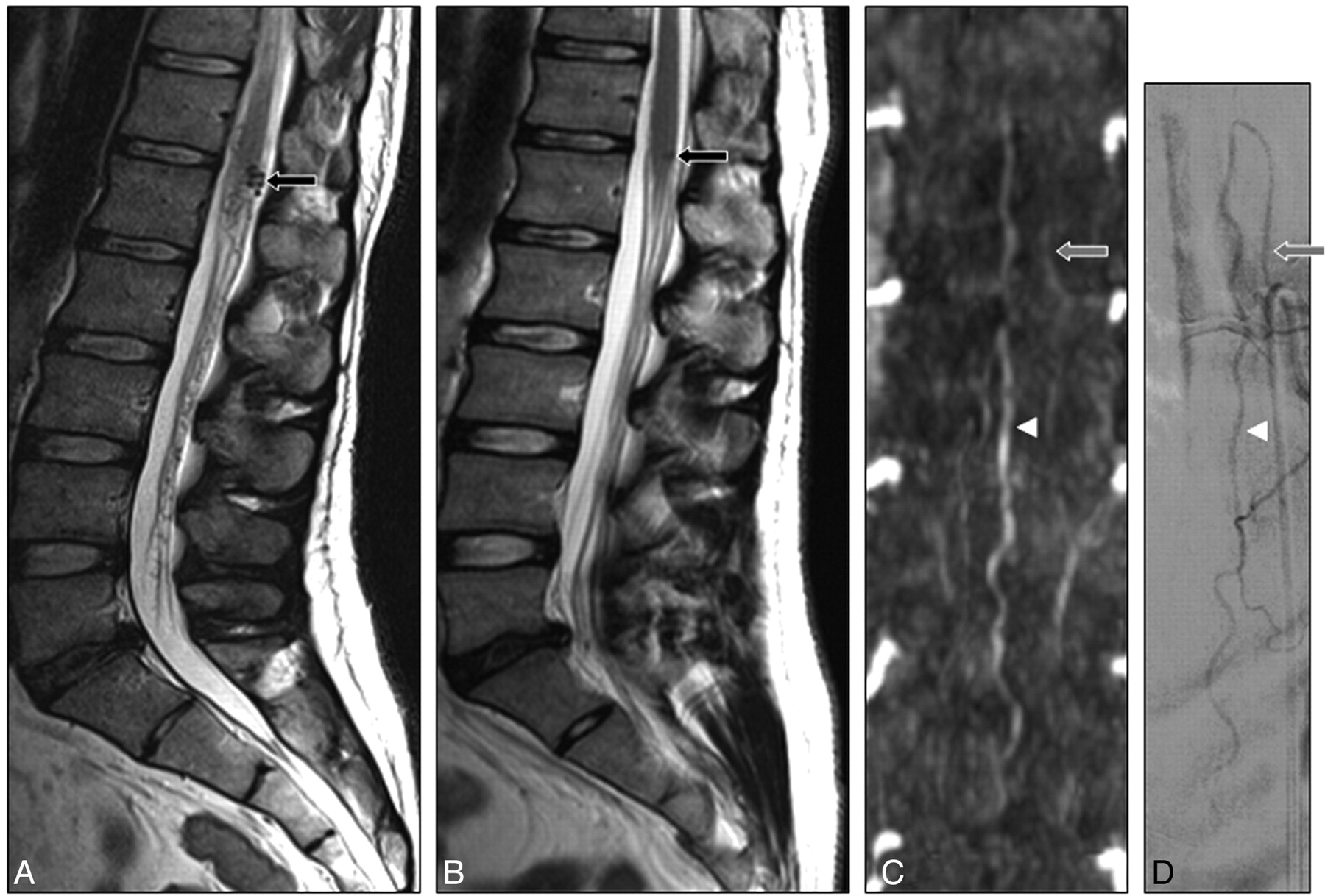
A and B, Pre- (A) and postangiography (B) T2-weighted MR images showing complete disappearance of the tortuous enlarged veins and a small remnant of the AVM (black arrow, B). C, High-resolution MR angiogram demonstrates the radiculomedullary artery at T9 left (gray arrow) with a patent anterior spinal artery (white arrowhead). D, Spinal angiogram confirms persistent obliteration of the AVM with a patent anterior spinal artery (white arrowhead).

Discussion

Spontaneous obliteration of central nervous system AVMs has occasionally been described.6–8 Most of these cases were cerebral AVMs that thrombosed after symptomatic intracerebral or subarachnoid hemorrhage. This spontaneous thrombosis of a cerebral AVM may be explained by reduction of blood flow as a result of compression on the proximal draining veins by the hematoma or edema, feeding artery occlusion through vasospasm, thromboembolism, or systemic coagulation disorders.9

Spontaneous obliteration of spinal AVMs has been described in only 3 cases. One spinal AVM at the T11–12 level thrombosed after hemorrhage. However, in this case, a previous angiogram was not available and surgical exploration was not performed.2 The second case involved complete thrombosis of a perimedullary spinal cord AVM at the T11-L1 level. The thrombosis probably occurred soon after diagnostic angiography and was associated with neurologic deterioration. MR imaging and angiography performed 1 week later revealed complete occlusion of the lesion. Thrombosis of the dominant draining vein triggered by contrast injection was believed to be the most probable cause of the observed phenomenon.9 The third case was an angiographically confirmed spinal AVM at the T12-L1 level that was left untreated. The patient returned 3 years later with sudden paraparesis and bladder retention. Repeat angiography demonstrated complete thrombosis of the AVM, also at follow-up. The patient did not recover completely. It was thought that thrombosis of the draining veins, triggered by the interaction of genetic and hemodynamic factors, was the most probable cause.1

Obliteration during angiography with confirmation 3 months later, as in our case, has not been described previously, to our knowledge. The mechanism that led to thrombosis of the AVM remains unclear. Possible mechanical explanations are thromboembolism, dissection of the vessel wall, and vessel spasm. However, in our case, dissection of the vessel wall or vasospasm did not occur, and there were no thrombi visualized on the angiogram. Nonionic contrast agents, as used in our patient, can induce coagulation via activation of platelets, aggregation of red blood cells, changes in the endothelium, and changes of the osmolality and ionic strength.3,10 We believe this toxic effect of the contrast agent is the most likely explanation for the acute thrombosis. The patient's symptoms resolved soon after obliteration of the AVM, unlike in the 2 previously documented cases. Apparently, the thrombosis of the AVM remained restricted to the nidus itself with unaffected patency of the anterior spinal axis.

It is suggested that spontaneously thrombosed AVMs may recanalize and that follow-up angiographic evaluation is recommended, preferably after 2 months or later.7 We performed follow-up angiography at 3 and 6 months, which showed persistent complete occlusion of the AVM by both MR and conventional angiography.

References

  1. 1.↵
    1. Panciani PP,
    2. Fontanella M,
    3. Crobeddu E,
    4. et al
    . Spontaneous occlusion of a spinal arteriovenous malformation: is treatment always necessary? J Neurosurg Spine 2010;12:397–401
    CrossRefPubMed
  2. 2.↵
    1. Chun JY,
    2. Gulati M,
    3. Halbach V,
    4. et al
    . Thrombosis of a spinal arteriovenous malformation after hemorrhage: case report. Surg Neurol 2004;61:92–94
    CrossRefPubMed
  3. 3.↵
    1. Hay KL,
    2. Bull BS.
    . Factors influencing the activation of platelets by nonionic contrast medium. J Vasc Interv Radiol 1996;7:401–07
    CrossRefPubMed
  4. 4.↵
    1. Mull M,
    2. Nijenhuis RJ,
    3. Backes WH,
    4. et al
    . Value and limitations of contrast-enhanced MR angiography in spinal arteriovenous malformations and dural arteriovenous fistulas. AJNR Am J Neuroradiol 2007;28:1249–58
    Abstract/FREE Full Text
  5. 5.↵
    1. Nijenhuis RJ,
    2. Mull M,
    3. Thron AK,
    4. et al
    . MR angiography of the Adamkiewicz artery validated by digital subtraction angiography. AJNR Am J Neuroradiol 2006;27:1565–72
    Abstract/FREE Full Text
  6. 6.↵
    1. Konus OL,
    2. Ilgit ET,
    3. Ozdemir A,
    4. et al
    . Spontaneous thrombosis of a vein of Galen aneurysmal malforation: possible effect of contrast media. Eur Radiol 2000;10:1456–58
    CrossRefPubMed
  7. 7.↵
    1. Krapf H,
    2. Siekmann R,
    3. Freudenstein D,
    4. et al
    . Spontaneous occlusion of a cerebral arteriovenous malformation: angiography and MR imaging follow-up and review of the literature. AJNR Am J Neuroradiol 2001;22:1556–60
    Abstract/FREE Full Text
  8. 8.↵
    1. Lee SK,
    2. Vilela P,
    3. Willinsky R,
    4. et al
    . Spontaneous regression of cerebral arteriovenous malformation: clinical and angiographic analysis with review of the literature. Neuroradiology 2002;44:11–16
    CrossRefPubMed
  9. 9.↵
    1. Gupta V,
    2. Rizvi T,
    3. Garg A,
    4. et al
    . Postangiographic thrombosis of a spinal arteriovenous malformation. J Neurosurg Spine 2005;22:486–90
  10. 10.↵
    1. Eloy R
    . Thromboembolic Risks in Angiography: Role of Iodinated Contrast Media. Berlin, Germany: Springer-Verlag; 1990
  • Received September 1, 2010.
  • Accepted after revision October 15, 2010.
  • © 2012 by American Journal of Neuroradiology
View Abstract
PreviousNext
Back to top

In this issue

American Journal of Neuroradiology: 33 (1)
American Journal of Neuroradiology
Vol. 33, Issue 1
1 Jan 2012
  • Table of Contents
  • Index by author
Advertisement
Print
Download PDF
Email Article

Thank you for your interest in spreading the word on American Journal of Neuroradiology.

NOTE: We only request your email address so that the person you are recommending the page to knows that you wanted them to see it, and that it is not junk mail. We do not capture any email address.

Enter multiple addresses on separate lines or separate them with commas.
Contrast Agent Induced Obliteration of a Spinal Arteriovenous Malformation
(Your Name) has sent you a message from American Journal of Neuroradiology
(Your Name) thought you would like to see the American Journal of Neuroradiology web site.
CAPTCHA
This question is for testing whether or not you are a human visitor and to prevent automated spam submissions.
Cite this article
R.J. Nijenhuis, M. Sluzewski, W.J. van Rooij
Contrast Agent Induced Obliteration of a Spinal Arteriovenous Malformation
American Journal of Neuroradiology Jan 2012, 33 (1) E1-E3; DOI: 10.3174/ajnr.A2436

Citation Manager Formats

  • BibTeX
  • Bookends
  • EasyBib
  • EndNote (tagged)
  • EndNote 8 (xml)
  • Medlars
  • Mendeley
  • Papers
  • RefWorks Tagged
  • Ref Manager
  • RIS
  • Zotero
0 Responses
Respond to this article
Share
Bookmark this article
Contrast Agent Induced Obliteration of a Spinal Arteriovenous Malformation
R.J. Nijenhuis, M. Sluzewski, W.J. van Rooij
American Journal of Neuroradiology Jan 2012, 33 (1) E1-E3; DOI: 10.3174/ajnr.A2436
del.icio.us logo Twitter logo Facebook logo Mendeley logo
  • Tweet Widget
  • Facebook Like
  • Google Plus One
Purchase

Jump to section

  • Article
    • Abstract
    • Abbreviations
    • Case Report
    • Discussion
    • References
  • Figures & Data
  • Info & Metrics
  • Responses
  • References
  • PDF

Related Articles

  • No related articles found.
  • PubMed
  • Google Scholar

Cited By...

  • No citing articles found.
  • Crossref (1)
  • Google Scholar

This article has been cited by the following articles in journals that are participating in Crossref Cited-by Linking.

  • Spontaneous regression of extradural high-flow vascular malformation in spinal arteriovenous metameric syndrome (SAMS): A unique case report
    Prasert Iampreechakul, Songpol Chuntaroj, Yodkhwan Wattanasen, Sunisa Hangsapruek, Punjama Lertbutsayanukul, Somkiet Siriwimonmas
    Surgical Neurology International 2023 14

More in this TOC Section

  • Atypical Diffusion-Restricted Lesion in 5-Fluorouracil Encephalopathy
  • Multimodal Imaging of Spike Propagation: A Technical Case Report
  • Dural Infantile Hemangioma Masquerading as a Skull Vault Lesion
Show more Case Reports

Similar Articles

Advertisement

Indexed Content

  • Current Issue
  • Accepted Manuscripts
  • Article Preview
  • Past Issues
  • Editorials
  • Editors Choice
  • Fellow Journal Club
  • Letters to the Editor

Cases

  • Case Collection
  • Archive - Case of the Week
  • Archive - Case of the Month
  • Archive - Classic Case

Special Collections

  • Special Collections

Resources

  • News and Updates
  • Turn around Times
  • Submit a Manuscript
  • Author Policies
  • Manuscript Submission Guidelines
  • Evidence-Based Medicine Level Guide
  • Publishing Checklists
  • Graphical Abstract Preparation
  • Imaging Protocol Submission
  • Submit a Case
  • Become a Reviewer/Academy of Reviewers
  • Get Peer Review Credit from Publons

Multimedia

  • AJNR Podcast
  • AJNR SCANtastic
  • Video Articles

About Us

  • About AJNR
  • Editorial Board
  • Not an AJNR Subscriber? Join Now
  • Alerts
  • Feedback
  • Advertise with us
  • Librarian Resources
  • Permissions
  • Terms and Conditions

American Society of Neuroradiology

  • Not an ASNR Member? Join Now

© 2025 by the American Society of Neuroradiology All rights, including for text and data mining, AI training, and similar technologies, are reserved.
Print ISSN: 0195-6108 Online ISSN: 1936-959X

Powered by HighWire