Advertisement

Main menu

  • Home
  • Content
    • Current Issue
    • Accepted Manuscripts
    • Article Preview
    • Past Issue Archive
    • AJNR Case Collection
    • Case of the Week Archive
    • Classic Case Archive
    • Case of the Month Archive
  • Special Collections
    • Spinal CSF Leak Articles (Jan 2020-June 2024)
    • 2024 AJNR Journal Awards
    • Most Impactful AJNR Articles
  • Multimedia
    • AJNR Podcast
    • AJNR Scantastics
    • Video Articles
  • For Authors
    • Submit a Manuscript
    • Author Policies
    • Fast publishing of Accepted Manuscripts
    • Graphical Abstract Preparation
    • Manuscript Submission Guidelines
    • Imaging Protocol Submission
    • Submit a Case for the Case Collection
  • About Us
    • About AJNR
    • Editorial Board
  • More
    • Become a Reviewer/Academy of Reviewers
    • Subscribers
    • Permissions
    • Alerts
    • Feedback
    • Advertisers
    • ASNR Home
  • Other Publications
    • ajnr

User menu

  • Alerts
  • Log in

Search

  • Advanced search
American Journal of Neuroradiology

American Journal of Neuroradiology

ASHNR American Society of Functional Neuroradiology ASHNR American Society of Pediatric Neuroradiology ASSR
  • Alerts
  • Log in

Advanced Search

  • Home
  • Content
    • Current Issue
    • Accepted Manuscripts
    • Article Preview
    • Past Issue Archive
    • AJNR Case Collection
    • Case of the Week Archive
    • Classic Case Archive
    • Case of the Month Archive
  • Special Collections
    • Spinal CSF Leak Articles (Jan 2020-June 2024)
    • 2024 AJNR Journal Awards
    • Most Impactful AJNR Articles
  • Multimedia
    • AJNR Podcast
    • AJNR Scantastics
    • Video Articles
  • For Authors
    • Submit a Manuscript
    • Author Policies
    • Fast publishing of Accepted Manuscripts
    • Graphical Abstract Preparation
    • Manuscript Submission Guidelines
    • Imaging Protocol Submission
    • Submit a Case for the Case Collection
  • About Us
    • About AJNR
    • Editorial Board
  • More
    • Become a Reviewer/Academy of Reviewers
    • Subscribers
    • Permissions
    • Alerts
    • Feedback
    • Advertisers
    • ASNR Home
  • Follow AJNR on Twitter
  • Visit AJNR on Facebook
  • Follow AJNR on Instagram
  • Join AJNR on LinkedIn
  • RSS Feeds

Welcome to the new AJNR, Updated Hall of Fame, and more. Read the full announcements.


AJNR is seeking candidates for the position of Associate Section Editor, AJNR Case Collection. Read the full announcement.


LetterLETTER

Imaging of Facial Fillers: Additional Insights

D.T. Ginat and C.J. Schatz
American Journal of Neuroradiology December 2012, 33 (11) E140-E141; DOI: https://doi.org/10.3174/ajnr.A3394
D.T. Ginat
aDepartment of Radiology Massachusetts General Hospital, Harvard Medical School Boston, Massachusetts
  • Find this author on Google Scholar
  • Find this author on PubMed
  • Search for this author on this site
C.J. Schatz
bBeverly Tower Wilshire Advanced Imaging Beverly Hills, California University of Southern California Keck School of Medicine Los Angeles, California
  • Find this author on Google Scholar
  • Find this author on PubMed
  • Search for this author on this site
  • Article
  • Figures & Data
  • Info & Metrics
  • Responses
  • References
  • PDF
Loading

We appreciate the interest that readers have expressed in our article “Imaging Features of Midface Injectable Fillers and Associated Complications,”1 and we are pleased to be regarded as pioneers in the field. However, we would like to clarify a few points and share additional insights on the subject.

Our review article certainly shows the utility of CT and MR imaging for depicting many of the early and late complications associated with facial fillers besides foreign-body granulomas.1 In particular, we have, on occasion, received requests to delineate the extent of filler material, especially when migration has occurred. Indeed, some of these cases are shown in our article.

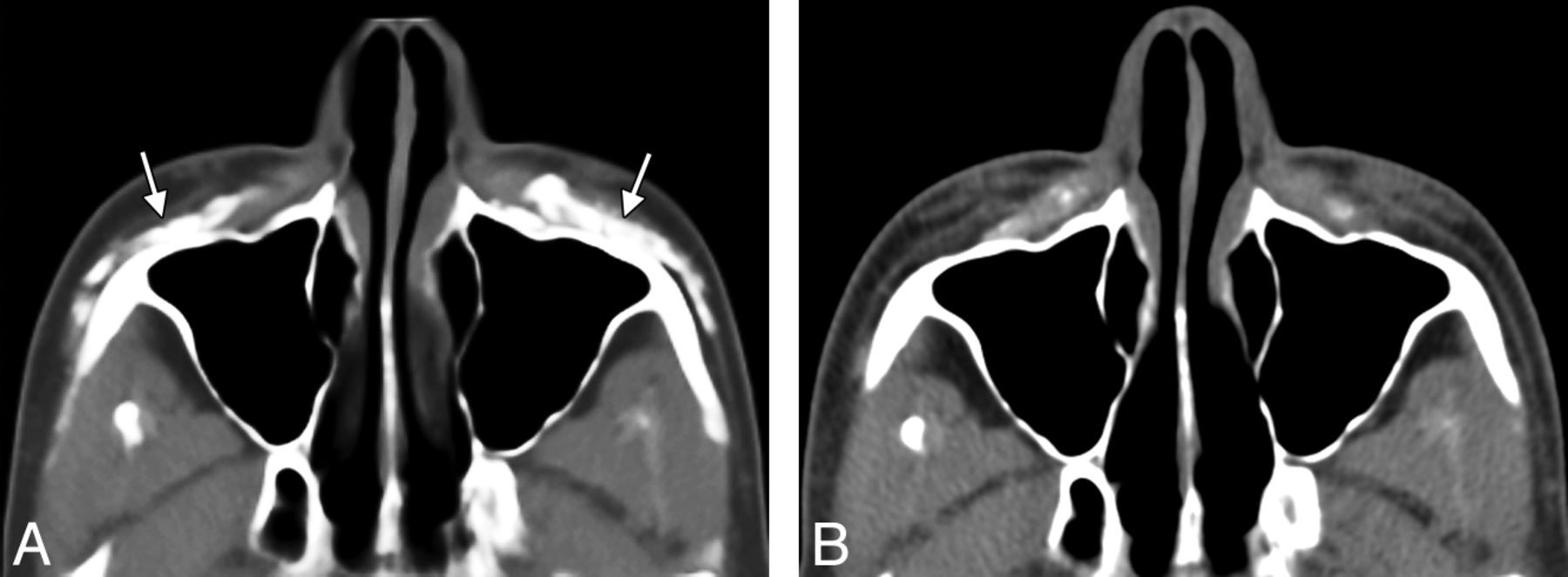
Although facial fillers may appear different from one patient to another and can change with time (Fig 1), ), certain fundamental imaging characteristics of facial fillers can be surmised. This is the case in many other areas of radiology in which pattern-recognition applies. Although imaging is not intended simply to differentiate the types of facial filler materials, some of these have rather distinctive appearances.

Fig 1.
  • Download figure
  • Open in new tab
  • Download powerpoint
Fig 1.

Axial CT image (A) shows hyperattenuated deposits of calcium hydroxylapatite within the bilateral cheeks (arrows). Axial CT image obtained 22 months (B) later shows significant interval decrease in the hyperattenuated deposits.

Although uncommon, scar formation is a known complication of facial fillers. This phenomenon has been previously described with silicone and Artecoll (Suneva Medical, San Diego, California), for example: “Siliconomas and scarring can occur after a variable delay after the silicone injections, ranging from 3 weeks to a decade. … If Artecoll (collagen and polymethylmethacrylate) is injected too superficially, it can lead to persistent itchiness, redness and even hypertrophic scarring that may need to be reduced by local corticosteroid injections.”2

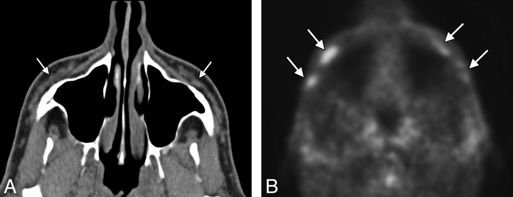
With regard to the hypermetabolism associated with injection of a calcium hydroxylapatite filler, this can be a source of false-positive findings on PET.3,4 We have also noted hypermetabolism associated with fillers other than calcium hydroxylapatite (Fig 2, suggestive of an inflammatory response that can be subclinical.

Fig 2.
  • Download figure
  • Open in new tab
  • Download powerpoint
Fig 2.

Axial CT image (A) shows nearly fluid-attenuation filler material within the bilateral cheeks (arrows). PET image (B) shows hypermetabolic foci within the cheek subcutaneous tissues corresponding to reported collagen-based fillers (arrows). However, the patient did not have symptoms related to this finding, and no biopsy was performed.

The study of facial fillers is an evolving multidisciplinary field with many questions remaining to be answered.

References

  1. 1.↵
    1. Ginat DT,
    2. Schatz CJ
    . Imaging features of midface injectable fillers and associated complications. AJNR Am J Neuroradiol 2012 Jul 26 [Epub ahead of print]
  2. 2.↵
    1. Lafaille P,
    2. Benedetto A
    . Fillers: contraindications, side effects and precautions. J Cutan Aesthet Surg 2010; 3: 16– 19
    CrossRefPubMed
  3. 3.↵
    1. Damrose EJ
    . Radiographic properties of injected calcium hydroxylapatite: potential false positive findings on positron emission tomography. J Laryngol Otol 2008; 122: 1394– 96
    CrossRefPubMed
  4. 4.↵
    1. Feeney JN,
    2. Fox JJ,
    3. Akhurst T
    . Radiological impact of the use of calcium hydroxylapatite dermal fillers. Clin Radiol 2009; 64: 897– 902
    CrossRefPubMed
  • © 2012 by American Journal of Neuroradiology
PreviousNext
Back to top

In this issue

American Journal of Neuroradiology: 33 (11)
American Journal of Neuroradiology
Vol. 33, Issue 11
1 Dec 2012
  • Table of Contents
  • Index by author
Advertisement
Print
Download PDF
Email Article

Thank you for your interest in spreading the word on American Journal of Neuroradiology.

NOTE: We only request your email address so that the person you are recommending the page to knows that you wanted them to see it, and that it is not junk mail. We do not capture any email address.

Enter multiple addresses on separate lines or separate them with commas.
Imaging of Facial Fillers: Additional Insights
(Your Name) has sent you a message from American Journal of Neuroradiology
(Your Name) thought you would like to see the American Journal of Neuroradiology web site.
CAPTCHA
This question is for testing whether or not you are a human visitor and to prevent automated spam submissions.
Cite this article
D.T. Ginat, C.J. Schatz
Imaging of Facial Fillers: Additional Insights
American Journal of Neuroradiology Dec 2012, 33 (11) E140-E141; DOI: 10.3174/ajnr.A3394

Citation Manager Formats

  • BibTeX
  • Bookends
  • EasyBib
  • EndNote (tagged)
  • EndNote 8 (xml)
  • Medlars
  • Mendeley
  • Papers
  • RefWorks Tagged
  • Ref Manager
  • RIS
  • Zotero
0 Responses
Respond to this article
Share
Bookmark this article
Imaging of Facial Fillers: Additional Insights
D.T. Ginat, C.J. Schatz
American Journal of Neuroradiology Dec 2012, 33 (11) E140-E141; DOI: 10.3174/ajnr.A3394
del.icio.us logo Twitter logo Facebook logo Mendeley logo
  • Tweet Widget
  • Facebook Like
  • Google Plus One
Purchase

Jump to section

  • Article
    • References
  • Figures & Data
  • Info & Metrics
  • Responses
  • References
  • PDF

Related Articles

  • No related articles found.
  • PubMed
  • Google Scholar

Cited By...

  • No citing articles found.
  • Crossref (9)
  • Google Scholar

This article has been cited by the following articles in journals that are participating in Crossref Cited-by Linking.

  • Injectable facial fillers: imaging features, complications, and diagnostic pitfalls at MRI and PET CT
    Pravin Mundada, Romain Kohler, Sana Boudabbous, Laurence Toutous Trellu, Alexandra Platon, Minerva Becker
    Insights into Imaging 2017 8 6
  • Complications After Facial Injections With Permanent Fillers: Important Limitations and Considerations of MRI Evaluation
    Jonathan A. Kadouch, Charlotte J. Tutein Nolthenius, Daniel J. Kadouch, Henk-Jan van der Woude, Refaat B. Karim, Rick Hoekzema
    Aesthetic Surgery Journal 2014 34 6
  • Penile girth augmentation by injectable fillers: a comprehensive review of imaging features and inflammatory complications
    Sherif A. Abo-Hedibah, Amr N. Badawi, Sameh A. Aly, Sherif R. M. Ismail, Ali H. Elmokadem
    Abdominal Radiology 2021 46 4
  • Imaging Features and Complications of Facial Cosmetic Procedures
    Andrea Meneses Soares de Sousa, Angelo Chelotti Duarte, Marcos Decnop, Daniel de Faria Guimarães, Carlos Alberto Ferreira Coelho Neto, Maíra de Oliveira Sarpi, Luis Gustavo Palhiari Duarte, Soraia Ale Souza, Larissa Freire Segato, Julia Diva Zavariz, Suresh K. Mukherji, Márcio Ricardo Taveira Garcia
    RadioGraphics 2023 43 12
  • ESR Essentials: pseudolesions in head and neck—practice recommendations by the European Society of Head and Neck Radiology
    Darka Hadnadjev Šimonji, Aleksandar Spasić, Maja Stankov
    European Radiology 2025
  • MRI Visualization and Distribution Patterns of Foreign Modeling Agents: A Brief Pictorial Review for Clinicians
    Leslie-Marisol Gonzalez-Hermosillo, Victor-Hugo Ramos-Pacheco, Daisy-Carolina Gonzalez-Hermosillo, Alicia-Maria-del-Consuelo Cervantes-Sanchez, Alejandro-Eduardo Vega-Gutierrez, Sergey K. Ternovoy, Ernesto Roldan-Valadez, Muthu Rama Krishnan Mookiah
    BioMed Research International 2021 2021 1
  • Non-ischemic complications of dermal fillers
    Preeya Mehta, Joy Li, Julie Woodward, Sandy Zhang-Nunes
    Plastic and Aesthetic Research 2023
  • Sequential MRI as a Diagnostic Tool for Follow‐Up of Hyaluronic Acid Dermal Filler, in a Woman Who Underwent Radiation Therapy for Oral Cancer
    Gloria Bettini, Ferdinando De Negri, Roberto Amore, Sergio Rexhep Tari, Antonio Scarano
    Clinical Case Reports 2025 13 4
  • Three-Dimensional Photography for Evaluating the Effectiveness of Botulinum Toxin Injection for Masseter Hypertrophy
    Dun-Wei Huang, Chung-Yu Lai, Jia-En Chen, Chin-Chieh Yi, Ya-Hsin Chen, Chih-Hsin Wang, Shyi-Gen Chen
    Aesthetic Plastic Surgery 2024 48 20

More in this TOC Section

  • Brain AVM’s Nidus: What if We Hadn’t Understood Anything?
  • Letter to the Editor regarding “Automated Volumetric Software in Dementia: Help or Hindrance to the Neuroradiologist?”
  • Reply:
Show more Letters

Similar Articles

Advertisement

Indexed Content

  • Current Issue
  • Accepted Manuscripts
  • Article Preview
  • Past Issues
  • Editorials
  • Editors Choice
  • Fellow Journal Club
  • Letters to the Editor

Cases

  • Case Collection
  • Archive - Case of the Week
  • Archive - Case of the Month
  • Archive - Classic Case

Special Collections

  • Special Collections

Resources

  • News and Updates
  • Turn around Times
  • Submit a Manuscript
  • Author Policies
  • Manuscript Submission Guidelines
  • Evidence-Based Medicine Level Guide
  • Publishing Checklists
  • Graphical Abstract Preparation
  • Imaging Protocol Submission
  • Submit a Case
  • Become a Reviewer/Academy of Reviewers
  • Get Peer Review Credit from Publons

Multimedia

  • AJNR Podcast
  • AJNR SCANtastic
  • Video Articles

About Us

  • About AJNR
  • Editorial Board
  • Not an AJNR Subscriber? Join Now
  • Alerts
  • Feedback
  • Advertise with us
  • Librarian Resources
  • Permissions
  • Terms and Conditions

American Society of Neuroradiology

  • Not an ASNR Member? Join Now

© 2025 by the American Society of Neuroradiology All rights, including for text and data mining, AI training, and similar technologies, are reserved.
Print ISSN: 0195-6108 Online ISSN: 1936-959X

Powered by HighWire