
Article Figures & Data
Figures
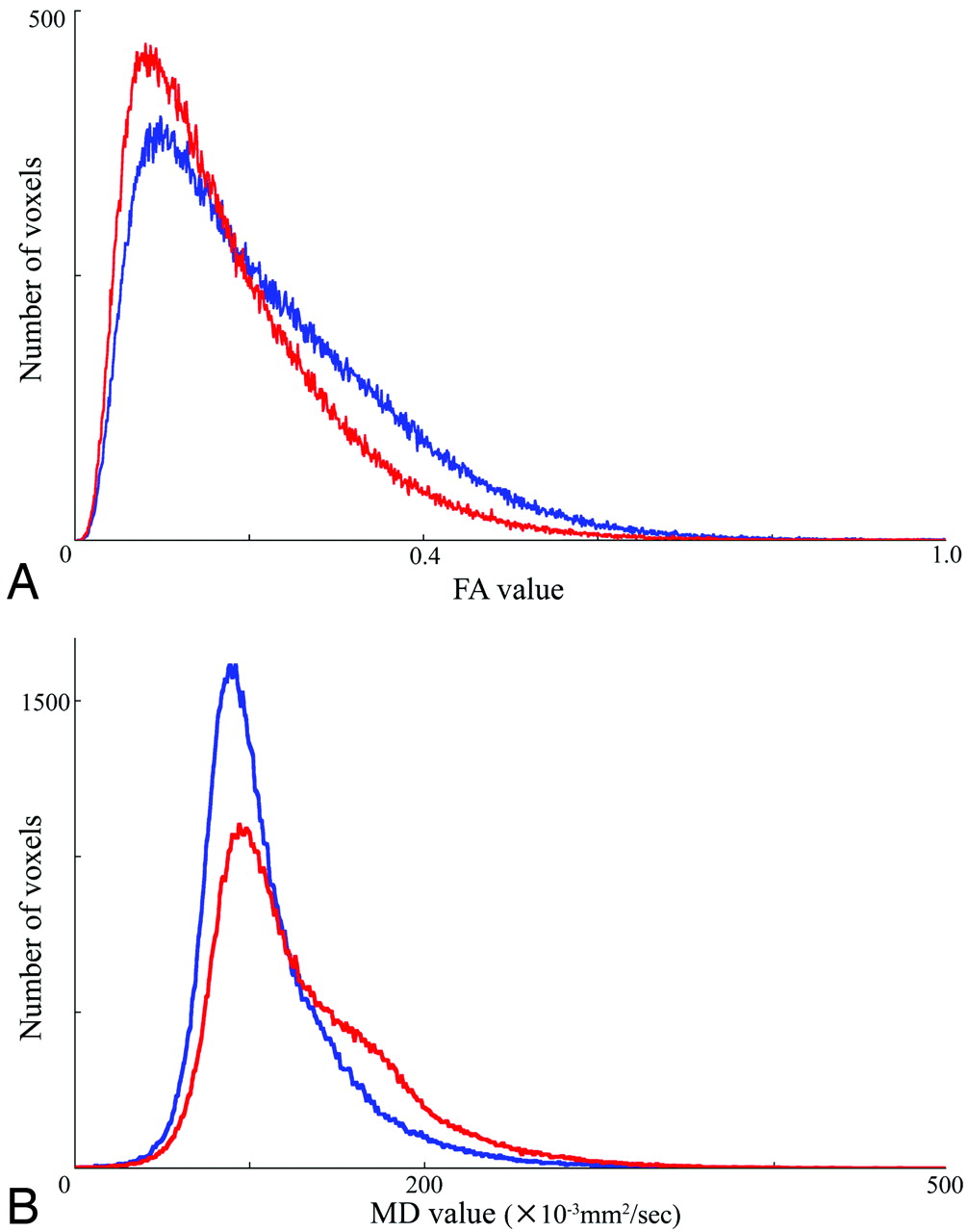
- Fig 1.
Representative FA (A) and MD (B) histograms of the HDWM. In both histograms, averaging the histograms of 4 patients with almost normal cognitive function (MMSE 29–30) is represented in blue, whereas averaging the histograms of 4 patients with dementia (MMSE 10–16) is represented in red.
- Fig 2.
Scatterplot of callosal mean FA values against MMSE scores in each patient.
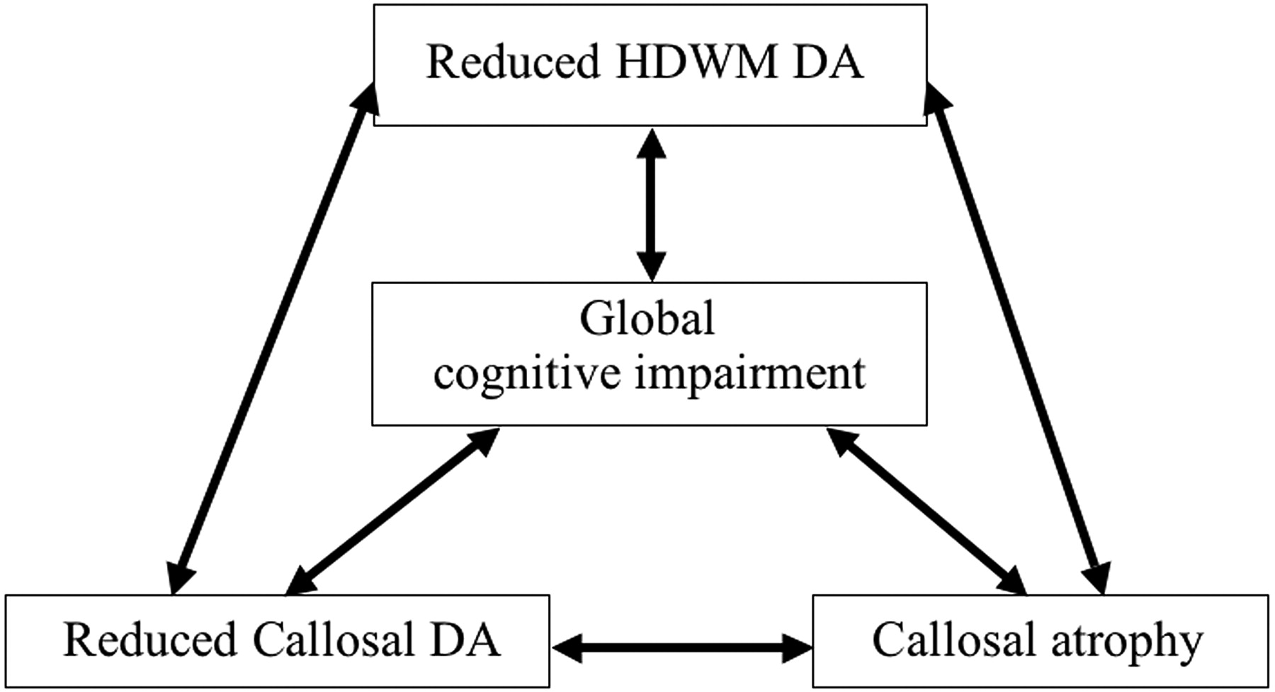
- Fig 3.
Schematic diagram of the intercorrelation between reduced HDWM DA, reduced callosal DA, callosal atrophy, and cognitive impairment.
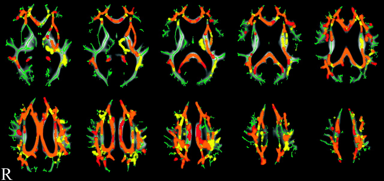
- Fig 4.
Result of voxel-based analysis of FA and MD maps by using TBSS. Orange shows where both FA reduction and MD increase had significant correlation with the MMSE score decline. Red or yellow shows where FA reduction or MD increase had significant correlation with the MMSE score decline, respectively. All results are adjusted for WML volume and white matter volume. Permutation-based inference on cluster size: t > 2, FWE-corrected P < .05; permutation number, 5000. Green is the mean FA skeleton, underneath the orange, red, and yellow. In the background is the mean FA image of the 24 patients in the study.


Tables
MMSE FAB VFT WMSR HDWM DA FA25 0.77b 0.76b 0.69b 0.77b Median FA 0.67b 0.73b 0.61c 0.62d Mean FA 0.74b 0.74b 0.66e 0.73f MD25 −0.75b −0.73b −0.72b −0.84b Median MD −0.72b −0.71b −0.70b −0.84b Mean MD −0.74b −0.67b −0.68f −0.85b Callosal mean FA 0.95b 0.89b 0.80b 0.75b Callosal mean MD −0.90b −0.72b −0.71b −0.85b Callosal volume 0.75b 0.83b 0.86b 0.63d - Table 2:
Correlation between HDWM DA or callosal parameters and cognitive scores evaluated with multiple regression analysesa
MMSE FAB VFT WMSR HDWM DA FA25 0.76b 0.79b 0.64c 0.72b Mean FA 0.71b 0.73b 0.56d 0.66c MD25 −0.99b −0.89b −0.59e −0.83b Mean MD −0.91b −0.78b −0.52f −0.86b Callosal mean FA 0.80b 0.85b 0.70b 0.73g Callosal mean MD −0.74b −0.66b −0.59g,h −0.82b Callosal volume 0.74b 0.8b 0.72b 0.63f -
↵a Figures are standardized regression coefficients between each of the HDWM DA or callosal parameters and each of the cognitive scores, adjusted for age, sex, WML volume, and white matter volume.
-
↵b Significant after Bonferroni correction (P < .05/28 = .0018).
-
↵c P = .002.
-
↵d P = .006.
-
↵e P = .02.
-
↵f P = .03.
-
↵g P = .003.
-
↵h White matter volume showed a weak contribution to the correlation between callosal MD and VFT (standard regression coefficient, 0.43; P =.046).
-
In this issue


Jump to section
Related Articles
Cited By...
- Leukoaraiosis and acute ischemic stroke: 90-day clinical outcome following endovascular recanalization, with proposed "L-ASPECTS"
- Using DTI to assess white matter microstructure in cerebral small vessel disease (SVD) in multicentre studies
- Tractography at 3T MRI of Corpus Callosum Tracts Crossing White Matter Hyperintensities