
Article Figures & Data
Figures
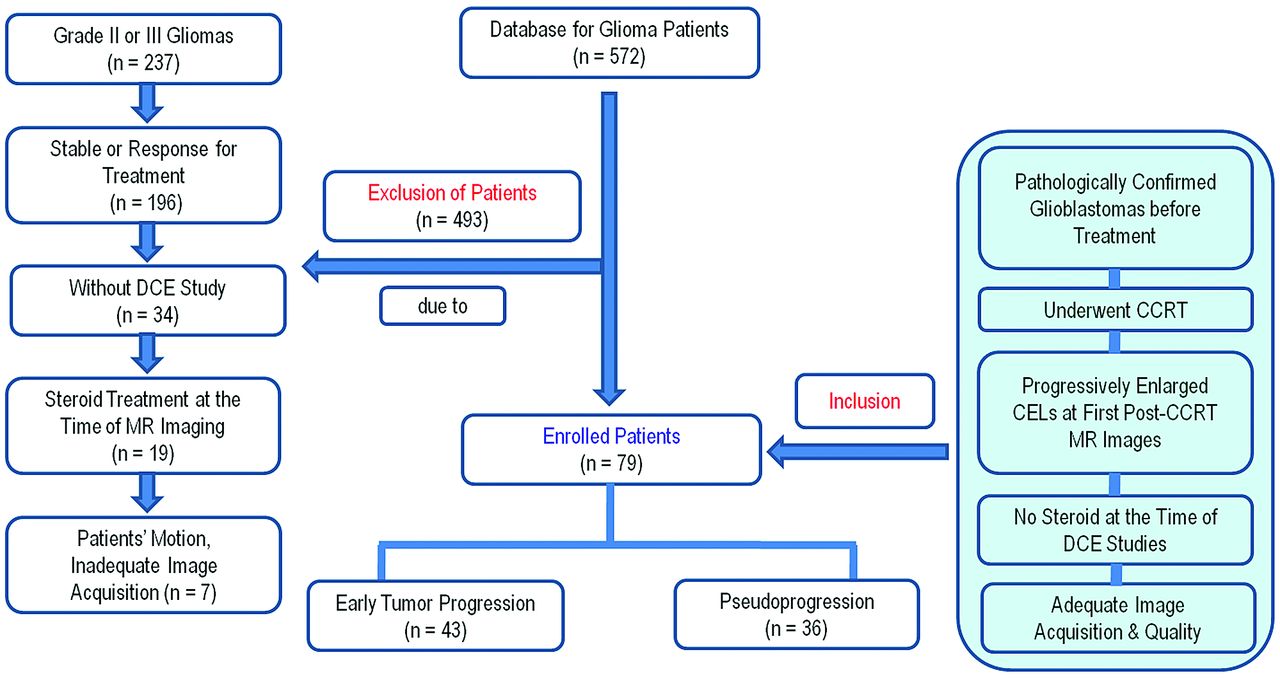
- Fig 1.
Flowchart of the study population. CEL, contrast-enhancing lesion.
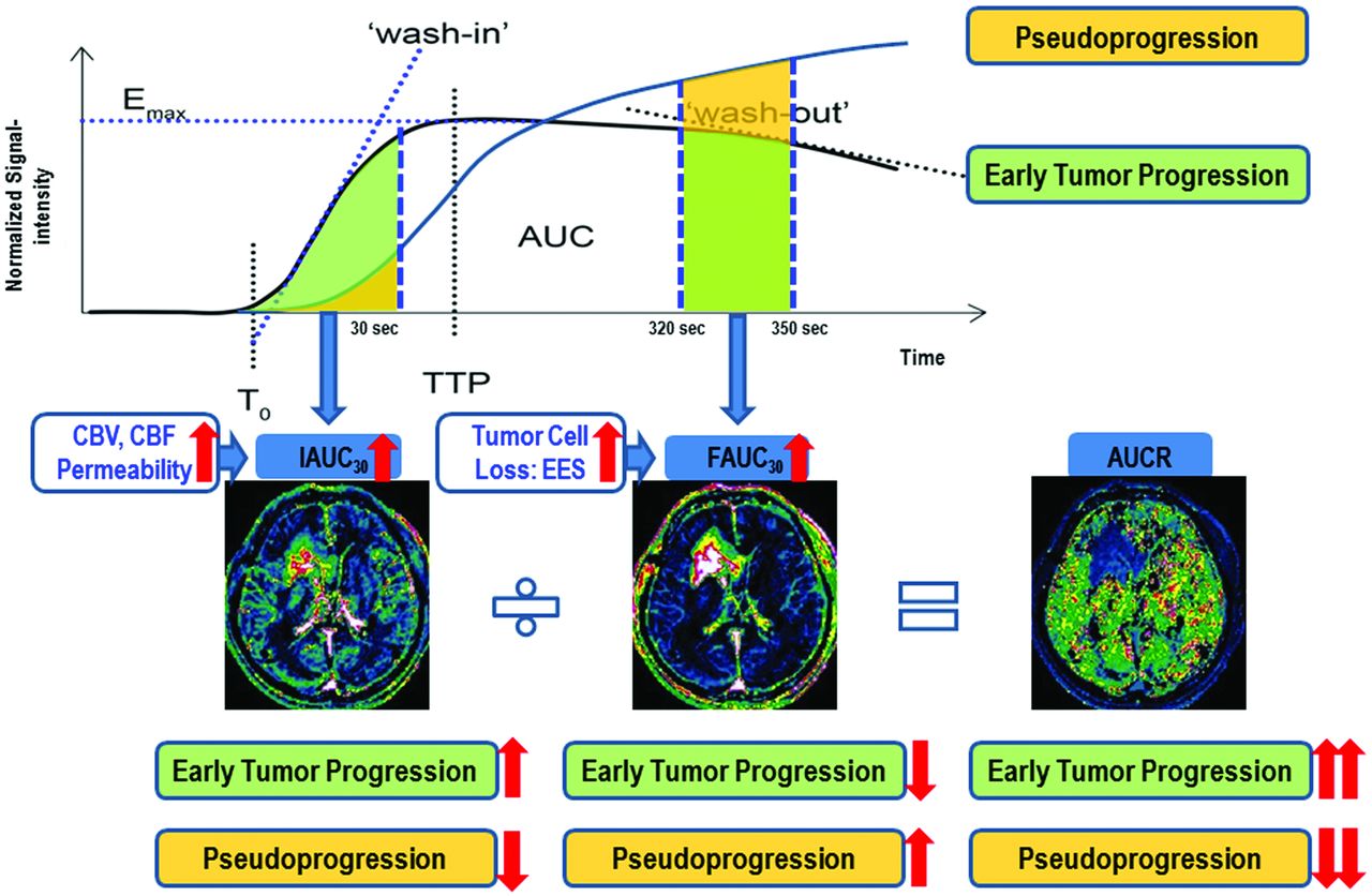
- Fig 2.
Illustration for calculating the AUCR from DCE perfusion MR imaging and the flowchart of our hypothesis.
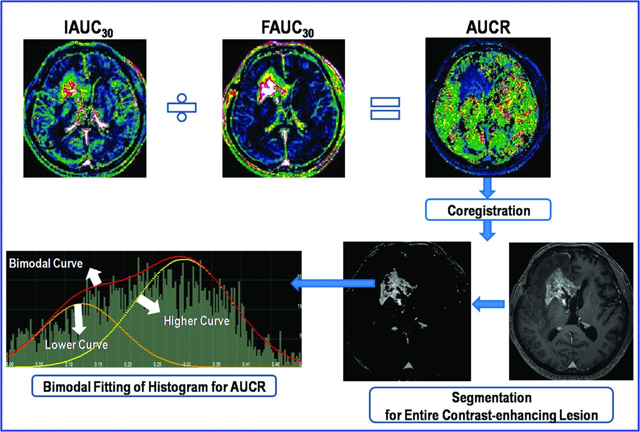
- Fig 3.
Illustration of the step for calculating the AUCR and its histogram.
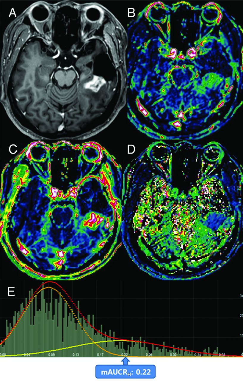
- Fig 5.
Images obtained in a 56-year-old man with posttreatment glioblastoma who had pseudoprogression. Contrast-enhanced T1-weighted imaging (A) obtained 3 weeks after concomitant chemoradiotherapy showed a necrotic, contrast-enhancing mass posterior to the surgical cavity of the left temporal lobe. The IAUC30 (B) and FAUC30 (C) maps derived from dynamic contrast-enhanced, T1-perfusion MR imaging. In B, a visual decrease of the IAUC30 value was noted in the entire contrast-enhancing lesion. The AUCR map (D) and its bimodal histogram (E) showed a decrease in the mean value of the higher curve, thus indicating pseudoprogression.
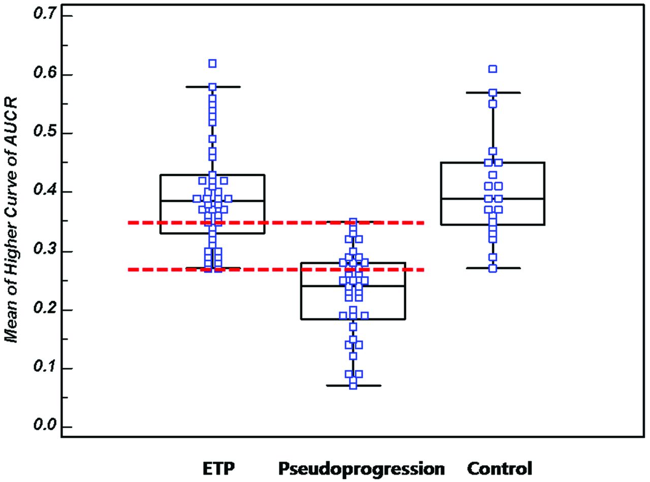
- Fig 6.
A box-and-whisker with scatterplots shows the mAUCRH of the ETP, pseudoprogression, and control groups. A clear difference between the ETP group and the pseudoprogression group can be seen (P < .0001); however, an overlap zone is visible between an mAUCRH of 0.27 and 0.35 (interval between dotted lines).
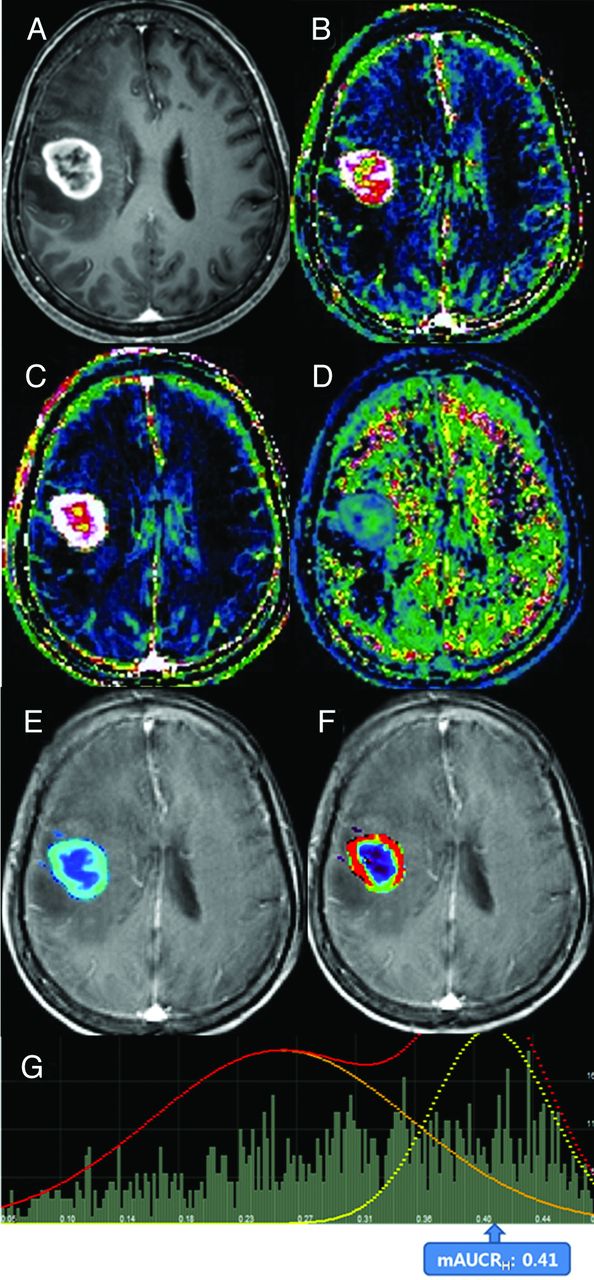
- Fig 7.
Images obtained in a 58-year-old woman with pathologically confirmed treatment-naïve glioblastoma. Contrast-enhanced, T1-weighted image obtained before surgery. A, The image showed a necrotic, contrast-enhancing mass in the right frontal lobe. IAUC30 (B) and FAUC30 (C) maps derived from dynamic contrast-enhanced, T1-perfusion MR imaging. B, A visual increase of the IAUC30 value was noted in the entire contrast-enhancing lesion. The AUCR (D), Ktrans (E), Ve (F) maps, and AUCR bimodal histogram (G) are shown. F, The distribution of visually high Ktrans corresponded with that of the IAUC30 map. G, An AUCR histogram showed increases in bimodal histogram parameters similar with those of ETP.


Tables
Variables Pseudoprogression ETP No. of male patients (%) 17 (45.9) 19 (45.2) No. of female patients (%) 20 (54.1) 23 (54.8) Age (y)a 48.5 ± 9.1 52.6 ± 8.5 Mean KPSa 93.0 ± 5.9 92.4 ± 6.3 Tumor volume (cm3)a 50.2 ± 17.1 55.9 ± 22.12 Surgical extent before CCRT Biopsy 3 6 Subtotal resection 17 17 Gross total resection 17 19 Mean radiation dose (at CCRT, Gy) 59.5 59.7 Mean interval between CCRT and new or enlarging contrast-enhancing lesion (d) 31.2 29.7 MGMT promoter status (methylated/unmethylated) 10/4 7/12 Note:—KPS indicates Karnofsky performance status; MGMT, O(6)-methylguanine methyltransferase.
↵a Data are mean ± SD.
- Table 2:
Multiple comparison test (P value) of the AUCR histogram parameters in the early tumor progression, pseudoprogression, and control groups
AUCR50 AUCR75 AUCR90 AUCRmode mAUCRH Pseudoprogression vs ETP group <.0001 <.0001 <.0001 <.0001 <.0001 Pseudoprogression vs control group <.0001 <.0001 <.0001 <.0001 <.0001 ETP vs control group .557 .572 .771 .752 .747 Note:—AUCR indicates area under the time signal-intensity curve ratio; AUCR50, 50 percentile cutoff value of AUCR; AUCR75, 75 percentile cutoff value of AUCR; AUCR90, 90 percentile cutoff value of AUCR; AUCRmode, AUCR at mode; mAUCRH, mean of the higher curve of AUCR.
- Table 3:
Diagnostic performance of the AUCR histogram parameters for differentiating ETP from pseudoprogression
Parameter Az Valueab Sensitivity (%) Specificity (%) PPV (%) NPV (%) Cutoff Value AUCR50 0.871 (0.757–0.939) 87.2 83.1 84.3 81.1 0.19 AUCR75 0.842 (0.741–0.922) 82.6 81.1 80.9 78.2 0.25 AUCR90 0.879 (0.772–0.949) 89.6 81.7 85.0 87.1 0.34 AUCRmode 0.791 (0.677–0.892) 73.1 79.7 79.1 72.5 0.16 mAUCRH 0.901 (0.791–0.976) 90.1 82.9 87.5 87.9 0.31


Jump to section
Related Articles
Cited By...
- Structural and practical identifiability of contrast transport models for DCE-MRI
- Response Assessment in Neuro-Oncology Criteria for Gliomas: Practical Approach Using Conventional and Advanced Techniques
- Detection of Local Recurrence in Patients with Head and Neck Squamous Cell Carcinoma Using Voxel-Based Color Maps of Initial and Final Area under the Curve Values Derived from DCE-MRI
- Quantitative Evaluation for Differentiating Malignant and Benign Thyroid Nodules Using Histogram Analysis of Grayscale Sonograms
- Differentiating Tumor Progression from Pseudoprogression in Patients with Glioblastomas Using Diffusion Tensor Imaging and Dynamic Susceptibility Contrast MRI
- ASFNR Recommendations for Clinical Performance of MR Dynamic Susceptibility Contrast Perfusion Imaging of the Brain
- Diffusion and Perfusion MRI to Differentiate Treatment-Related Changes Including Pseudoprogression from Recurrent Tumors in High-Grade Gliomas with Histopathologic Evidence