Advertisement

Main menu

  • Home
  • Content
    • Current Issue
    • Accepted Manuscripts
    • Article Preview
    • Past Issue Archive
    • AJNR Case Collection
    • Case of the Week Archive
    • Classic Case Archive
    • Case of the Month Archive
  • Special Collections
    • Spinal CSF Leak Articles (Jan 2020-June 2024)
    • 2024 AJNR Journal Awards
    • Most Impactful AJNR Articles
  • Multimedia
    • AJNR Podcast
    • AJNR Scantastics
    • Video Articles
  • For Authors
    • Submit a Manuscript
    • Author Policies
    • Fast publishing of Accepted Manuscripts
    • Graphical Abstract Preparation
    • Manuscript Submission Guidelines
    • Imaging Protocol Submission
    • Submit a Case for the Case Collection
  • About Us
    • About AJNR
    • Editorial Board
  • More
    • Become a Reviewer/Academy of Reviewers
    • Subscribers
    • Permissions
    • Alerts
    • Feedback
    • Advertisers
    • ASNR Home
  • Other Publications
    • ajnr

User menu

  • Alerts
  • Log in

Search

  • Advanced search
American Journal of Neuroradiology

American Journal of Neuroradiology

ASHNR American Society of Functional Neuroradiology ASHNR American Society of Pediatric Neuroradiology ASSR
  • Alerts
  • Log in

Advanced Search

  • Home
  • Content
    • Current Issue
    • Accepted Manuscripts
    • Article Preview
    • Past Issue Archive
    • AJNR Case Collection
    • Case of the Week Archive
    • Classic Case Archive
    • Case of the Month Archive
  • Special Collections
    • Spinal CSF Leak Articles (Jan 2020-June 2024)
    • 2024 AJNR Journal Awards
    • Most Impactful AJNR Articles
  • Multimedia
    • AJNR Podcast
    • AJNR Scantastics
    • Video Articles
  • For Authors
    • Submit a Manuscript
    • Author Policies
    • Fast publishing of Accepted Manuscripts
    • Graphical Abstract Preparation
    • Manuscript Submission Guidelines
    • Imaging Protocol Submission
    • Submit a Case for the Case Collection
  • About Us
    • About AJNR
    • Editorial Board
  • More
    • Become a Reviewer/Academy of Reviewers
    • Subscribers
    • Permissions
    • Alerts
    • Feedback
    • Advertisers
    • ASNR Home
  • Follow AJNR on Twitter
  • Visit AJNR on Facebook
  • Follow AJNR on Instagram
  • Join AJNR on LinkedIn
  • RSS Feeds

Welcome to the new AJNR, Updated Hall of Fame, and more. Read the full announcements.


AJNR is seeking candidates for the position of Associate Section Editor, AJNR Case Collection. Read the full announcement.


Getting new auth cookie, if you see this message a lot, tell someone!
Review ArticleReview Articles
Open Access

Inherited Cerebellar Ataxia in Childhood: A Pattern-Recognition Approach Using Brain MRI

L. Vedolin, G. Gonzalez, C.F. Souza, C. Lourenço and A.J. Barkovich
American Journal of Neuroradiology May 2013, 34 (5) 925-934; DOI: https://doi.org/10.3174/ajnr.A3055
L. Vedolin
aFrom the Neuroradiology Section (L.V.), Hospital Moinhos de Vento, Porto Alegre, Brazil
ePediatric Neuroradiology (L.V., G.G., A.J.B.), University of California, San Francisco, California.
  • Find this author on Google Scholar
  • Find this author on PubMed
  • Search for this author on this site
G. Gonzalez
bRadiology Department (G.G.), Hospital San Juan de Dios, Santiago, Chile
ePediatric Neuroradiology (L.V., G.G., A.J.B.), University of California, San Francisco, California.
  • Find this author on Google Scholar
  • Find this author on PubMed
  • Search for this author on this site
C.F. Souza
cGenetics Department (C.F.S.), Universidade Federal do Rio Grande do Sul, Porto Alegre, Brazil
  • Find this author on Google Scholar
  • Find this author on PubMed
  • Search for this author on this site
C. Lourenço
dGenetics Department (C.L.), Universidade de Sao Paulo, Ribeirao Preto, Brazil
  • Find this author on Google Scholar
  • Find this author on PubMed
  • Search for this author on this site
A.J. Barkovich
ePediatric Neuroradiology (L.V., G.G., A.J.B.), University of California, San Francisco, California.
  • Find this author on Google Scholar
  • Find this author on PubMed
  • Search for this author on this site
  • Article
  • Figures & Data
  • Supplemental
  • Info & Metrics
  • Responses
  • References
  • PDF
Loading

Article Figures & Data

Figures

  • Fig 1.
    • Download figure
    • Open in new tab
    • Download powerpoint
    Fig 1.

    Cerebellar atrophy. A, Coronal T2 image shows cortical thinning and loss of underlying white matter leading to enlarged fissures in the vermis and cerebellar hemispheres. B, Follow-up coronal T2 image 3 years later shows progressive cortical and white matter atrophy.
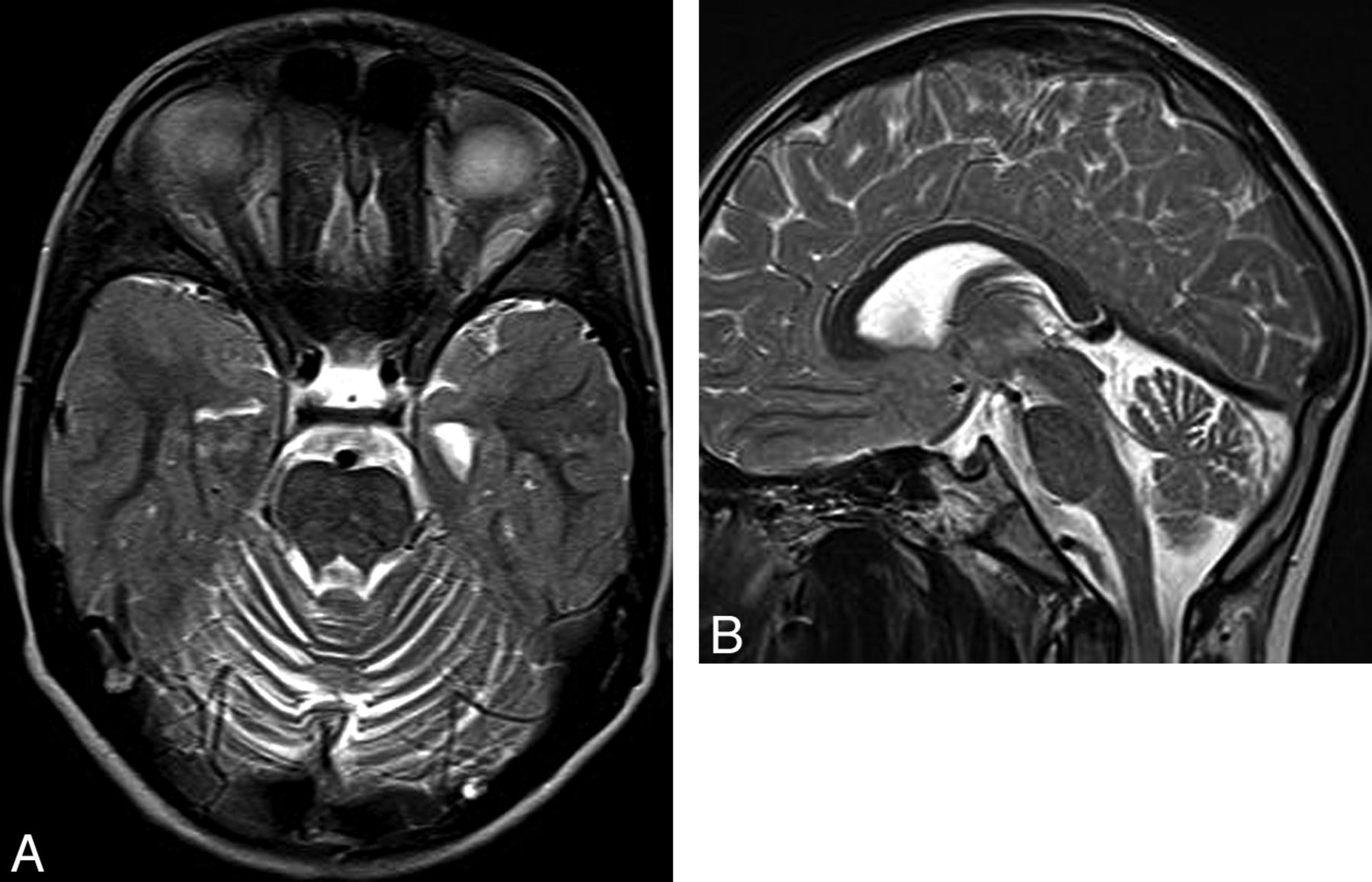
  • Fig 2.
    • Download figure
    • Open in new tab
    • Download powerpoint
    Fig 2.

    Ataxia-telangiectasia. A, Axial T2 image shows cortical thinning and fissure enlargement in the vermis and cerebellar hemispheres. B, Midline sagittal T2 image demonstrates an atrophic vermis but normal volume of the brain stem. Although nonspecific, this pattern of diffuse cerebellar atrophy suggests a degenerative disease.

  • Fig 3.
    • Download figure
    • Open in new tab
    • Download powerpoint
    Fig 3.

    Infantile neuronal ceroid lipofuscinosis. Coronal T1 image (A) shows cerebral and cerebellar atrophy. Coronal FLAIR image (B) demonstrates diffuse abnormal signal intensity affecting the cortex of both cerebellar hemispheres. Compare the signal intensity of the cerebellar cortex with occipital cortex.
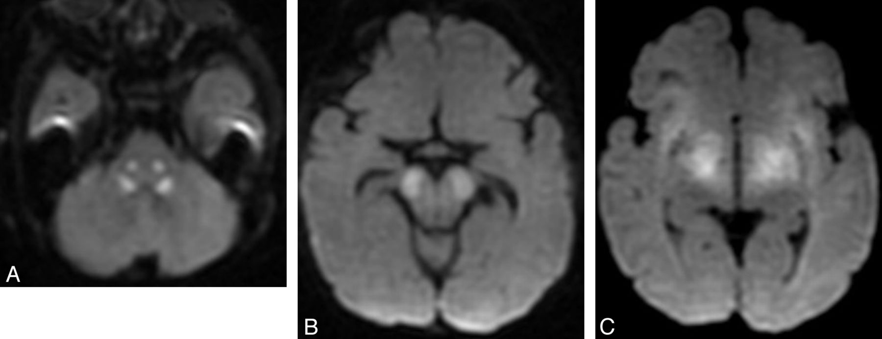
  • Fig 4.
    • Download figure
    • Open in new tab
    • Download powerpoint
    Fig 4.

    Acute stage of maple syrup disease. Axial T2 images (A and B) show diffuse signal change affecting deep cerebellar white matter (arrows) and midbrain (arrowheads). Trace from diffusion-weighted imaging shows reduced diffusivity in several regions of the midbrain (C).
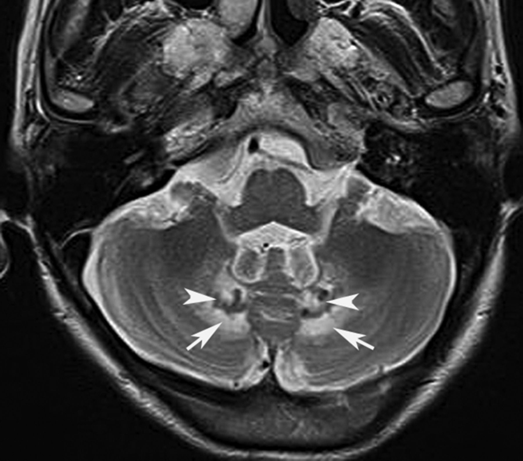
  • Fig 5.
    • Download figure
    • Open in new tab
    • Download powerpoint
    Fig 5.

    Cerebrotendinous xanthomatosis. Axial T2 image shows signal change affecting the deep cerebellar white matter (arrows). Notice the abnormal hypointensity in the gray matter of the cerebellar nuclei (arrowheads).

  • Fig 6.
    • Download figure
    • Open in new tab
    • Download powerpoint
    Fig 6.

    Hypomyelination with congenital cataracts. Axial FLAIR images (A and B) demonstrate diffuse signal-intensity abnormality (arrows) in the cerebellar (A) and cerebral (B) white matter.

  • Fig 7.
    • Download figure
    • Open in new tab
    • Download powerpoint
    Fig 7.

    Adrenoleukodystrophy (A and B) and peroxisomal acyl-coenzyme A oxidase deficiency (C). Axial T2 (A) shows pontine corticospinal tract lesions (arrows), and postcontrast axial T1 (B) demonstrates bilateral and symmetric white matter demyelination and inflammation (enhancement, arrowheads) involving the parietal lobes and splenium of the corpus callosum. Cerebellar white matter and pontine corticospinal tract lesions (C) are also shown in a patient with peroxisomal acyl-coenzyme A oxidase deficiency.
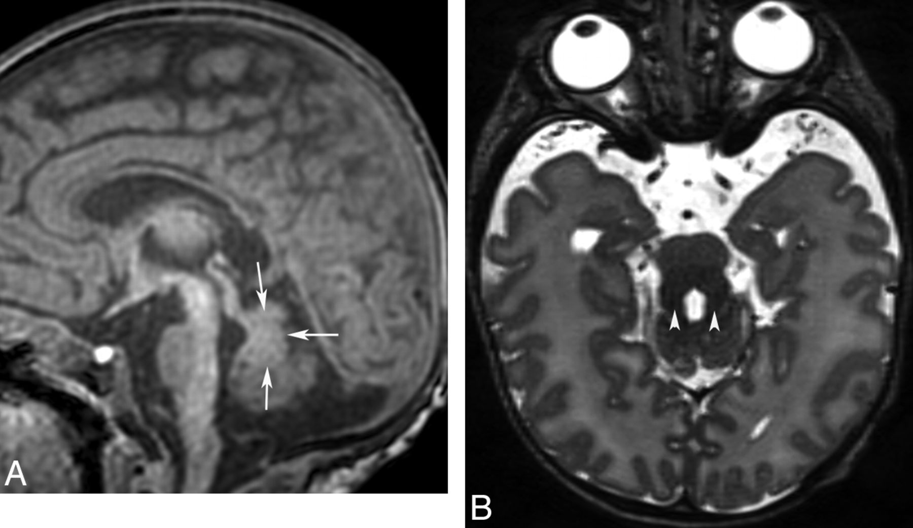
  • Fig 8.
    • Download figure
    • Open in new tab
    • Download powerpoint
    Fig 8.

    Mitochondrial disorder. Sagittal T1 (A) and axial FLAIR (B) images show abnormal signal intensity (arrowheads) in the dorsal midbrain.
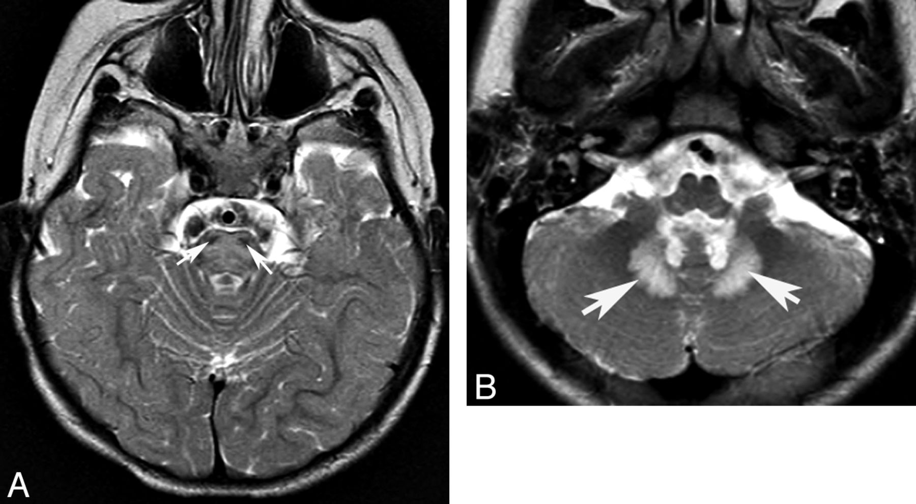
  • Fig 9.
    • Download figure
    • Open in new tab
    • Download powerpoint
    Fig 9.

    Complex II mitochondriopathy. Axial T2 images demonstrate hyperintense signal in the pons. (A, note sparing of corticospinal tracts, white arrowheads) and hilum of the dentate nucleus (B).

  • Fig 10.
    • Download figure
    • Open in new tab
    • Download powerpoint
    Fig 10.

    Diffusivity changes in mitochondrial disorder. Axial DWI images show brain stem reduced diffusion in the central tegmental tracts (A), cerebral peduncles (B), and subthalamic regions. Single-voxel proton MR spectroscopy (C) from the same patient shows reduced NAA and markedly elevated lactate (doublet at 1.33 cpm).
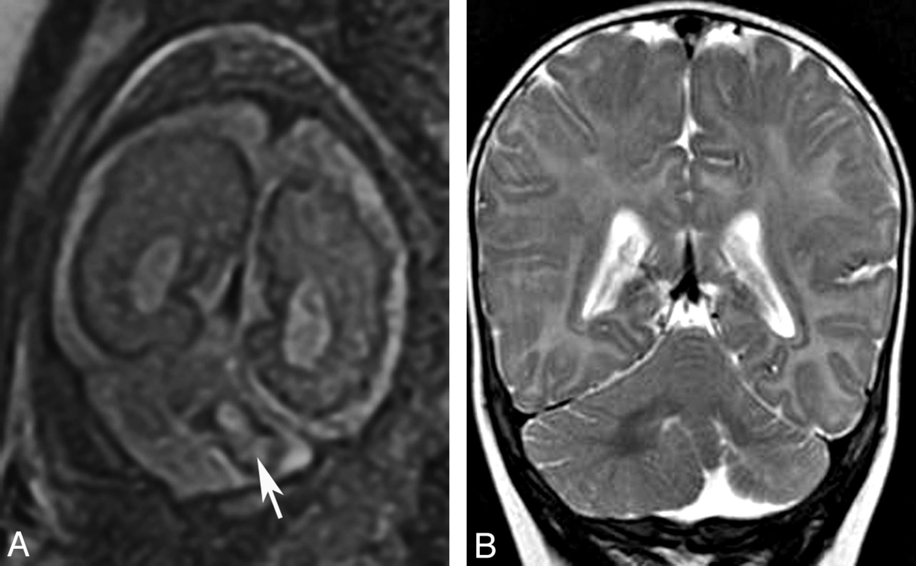
  • Fig 11.
    • Download figure
    • Open in new tab
    • Download powerpoint
    Fig 11.

    Localized cerebellar hypoplasia. Fetal coronal T2 image (A) shows a left-sided cerebellar hematoma (arrow). Postnatal coronal T2 image (B) demonstrates hypoplasia of the left cerebellar hemisphere.
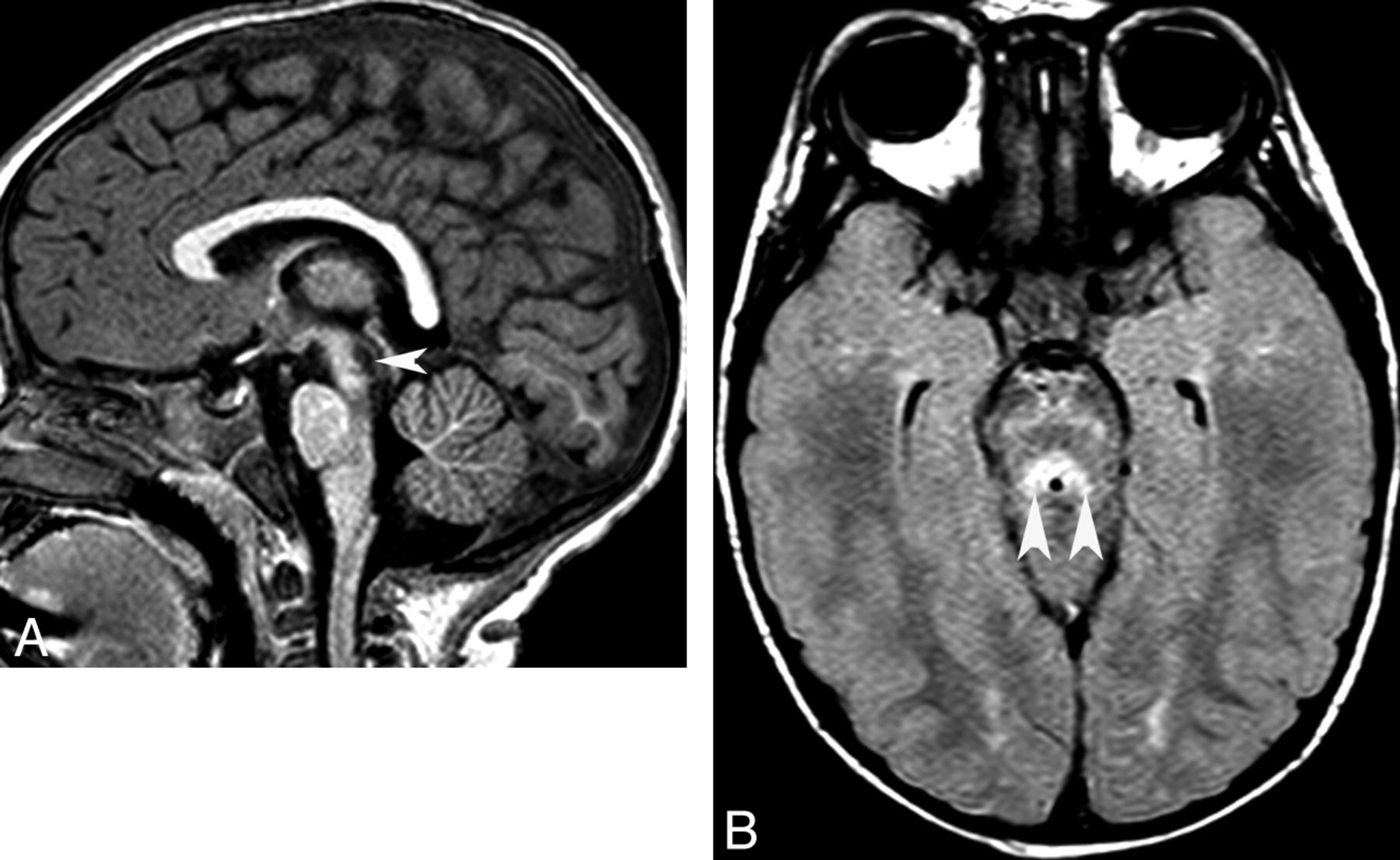
  • Fig 12.
    • Download figure
    • Open in new tab
    • Download powerpoint
    Fig 12.

    Cerebellar hypoplasia. Sagittal T1 image of a neonate shows severe brain stem and vermis hypoplasia. The superior vermis is below the intercollicular sulcus and above the obex. Note severe supratentorial atrophy, suggesting prenatal onset of volume loss.

  • Fig 13.
    • Download figure
    • Open in new tab
    • Download powerpoint
    Fig 13.

    Congenital disorder of glycosylation (type Ia). Coronal T2 image shows a small cerebellum with very small cerebellar folia and enlarged fissures. This pattern probably reflects a process that begins in utero (hypoplasia) and progresses in infancy (atrophy).

  • Fig 14.
    • Download figure
    • Open in new tab
    • Download powerpoint
    Fig 14.

    Joubert syndrome and related disorders (ciliopathies). Sagittal T1 (A) and axial T2 (B) images show a small dysgenetic vermis (arrows) and large horizontal superior cerebellar peduncles (arrowheads, molar tooth sign).

  • Fig 15.
    • Download figure
    • Open in new tab
    • Download powerpoint
    Fig 15.

    Rhomboencephalosynapsis. Axial T2 (A) and coronal T2 (B) images show continuity of the deep cerebellar white matter across the midline due to complete absence of the vermis.

  • Fig 16.
    • Download figure
    • Open in new tab
    • Download powerpoint
    Fig 16.

    Cerebellar cortical dysgenesis. Axial T1 images (A and B) and a coronal T1 image (C) demonstrate abnormal cerebellar foliation. The cause of this is usually unknown. Hydrocephalus is also present (C).

  • Fig 17.
    • Download figure
    • Open in new tab
    • Download powerpoint
    Fig 17.

    Pontine tegmental cap dysplasia. Sagittal T2 image (A) shows a rounded “cap” over the dorsal pons (arrow). Axial T1 image (B) demonstrates a small pons and a flat anterior border of the forth ventricle secondary to transversely oriented axons.

PreviousNext
Back to top

In this issue

American Journal of Neuroradiology: 34 (5)
American Journal of Neuroradiology
Vol. 34, Issue 5
1 May 2013
  • Table of Contents
  • Index by author
Advertisement
Print
Download PDF
Email Article

Thank you for your interest in spreading the word on American Journal of Neuroradiology.

NOTE: We only request your email address so that the person you are recommending the page to knows that you wanted them to see it, and that it is not junk mail. We do not capture any email address.

Enter multiple addresses on separate lines or separate them with commas.
Inherited Cerebellar Ataxia in Childhood: A Pattern-Recognition Approach Using Brain MRI
(Your Name) has sent you a message from American Journal of Neuroradiology
(Your Name) thought you would like to see the American Journal of Neuroradiology web site.
CAPTCHA
This question is for testing whether or not you are a human visitor and to prevent automated spam submissions.
Cite this article
L. Vedolin, G. Gonzalez, C.F. Souza, C. Lourenço, A.J. Barkovich
Inherited Cerebellar Ataxia in Childhood: A Pattern-Recognition Approach Using Brain MRI
American Journal of Neuroradiology May 2013, 34 (5) 925-934; DOI: 10.3174/ajnr.A3055

Citation Manager Formats

  • BibTeX
  • Bookends
  • EasyBib
  • EndNote (tagged)
  • EndNote 8 (xml)
  • Medlars
  • Mendeley
  • Papers
  • RefWorks Tagged
  • Ref Manager
  • RIS
  • Zotero
0 Responses
Respond to this article
Share
Bookmark this article
Inherited Cerebellar Ataxia in Childhood: A Pattern-Recognition Approach Using Brain MRI
L. Vedolin, G. Gonzalez, C.F. Souza, C. Lourenço, A.J. Barkovich
American Journal of Neuroradiology May 2013, 34 (5) 925-934; DOI: 10.3174/ajnr.A3055
del.icio.us logo Twitter logo Facebook logo Mendeley logo
  • Tweet Widget
  • Facebook Like
  • Google Plus One
Purchase

Jump to section

  • Article
    • Abstract
    • ABBREVIATIONS:
    • Degenerative Pattern of CAC
    • Malformative Pattern of CAC
    • Conclusions
    • References
  • Figures & Data
  • Supplemental
  • Info & Metrics
  • Responses
  • References
  • PDF

Related Articles

  • PubMed
  • Google Scholar

Cited By...

  • No citing articles found.
  • Crossref (24)
  • Google Scholar

This article has been cited by the following articles in journals that are participating in Crossref Cited-by Linking.

  • Imaging of Creutzfeldt-Jakob Disease: Imaging Patterns and Their Differential Diagnosis
    Diego Cardoso Fragoso, Augusto Lio da Mota Gonçalves Filho, Felipe Torres Pacheco, Bernardo Rodi Barros, Ingrid Aguiar Littig, Renato Hoffmann Nunes, Antônio Carlos Martins Maia Júnior, Antonio J. da Rocha
    RadioGraphics 2017 37 1
  • A nationwide survey of PMM2-CDG in Italy: high frequency of a mild neurological variant associated with the L32R mutation
    Rita Barone, M. Carrozzi, R. Parini, R. Battini, D. Martinelli, M. Elia, M. Spada, F. Lilliu, G. Ciana, A. Burlina, V. Leuzzi, M. Leoni, L. Sturiale, G. Matthijs, J. Jaeken, M. Di Rocco, D. Garozzo, A. Fiumara
    Journal of Neurology 2015 262 1
  • The Cerebellum Looks Normal in Friedreich Ataxia
    M. Mascalchi
    American Journal of Neuroradiology 2013 34 2
  • Reliability of phenotypic early‐onset ataxia assessment: a pilot study
    Tjitske F Lawerman, Rick Brandsma, Joke T van Geffen, Roelineke J Lunsing, Huibert Burger, Marina A J Tijssen, Jeroen J de Vries, Tom J de Koning, Deborah A Sival
    Developmental Medicine & Child Neurology 2016 58 1
  • Brain Imaging and Genetic Risk in the Pediatric Population, Part 1
    Maria Gabriela Longo, Filippo Vairo, Carolina Fischinger Souza, Roberto Giugliani, Leonardo Modesti Vedolin
    Neuroimaging Clinics of North America 2015 25 1
  • ACR Appropriateness Criteria® Ataxia
    Amy F. Juliano, Bruno Policeni, Vikas Agarwal, Judah Burns, Julie Bykowski, H. Benjamin Harvey, Jenny K. Hoang, Christopher H. Hunt, Tabassum A. Kennedy, Gul Moonis, Jeffrey S. Pannell, Matthew S. Parsons, William J. Powers, Joshua M. Rosenow, Jason W. Schroeder, Konstantin Slavin, Matthew T. Whitehead, Amanda S. Corey
    Journal of the American College of Radiology 2019 16 5
  • Early Onset Ataxia with Comorbid Dystonia: Clinical, Anatomical and Biological Pathway Analysis Expose Shared Pathophysiology
    Deborah A. Sival, Martinica Garofalo, Rick Brandsma, Tom A. Bokkers, Marloes van den Berg, Tom J. de Koning, Marina A. J. Tijssen, Dineke S. Verbeek
    Diagnostics 2020 10 12
  • Main inherited neurodegenerative cerebellar ataxias, how to recognize them using magnetic resonance imaging?
    D. Heidelberg, S. Ronsin, F. Bonneville, S. Hannoun, C. Tilikete, F. Cotton
    Journal of Neuroradiology 2018 45 5
  • Role of neuroimaging in the diagnosis of hereditary cerebellar ataxias in childhood
    Giulia Perucca, Nicolas Leboucq, Agathe Roubertie, François Rivier, Nicolas Menjot, Consuelo Valentini, Alain Bonafe
    Journal of Neuroradiology 2016 43 3
  • Combined Genome, Transcriptome and Metabolome Analysis in the Diagnosis of Childhood Cerebellar Ataxia
    Ana Ching-López, Luis Javier Martinez-Gonzalez, Luisa Arrabal, Jorge Sáiz, Ángela Gavilán, Coral Barbas, Jose Antonio Lorente, Susana Roldán, Maria José Sánchez, Purificacion Gutierrez-Ríos
    International Journal of Molecular Sciences 2021 22 6

More in this TOC Section

  • Ultra-High-Field MR Neuroimaging
  • Mechanisms of Healing in Coiled Intracranial Aneurysms: A Review of the Literature
  • Armies of Pestilence: CNS Infections as Potential Weapons of Mass Destruction
Show more Review Articles

Similar Articles

Advertisement

Indexed Content

  • Current Issue
  • Accepted Manuscripts
  • Article Preview
  • Past Issues
  • Editorials
  • Editors Choice
  • Fellow Journal Club
  • Letters to the Editor

Cases

  • Case Collection
  • Archive - Case of the Week
  • Archive - Case of the Month
  • Archive - Classic Case

Special Collections

  • Special Collections

Resources

  • News and Updates
  • Turn around Times
  • Submit a Manuscript
  • Author Policies
  • Manuscript Submission Guidelines
  • Evidence-Based Medicine Level Guide
  • Publishing Checklists
  • Graphical Abstract Preparation
  • Imaging Protocol Submission
  • Submit a Case
  • Become a Reviewer/Academy of Reviewers
  • Get Peer Review Credit from Publons

Multimedia

  • AJNR Podcast
  • AJNR SCANtastic
  • Video Articles

About Us

  • About AJNR
  • Editorial Board
  • Not an AJNR Subscriber? Join Now
  • Alerts
  • Feedback
  • Advertise with us
  • Librarian Resources
  • Permissions
  • Terms and Conditions

American Society of Neuroradiology

  • Not an ASNR Member? Join Now

© 2025 by the American Society of Neuroradiology All rights, including for text and data mining, AI training, and similar technologies, are reserved.
Print ISSN: 0195-6108 Online ISSN: 1936-959X

Powered by HighWire