Advertisement

Main menu

  • Home
  • Content
    • Current Issue
    • Accepted Manuscripts
    • Article Preview
    • Past Issue Archive
    • AJNR Case Collection
    • Case of the Week Archive
    • Classic Case Archive
    • Case of the Month Archive
  • Special Collections
    • Spinal CSF Leak Articles (Jan 2020-June 2024)
    • 2024 AJNR Journal Awards
    • Most Impactful AJNR Articles
  • Multimedia
    • AJNR Podcast
    • AJNR Scantastics
    • Video Articles
  • For Authors
    • Submit a Manuscript
    • Author Policies
    • Fast publishing of Accepted Manuscripts
    • Graphical Abstract Preparation
    • Manuscript Submission Guidelines
    • Imaging Protocol Submission
    • Submit a Case for the Case Collection
  • About Us
    • About AJNR
    • Editorial Board
  • More
    • Become a Reviewer/Academy of Reviewers
    • Subscribers
    • Permissions
    • Alerts
    • Feedback
    • Advertisers
    • ASNR Home
  • Other Publications
    • ajnr

User menu

  • Alerts
  • Log in

Search

  • Advanced search
American Journal of Neuroradiology

American Journal of Neuroradiology

ASHNR American Society of Functional Neuroradiology ASHNR American Society of Pediatric Neuroradiology ASSR
  • Alerts
  • Log in

Advanced Search

  • Home
  • Content
    • Current Issue
    • Accepted Manuscripts
    • Article Preview
    • Past Issue Archive
    • AJNR Case Collection
    • Case of the Week Archive
    • Classic Case Archive
    • Case of the Month Archive
  • Special Collections
    • Spinal CSF Leak Articles (Jan 2020-June 2024)
    • 2024 AJNR Journal Awards
    • Most Impactful AJNR Articles
  • Multimedia
    • AJNR Podcast
    • AJNR Scantastics
    • Video Articles
  • For Authors
    • Submit a Manuscript
    • Author Policies
    • Fast publishing of Accepted Manuscripts
    • Graphical Abstract Preparation
    • Manuscript Submission Guidelines
    • Imaging Protocol Submission
    • Submit a Case for the Case Collection
  • About Us
    • About AJNR
    • Editorial Board
  • More
    • Become a Reviewer/Academy of Reviewers
    • Subscribers
    • Permissions
    • Alerts
    • Feedback
    • Advertisers
    • ASNR Home
  • Follow AJNR on Twitter
  • Visit AJNR on Facebook
  • Follow AJNR on Instagram
  • Join AJNR on LinkedIn
  • RSS Feeds

Welcome to the new AJNR, Updated Hall of Fame, and more. Read the full announcements.


AJNR is seeking candidates for the position of Associate Section Editor, AJNR Case Collection. Read the full announcement.


Getting new auth cookie, if you see this message a lot, tell someone!
Research ArticleBrain
Open Access

Secondary Degeneration Detected by Combining Voxel-Based Morphometry and Tract-Based Spatial Statistics in Subcortical Strokes with Different Outcomes in Hand Function

D. Yin, X. Yan, M. Fan, Y. Hu, W. Men, L. Sun and F. Song
American Journal of Neuroradiology July 2013, 34 (7) 1341-1347; DOI: https://doi.org/10.3174/ajnr.A3410
D. Yin
aFrom the Shanghai Key Laboratory of Magnetic Resonance (D.Y., X.Y., M.F., W.M.), Department of Physics, East China Normal University, Shanghai, China
  • Find this author on Google Scholar
  • Find this author on PubMed
  • Search for this author on this site
X. Yan
aFrom the Shanghai Key Laboratory of Magnetic Resonance (D.Y., X.Y., M.F., W.M.), Department of Physics, East China Normal University, Shanghai, China
  • Find this author on Google Scholar
  • Find this author on PubMed
  • Search for this author on this site
M. Fan
aFrom the Shanghai Key Laboratory of Magnetic Resonance (D.Y., X.Y., M.F., W.M.), Department of Physics, East China Normal University, Shanghai, China
  • Find this author on Google Scholar
  • Find this author on PubMed
  • Search for this author on this site
Y. Hu
bDepartment of Rehabilitation Medicine (Y.H., L.S., F.S.), Huashan Hospital, Fudan University, Shanghai, China
cState Key Laboratory of Medical Neurobiology (Y.H.), Fudan University, Shanghai, China.
  • Find this author on Google Scholar
  • Find this author on PubMed
  • Search for this author on this site
W. Men
aFrom the Shanghai Key Laboratory of Magnetic Resonance (D.Y., X.Y., M.F., W.M.), Department of Physics, East China Normal University, Shanghai, China
  • Find this author on Google Scholar
  • Find this author on PubMed
  • Search for this author on this site
L. Sun
bDepartment of Rehabilitation Medicine (Y.H., L.S., F.S.), Huashan Hospital, Fudan University, Shanghai, China
  • Find this author on Google Scholar
  • Find this author on PubMed
  • Search for this author on this site
F. Song
bDepartment of Rehabilitation Medicine (Y.H., L.S., F.S.), Huashan Hospital, Fudan University, Shanghai, China
  • Find this author on Google Scholar
  • Find this author on PubMed
  • Search for this author on this site
  • Article
  • Figures & Data
  • Supplemental
  • Info & Metrics
  • Responses
  • References
  • PDF
Loading

Article Figures & Data

Figures

  • Tables
  • Fig 1.
    • Download figure
    • Open in new tab
    • Download powerpoint
    Fig 1.

    T2-weighted images show the largest lesion (red arrow) of each patient at the axial section. The locations and extent of the lesions are quite consistent across all of the patients. Arabic numerals denote the patient numbers.
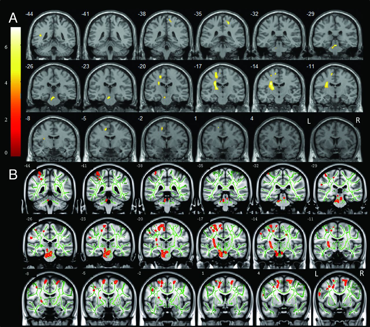
  • Fig 2.
    • Download figure
    • Open in new tab
    • Download powerpoint
    Fig 2.

    Significantly decreased FA was detected by VBM analysis (A) and TBSS analysis (B) in CPH compared with PPH. The threshold was set at a combined cutoff value of P < .001 and a minimal cluster size of 20 voxels (for VBM analysis) or P < .001 (for TBSS analysis). Color scale = t values; L = left; R = right. Arabic numeral indicates the location of each coronal section. Lesions were located on the left side.

Tables

  • Figures
    • View popup
    Table 1:

    Clinical and demographic data of 23 patients with subcortical stroke

    Pt No.SexAge (y)Location of LesionTime after Stroke (months)Lesion Volume (mL)FMA Scores (hand+wrist)
    PPH Group
        01M60L, IC, Tha5335.3212
        02F48L, IC, BG239.386
        03M76L, IC2162.1822
        04M60L, IC, BG3616.486
        05M71L, IC, Tha2217.6411
        06M63L, IC, BG, Th322.1413
        07M54L, IC, Tha312.3523
        08M60L, IC, Tha1126.9623
        09M65L, IC, Th128.8523
        10M53L, IC, BG, Tha22125.1015
        11M65L, IC, BGa681.7820
        12M56L, IC710.5511
    CPH Group
        01M67L, IC1712.332
        02M57L, IC, Th1923.730
        03F75L, IC, CR247.804
        04M63L, BG, IC, Tha1661.521
        05F65L, IC, Th1729.591
        06F68L, BG, IC, Tha6225.510
        07M68L, IC, Tha4732.731
        08M62L, BG, IC, Tha12156.140
        09M53L, BG, IC, Tha8655.531
        10F50L, BG, IC, Tha1318.660
        11M62L, IC, Th619.164
    • Note:—M indicates male; F, female; L, left; R, right; BG, basal ganglia; IC, internal capsule; Th, thalamus; CR, coronal radiata; FMA, Fugl-Meyer Assessment.

    • ↵a The characteristics of the lesion are hemorrhagic; others are ischemic.

    • View popup
    Table 2:

    VBM analysis of FA maps between the CPH group and the PPH group

    RegionsMNI CoordinatesPeak t ScoreNumber of VoxelsVolume (mL)
    xyz
    Brain stem (IL)−2−25−215.53220660
    Sublobar (internal capsule/basal ganglia/thalamus) (IL)−25−14276.955561668
    Superior temporal gyrus (IL)−42−45154.8168204
    Precentral gyrus (IL)−20−18544.92122366
    Medial frontal gyrus (cingulate gyrus) (IL)−13−4515.0562186
    Supplementary motor area (IL)−11−12605.2354162
    Postcentral gyrus (CL)18−36575.6755165
    • Note:—The threshold was set at a combined cutoff value of P < .001 (uncorrected) and a minimal cluster size of 20 voxels. IL indicates ipsilesional; CL, contralesional; MNI, Montreal Neurological Institute.

PreviousNext
Back to top

In this issue

American Journal of Neuroradiology: 34 (7)
American Journal of Neuroradiology
Vol. 34, Issue 7
1 Jul 2013
  • Table of Contents
  • Index by author
Advertisement
Print
Download PDF
Email Article

Thank you for your interest in spreading the word on American Journal of Neuroradiology.

NOTE: We only request your email address so that the person you are recommending the page to knows that you wanted them to see it, and that it is not junk mail. We do not capture any email address.

Enter multiple addresses on separate lines or separate them with commas.
Secondary Degeneration Detected by Combining Voxel-Based Morphometry and Tract-Based Spatial Statistics in Subcortical Strokes with Different Outcomes in Hand Function
(Your Name) has sent you a message from American Journal of Neuroradiology
(Your Name) thought you would like to see the American Journal of Neuroradiology web site.
CAPTCHA
This question is for testing whether or not you are a human visitor and to prevent automated spam submissions.
Cite this article
D. Yin, X. Yan, M. Fan, Y. Hu, W. Men, L. Sun, F. Song
Secondary Degeneration Detected by Combining Voxel-Based Morphometry and Tract-Based Spatial Statistics in Subcortical Strokes with Different Outcomes in Hand Function
American Journal of Neuroradiology Jul 2013, 34 (7) 1341-1347; DOI: 10.3174/ajnr.A3410

Citation Manager Formats

  • BibTeX
  • Bookends
  • EasyBib
  • EndNote (tagged)
  • EndNote 8 (xml)
  • Medlars
  • Mendeley
  • Papers
  • RefWorks Tagged
  • Ref Manager
  • RIS
  • Zotero
0 Responses
Respond to this article
Share
Bookmark this article
Secondary Degeneration Detected by Combining Voxel-Based Morphometry and Tract-Based Spatial Statistics in Subcortical Strokes with Different Outcomes in Hand Function
D. Yin, X. Yan, M. Fan, Y. Hu, W. Men, L. Sun, F. Song
American Journal of Neuroradiology Jul 2013, 34 (7) 1341-1347; DOI: 10.3174/ajnr.A3410
del.icio.us logo Twitter logo Facebook logo Mendeley logo
  • Tweet Widget
  • Facebook Like
  • Google Plus One
Purchase

Jump to section

  • Article
    • Abstract
    • ABBREVIATIONS:
    • Materials and Methods
    • Results
    • Discussion
    • Conclusions
    • Acknowledgments
    • Footnotes
    • REFERENCES
  • Figures & Data
  • Supplemental
  • Info & Metrics
  • Responses
  • References
  • PDF

Related Articles

  • PubMed
  • Google Scholar

Cited By...

  • Intravenous Bone Marrow Stem Cell Grafts Preferentially Migrate to Spleen and Abrogate Chronic Inflammation in Stroke
  • Crossref (33)
  • Google Scholar

This article has been cited by the following articles in journals that are participating in Crossref Cited-by Linking.

  • White matter lesional predictors of chronic visual neglect: a longitudinal study
    Marine Lunven, Michel Thiebaut De Schotten, Clémence Bourlon, Christophe Duret, Raffaella Migliaccio, Gilles Rode, Paolo Bartolomeo
    Brain 2015 138 3
  • Intravenous Bone Marrow Stem Cell Grafts Preferentially Migrate to Spleen and Abrogate Chronic Inflammation in Stroke
    Sandra A. Acosta, Naoki Tajiri, Jaclyn Hoover, Yuji Kaneko, Cesar V. Borlongan
    Stroke 2015 46 9
  • Post-stroke remodeling processes in animal models and humans
    Carla Cirillo, Nabila Brihmat, Evelyne Castel-Lacanal, Alice Le Friec, Marianne Barbieux-Guillot, Nicolas Raposo, Jérémie Pariente, Alain Viguier, Marion Simonetta-Moreau, Jean-François Albucher, Jean-Marc Olivot, Franck Desmoulin, Philippe Marque, François Chollet, Isabelle Loubinoux
    Journal of Cerebral Blood Flow & Metabolism 2020 40 1
  • Altered topological properties of the cortical motor-related network in patients with subcortical stroke revealed by graph theoretical analysis
    Dazhi Yin, Fan Song, Dongrong Xu, Limin Sun, Weiwei Men, Lili Zang, Xu Yan, Mingxia Fan
    Human Brain Mapping 2014 35 7
  • Decreased Functional Connectivity of Homotopic Brain Regions in Chronic Stroke Patients: A Resting State fMRI Study
    Chaozheng Tang, Zhiyong Zhao, Chuang Chen, Xiaohui Zheng, Fenfen Sun, Xiaoli Zhang, Jing Tian, Mingxia Fan, Yi Wu, Jie Jia, James A. Duce
    PLOS ONE 2016 11 4
  • Cortical atrophy and transcallosal diaschisis following isolated subcortical stroke
    Bastian Cheng, Philipp Dietzmann, Robert Schulz, Marlene Boenstrup, Lutz Krawinkel, Jens Fiehler, Christian Gerloff, Götz Thomalla
    Journal of Cerebral Blood Flow & Metabolism 2020 40 3
  • Frequency‐specific alterations of regional homogeneity in subcortical stroke patients with different outcomes in hand function
    Zhiyong Zhao, Chaozheng Tang, Dazhi Yin, Jie Wu, Jiayu Gong, Limin Sun, Jie Jia, Dongrong Xu, Mingxia Fan
    Human Brain Mapping 2018 39 11
  • Automatic quantitative analysis of experimental primary and secondary retinal neurodegeneration: implications for optic neuropathies
    B M Davis, L Guo, J Brenton, L Langley, E M Normando, M F Cordeiro
    Cell Death Discovery 2016 2 1
  • Associations between neuroanatomical abnormality and motor symptoms in paroxysmal kinesigenic dyskinesia
    Hong-Fu Li, Liqin Yang, Dazhi Yin, Wan-Jin Chen, Gong-Lu Liu, Wang Ni, Ning Wang, Wenwen Yu, Zhi-Ying Wu, Zheng Wang
    Parkinsonism & Related Disorders 2019 62
  • Analysis of correlation between white matter changes and functional responses in thalamic stroke: a DTI &amp; EEG study
    Adil Deniz Duru, Dilek Göksel Duru, Sami Yumerhodzha, Nerses Bebek
    Brain Imaging and Behavior 2016 10 2

More in this TOC Section

  • Statin Therapy Does Not Affect the Radiographic and Clinical Profile of Patients with TIA and Minor Stroke
  • Optimal MRI Sequence for Identifying Occlusion Location in Acute Stroke: Which Value of Time-Resolved Contrast-Enhanced MRA?
  • SWI or T2*: Which MRI Sequence to Use in the Detection of Cerebral Microbleeds? The Karolinska Imaging Dementia Study
Show more Brain

Similar Articles

Advertisement

Indexed Content

  • Current Issue
  • Accepted Manuscripts
  • Article Preview
  • Past Issues
  • Editorials
  • Editors Choice
  • Fellow Journal Club
  • Letters to the Editor

Cases

  • Case Collection
  • Archive - Case of the Week
  • Archive - Case of the Month
  • Archive - Classic Case

Special Collections

  • Special Collections

Resources

  • News and Updates
  • Turn around Times
  • Submit a Manuscript
  • Author Policies
  • Manuscript Submission Guidelines
  • Evidence-Based Medicine Level Guide
  • Publishing Checklists
  • Graphical Abstract Preparation
  • Imaging Protocol Submission
  • Submit a Case
  • Become a Reviewer/Academy of Reviewers
  • Get Peer Review Credit from Publons

Multimedia

  • AJNR Podcast
  • AJNR SCANtastic
  • Video Articles

About Us

  • About AJNR
  • Editorial Board
  • Not an AJNR Subscriber? Join Now
  • Alerts
  • Feedback
  • Advertise with us
  • Librarian Resources
  • Permissions
  • Terms and Conditions

American Society of Neuroradiology

  • Not an ASNR Member? Join Now

© 2025 by the American Society of Neuroradiology All rights, including for text and data mining, AI training, and similar technologies, are reserved.
Print ISSN: 0195-6108 Online ISSN: 1936-959X

Powered by HighWire