
Article Figures & Data
Figures
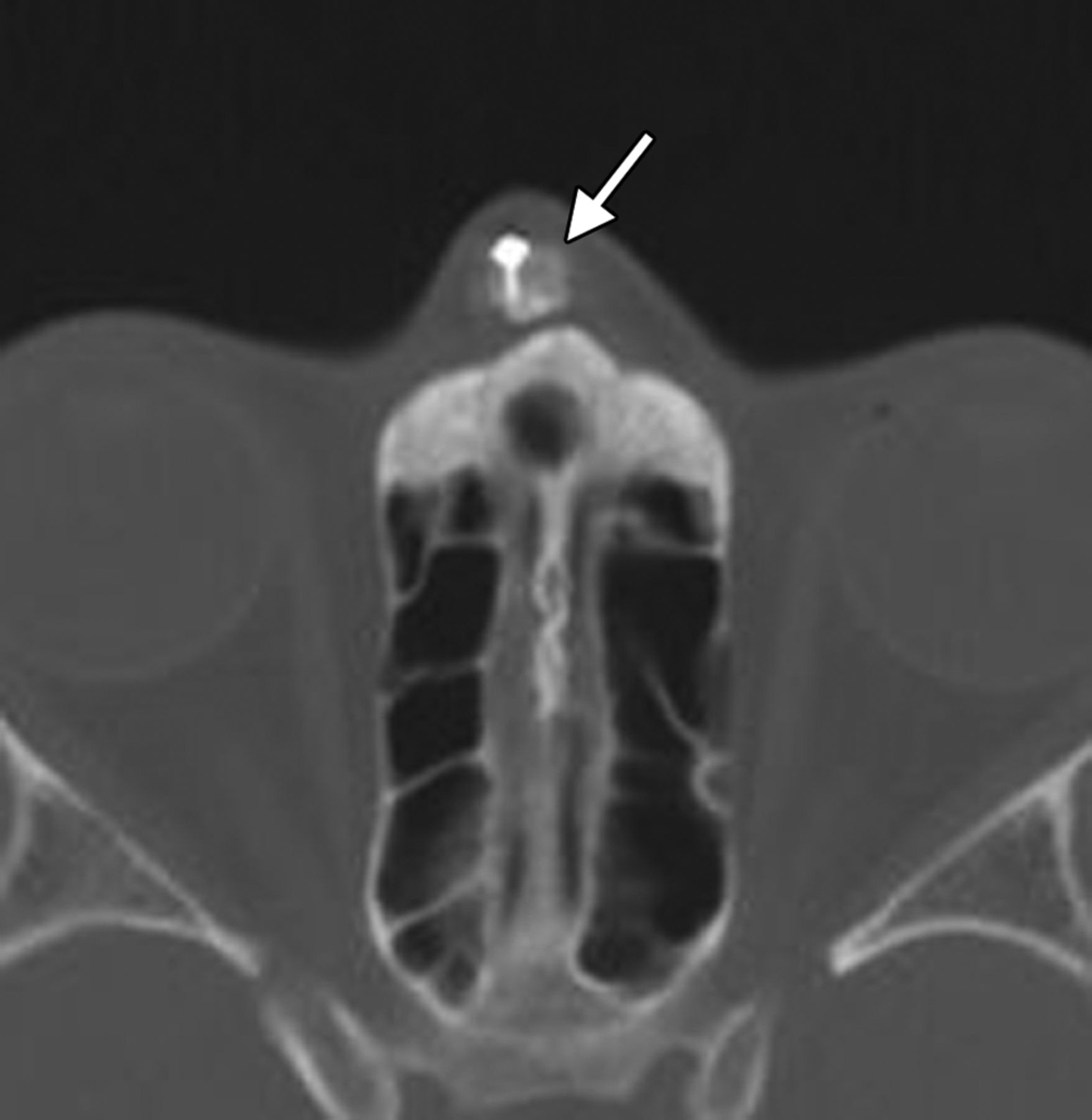
- Fig 1.
Radix implant. Axial CT image shows a bone graft positioned at the level of the nasal radix, secured by a metal plate and screws (arrow).
- Fig 2.
Dorsal nasal implant. Sagittal CT image shows a hyperattenuated expanded polytetrafluoroethylene nasal dorsum implant (arrow).
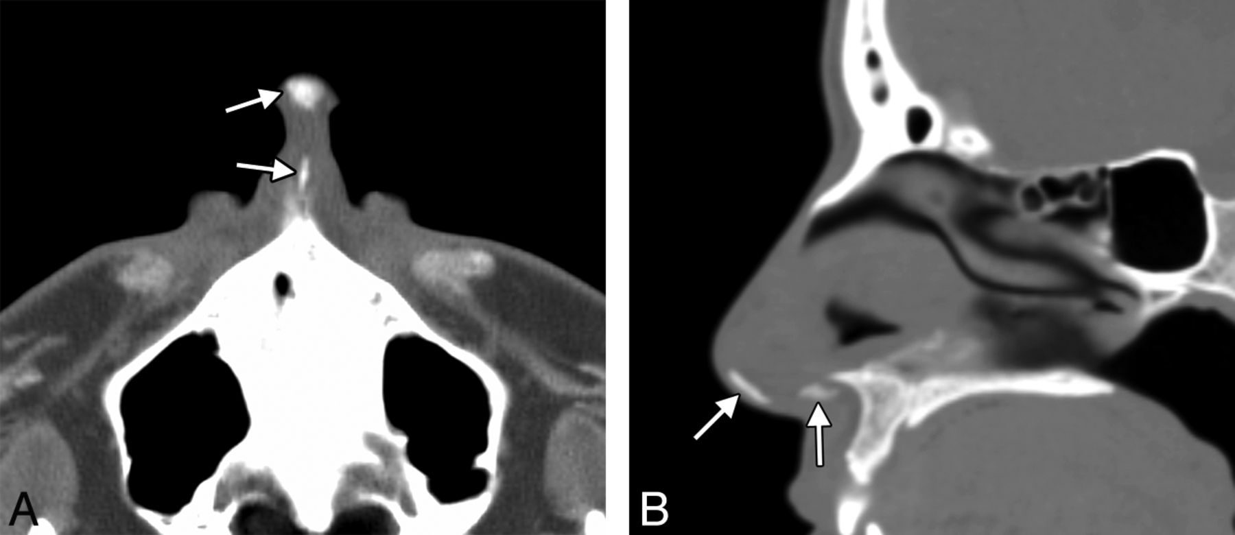
- Fig 3.
Tip augmentation with a columellar strut graft. Axial (A) and sagittal (B) CT images show hyperattenuated grafts within the infratip lobule and columella (arrows).
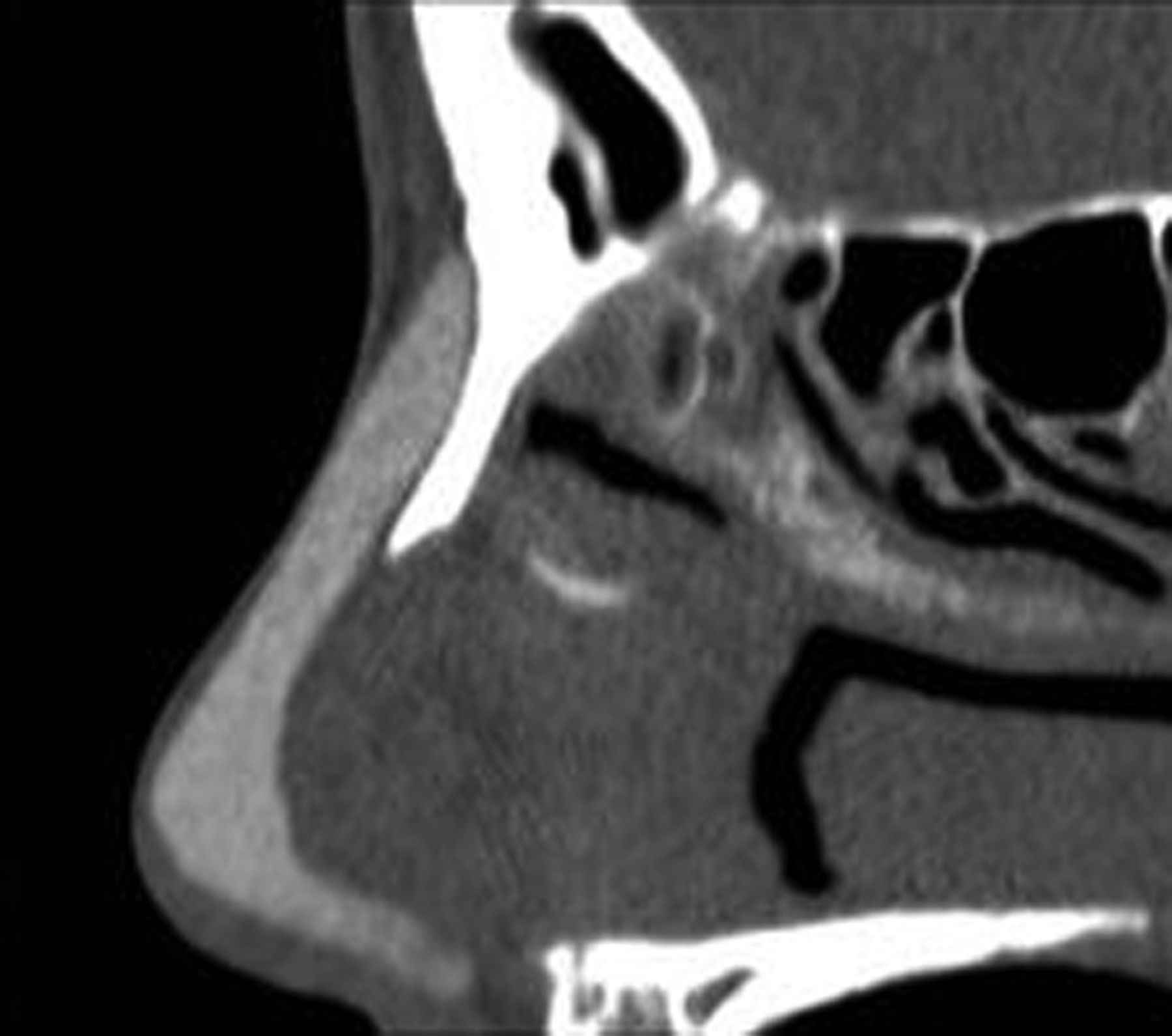
- Fig 4.
Silicone L-strut implant. Sagittal CT image shows the hyperattenuated L-shaped Silicone implant extending from the nasal dorsum to the columella.
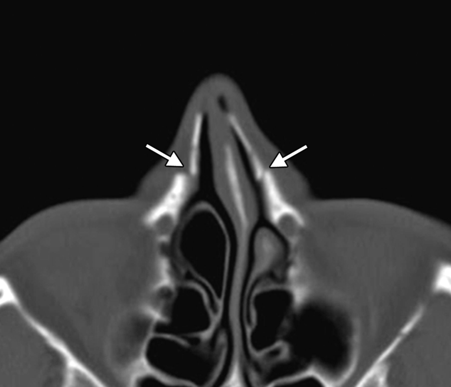
- Fig 5.
Lateral osteotomy. Axial CT image shows defects in the bilateral nasal processes of the maxillae (arrows). The lateral nasal walls are displaced medially (in-fractures).
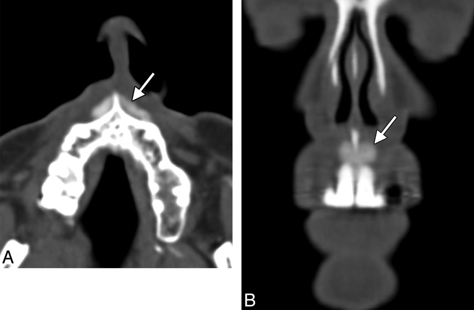
- Fig 6.
Premaxillary implant. Axial (A) and coronal (B) CT images show a hyperattenuated strip of silicone (arrows) positioned in the midline anterior to the nasal spine of the maxilla.
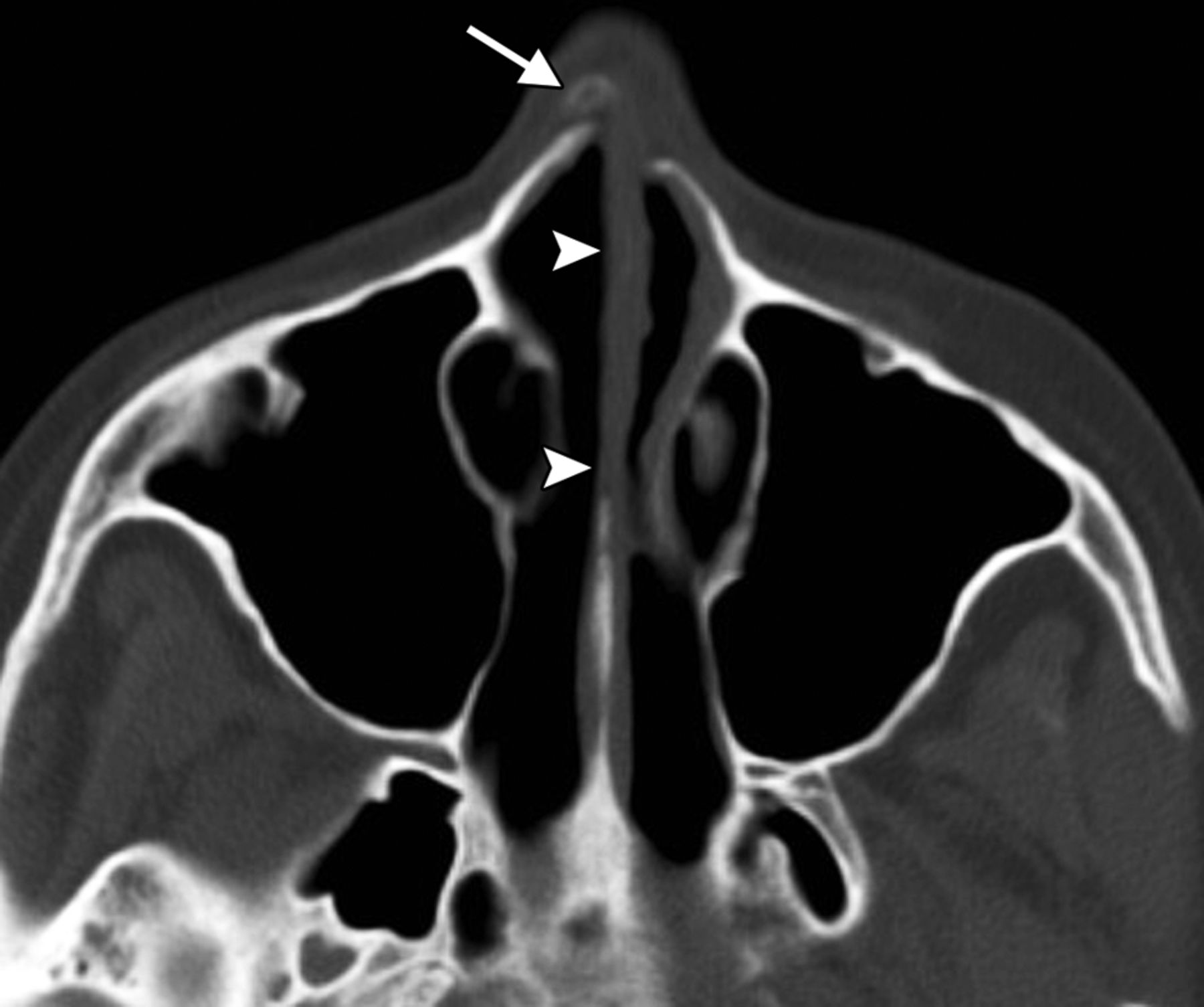
- Fig 7.
Septorhinoplasty. Axial CT image shows a very straight and thin nasal septum (arrowheads). A bone graft is present within the nasal dorsum (arrow).
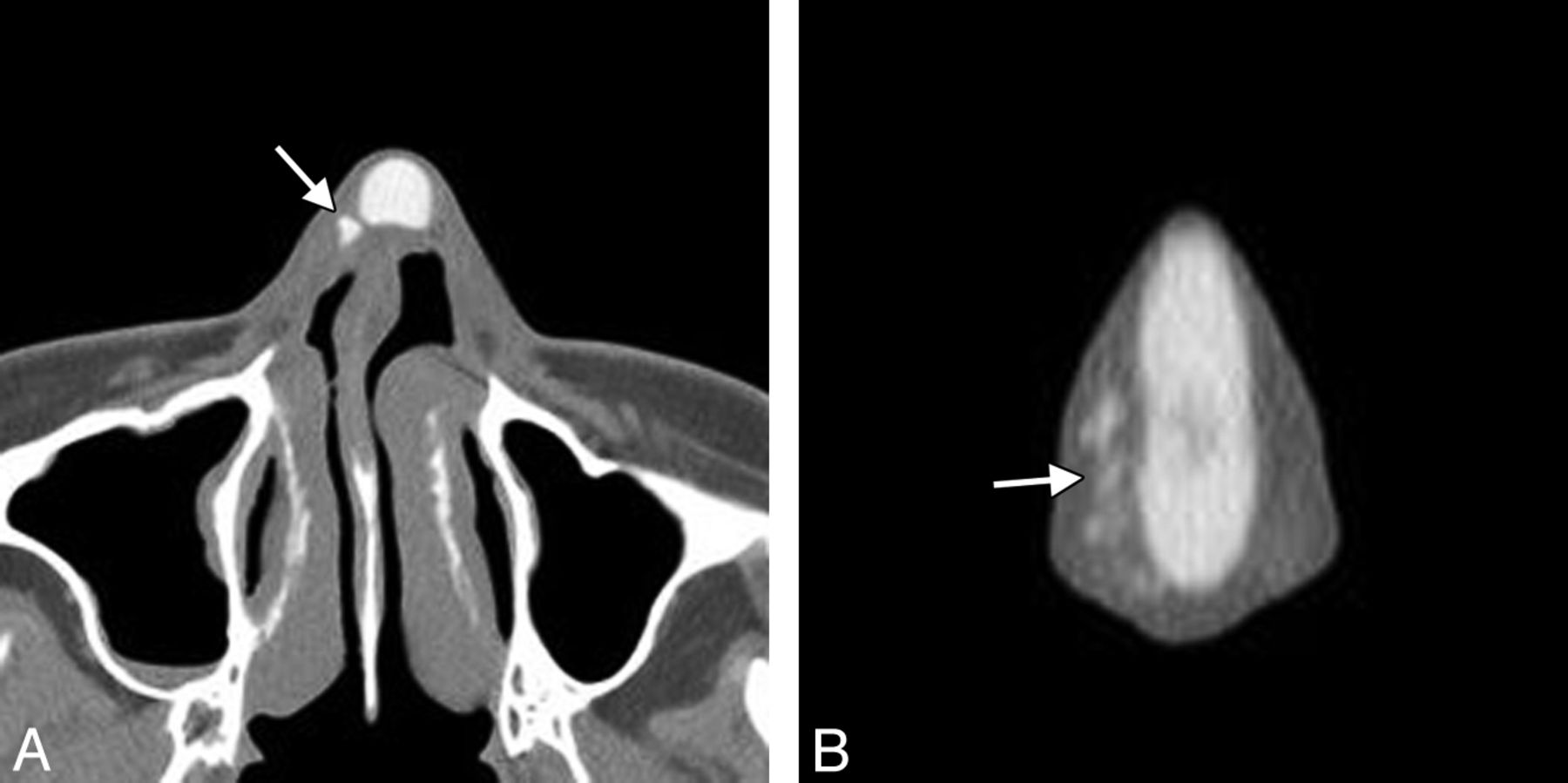
- Fig 8.
Rhinoplasty with filler. Axial (A) and coronal (B) CT images show hyperattenuated hydroxylapatite filler (arrows) to the right of the Silastic nasal dorsum implant. Note the deviated nasal septum.
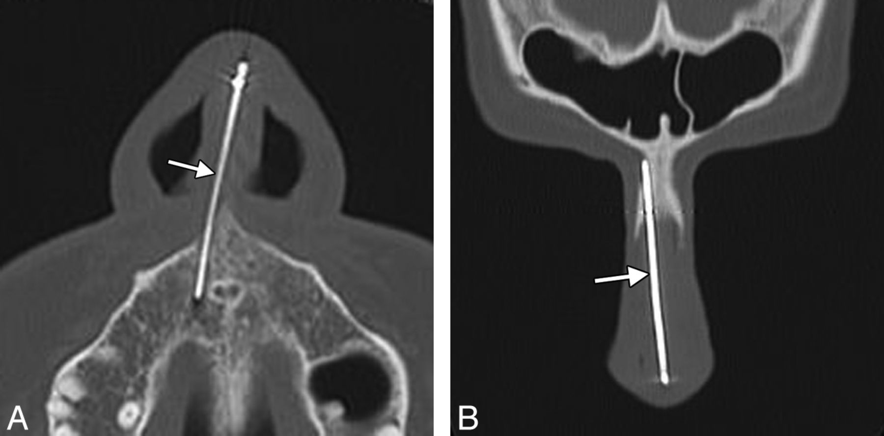
- Fig 9.
Kirschner wire strut. Axial (A) and coronal (B) CT images show a metallic wire (arrow) extending along the nasal dorsum and nasal base, where it inserts into the maxilla, lateral to the incisive canal.
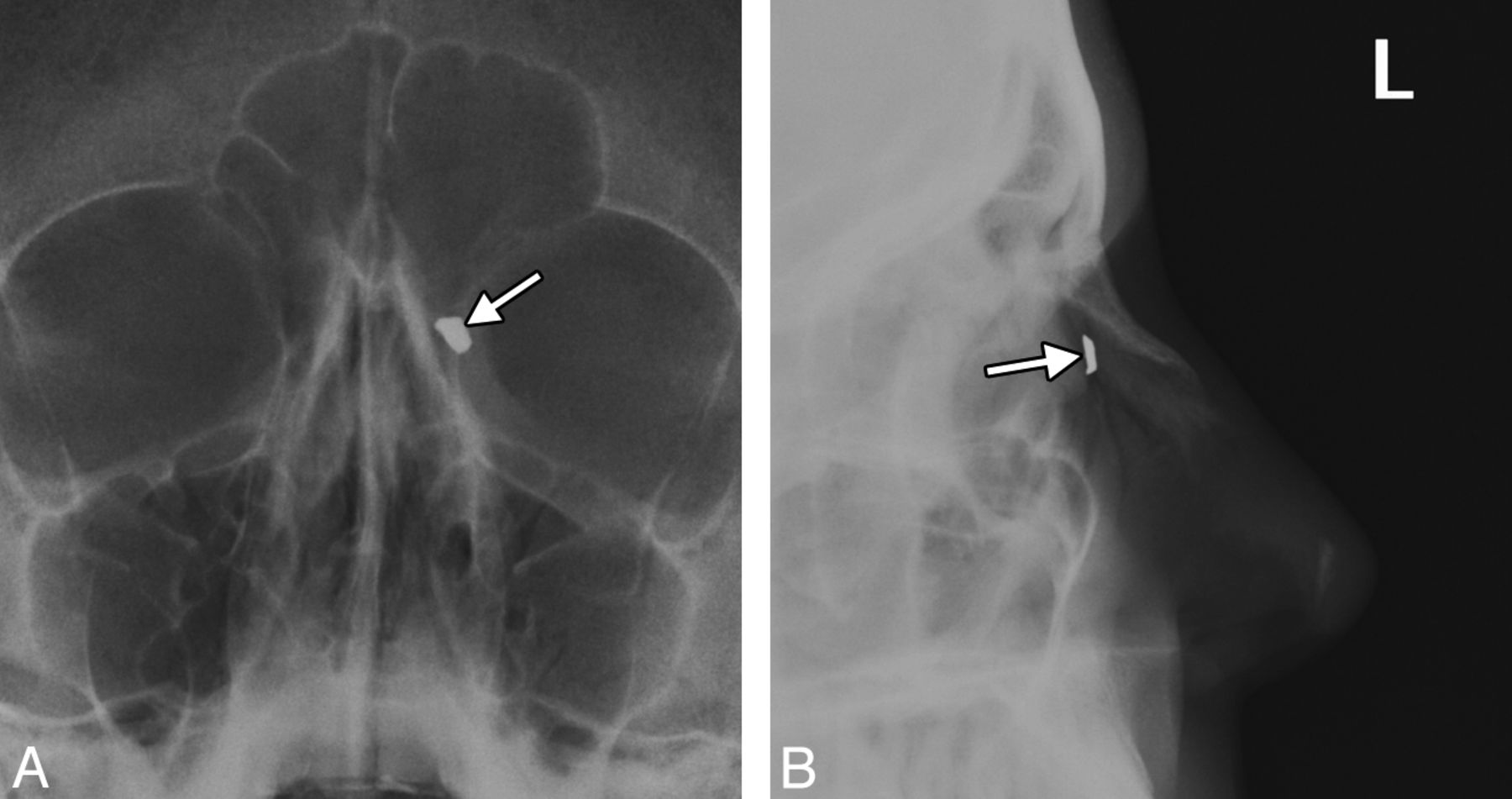
- Fig 10.
Retained osteotome fragment. Frontal (A) and lateral (B) radiographs show the retained metallic fragment (arrows) at the left lateral osteotomy site. Note the nasal-tip bone graft.
- Fig 11.
Implant infection. Sagittal postcontrast CT image shows a small fluid collection and associated inflammatory changes (arrow) in the nasal dorsum surrounding the implant.
- Fig 12.
Implant extrusion. Sagittal CT image shows a porous polyethylene dorsal nasal implant (arrow) projecting through a cutaneous defect.
- Fig 13.
Nerve impingement. The patient presented with dysesthesias in the maxillary nerve distribution after rhinoplasty. Sagittal CT image shows that the K-wire traverses the incisive canal at the expected location of the nasopalatine nerve (arrow).
- Fig 14.
Deformed K-wire. The patient presented with trauma to the nose. Submentovertex radiograph shows a bend in the K-wire (arrow).
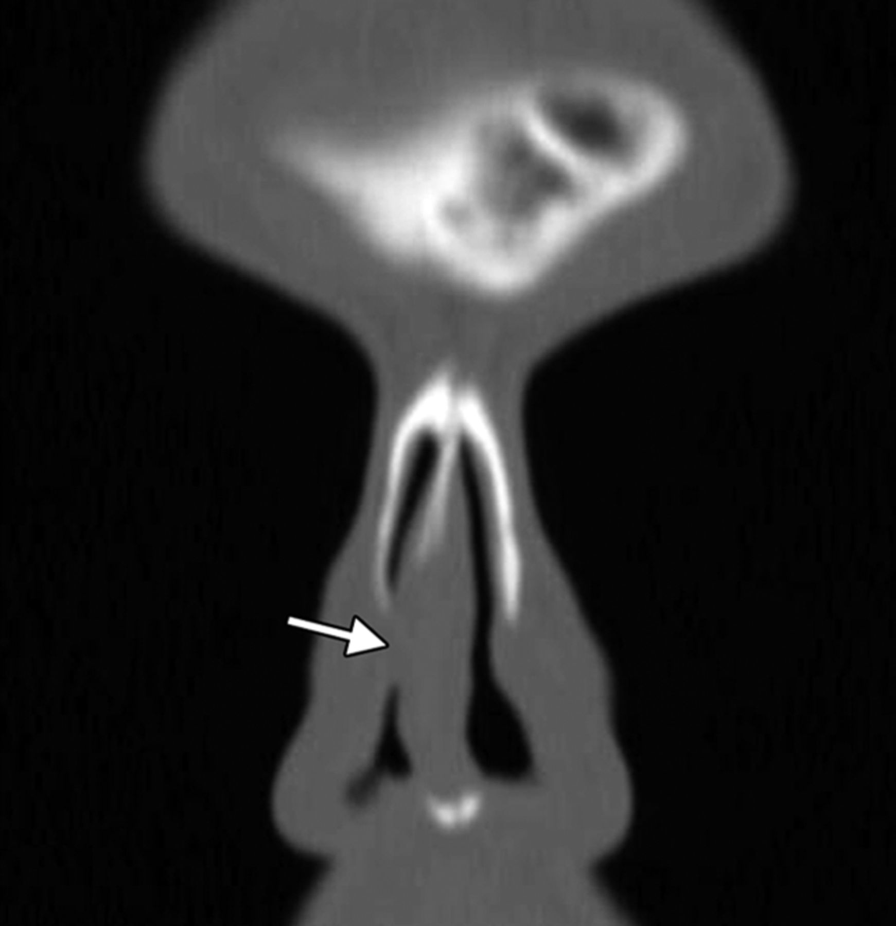
- Fig 15.
Nasal valve collapse. Coronal CT image shows stenosis of the right nasal valve (arrow). The left nasal valve remains patent.


Jump to section
Related Articles
Cited By...
- No citing articles found.