Advertisement

Main menu

  • Home
  • Content
    • Current Issue
    • Accepted Manuscripts
    • Article Preview
    • Past Issue Archive
    • AJNR Case Collection
    • Case of the Week Archive
    • Classic Case Archive
    • Case of the Month Archive
  • Special Collections
    • Spinal CSF Leak Articles (Jan 2020-June 2024)
    • 2024 AJNR Journal Awards
    • Most Impactful AJNR Articles
  • Multimedia
    • AJNR Podcast
    • AJNR Scantastics
    • Video Articles
  • For Authors
    • Submit a Manuscript
    • Author Policies
    • Fast publishing of Accepted Manuscripts
    • Graphical Abstract Preparation
    • Manuscript Submission Guidelines
    • Imaging Protocol Submission
    • Submit a Case for the Case Collection
  • About Us
    • About AJNR
    • Editorial Board
  • More
    • Become a Reviewer/Academy of Reviewers
    • Subscribers
    • Permissions
    • Alerts
    • Feedback
    • Advertisers
    • ASNR Home
  • Other Publications
    • ajnr

User menu

  • Alerts
  • Log in

Search

  • Advanced search
American Journal of Neuroradiology

American Journal of Neuroradiology

ASHNR American Society of Functional Neuroradiology ASHNR American Society of Pediatric Neuroradiology ASSR
  • Alerts
  • Log in

Advanced Search

  • Home
  • Content
    • Current Issue
    • Accepted Manuscripts
    • Article Preview
    • Past Issue Archive
    • AJNR Case Collection
    • Case of the Week Archive
    • Classic Case Archive
    • Case of the Month Archive
  • Special Collections
    • Spinal CSF Leak Articles (Jan 2020-June 2024)
    • 2024 AJNR Journal Awards
    • Most Impactful AJNR Articles
  • Multimedia
    • AJNR Podcast
    • AJNR Scantastics
    • Video Articles
  • For Authors
    • Submit a Manuscript
    • Author Policies
    • Fast publishing of Accepted Manuscripts
    • Graphical Abstract Preparation
    • Manuscript Submission Guidelines
    • Imaging Protocol Submission
    • Submit a Case for the Case Collection
  • About Us
    • About AJNR
    • Editorial Board
  • More
    • Become a Reviewer/Academy of Reviewers
    • Subscribers
    • Permissions
    • Alerts
    • Feedback
    • Advertisers
    • ASNR Home
  • Follow AJNR on Twitter
  • Visit AJNR on Facebook
  • Follow AJNR on Instagram
  • Join AJNR on LinkedIn
  • RSS Feeds

Welcome to the new AJNR, Updated Hall of Fame, and more. Read the full announcements.


AJNR is seeking candidates for the position of Associate Section Editor, AJNR Case Collection. Read the full announcement.


Research ArticleBrain

The Impact of Arterial Collateralization on Outcome after Intra-Arterial Therapy for Acute Ischemic Stroke

S. Seeta Ramaiah, L. Churilov, P. Mitchell, R. Dowling and B. Yan
American Journal of Neuroradiology April 2014, 35 (4) 667-672; DOI: https://doi.org/10.3174/ajnr.A3862
S. Seeta Ramaiah
aFrom the Melbourne Brain Centre (S.S.R., B.Y.)
  • Find this author on Google Scholar
  • Find this author on PubMed
  • Search for this author on this site
L. Churilov
cDepartment of Mathematics and Statistics (L.C.)
dNational Stroke Research Institute (L.C.), Florey Neuroscience Institutes, Austin Health, University of Melbourne, Parkville, Victoria, Australia.
  • Find this author on Google Scholar
  • Find this author on PubMed
  • Search for this author on this site
P. Mitchell
bNeurointervention Service (P.M., R.D., B.Y.), Department of Radiology, Royal Melbourne Hospital, Parkville, Victoria, Australia
  • Find this author on Google Scholar
  • Find this author on PubMed
  • Search for this author on this site
R. Dowling
bNeurointervention Service (P.M., R.D., B.Y.), Department of Radiology, Royal Melbourne Hospital, Parkville, Victoria, Australia
  • Find this author on Google Scholar
  • Find this author on PubMed
  • Search for this author on this site
B. Yan
aFrom the Melbourne Brain Centre (S.S.R., B.Y.)
bNeurointervention Service (P.M., R.D., B.Y.), Department of Radiology, Royal Melbourne Hospital, Parkville, Victoria, Australia
  • Find this author on Google Scholar
  • Find this author on PubMed
  • Search for this author on this site
  • Article
  • Figures & Data
  • Info & Metrics
  • Responses
  • References
  • PDF
Loading

Abstract

BACKGROUND AND PURPOSE: Although intra-arterial therapy for acute ischemic stroke is associated with superior recanalization rates, improved clinical outcomes are inconsistently observed following successful recanalization. There is emerging concern that unfavorable arterial collateralization, though unproven, predetermines poor outcome. We hypothesized that poor leptomeningeal collateralization, assessed by preprocedural CTA, is associated with poor outcome in patients with acute ischemic stroke undergoing intra-arterial therapy.

MATERIALS AND METHODS: We retrospectively analyzed patients with acute ischemic stroke with intracranial ICA and/or MCA occlusions who received intra-arterial therapy. The collaterals were graded on CTA. Univariate and multivariate analyses were used to investigate the association between the dichotomized leptomeningeal collateral score and functional outcomes at 3-months mRS ≤2, mortality, and intracranial hemorrhages.

RESULTS: Eighty-seven patients were included. The median age was 66 years (interquartile range, 54–76 years) and the median NIHSS score at admission was 18 (interquartile range, 14–20). The leptomeningeal collateral score 3 was found to have significant association with the good functional outcome at 3 months: OR = 3.13; 95% CI, 1.25–7.825; P = .016. This association remained significant when adjusted for the use of IV tissue plasminogen activator: alone, OR = 2.998; 95% CI, 1.154–7.786; P = .024; and for IV tissue plasminogen activator and other confounders (age, baseline NIHSS score, and Thrombolysis in Cerebral Infarction grades), OR = 2.985; 95% CI, 1.027–8.673; P = .045.

CONCLUSIONS: We found that poor arterial collateralization, defined as a collateral score of <3, was associated with poor outcome, after adjustment for recanalization success. We recommend that future studies include collateral scores as one of the predictors of functional outcome.

ABBREVIATIONS:

IAT
intra-arterial therapy
IQR
interquartile range

Intravenous tissue plasminogen activator is the only proved reperfusion therapy for acute ischemic stroke. However, a narrow therapeutic time window (<4.5 hours) limits its use because the clinical effectiveness is critically time-dependent.1⇓–3 In addition, recanalization rates with IV-tPA are low in the setting of large-artery occlusion, (eg, ICA occlusion <10%).4⇓–6 Intra-arterial therapy (IAT) has higher recanalization rates than intravenous thrombolysis, but this result has not been matched by concordant improvement in clinical outcomes.7⇓–9 Two recent randomized trials comparing IAT with IV-tPA, the Interventional Management of Stroke III trial and the Local versus Systemic Thrombolysis for Acute Ischemic Stroke trial, did not demonstrate superiority.10,11

Inadequate arterial collateralization is a possible mechanism to explain the mismatch between recanalization success and clinical outcome, apart from the presence of an already infarcted ischemic core and an incomplete microcirculatory reperfusion after focal cerebral ischemia.12,13 A favorable arterial collateralization as determined by a robust leptomeningeal anastomoses profile may enhance recanalization, improve downstream reperfusion, reduce the extent of infarct core and ischemic lesion growth, decrease hemorrhagic transformation, and improve outcome postrevascularization.14⇓–16

The leptomeningeal collateral scoring system based on CTA correlates with clinical outcome.17⇓⇓⇓–21 However, its role in IAT is unclear. We hypothesized that a poor leptomeningeal CTA score predicts clinical futility in patients undergoing IAT independent of recanalization status.

Materials and Methods

Study Design and Patient Cohort

This was a single-center retrospective review of 104 patients with acute ischemic stroke with ICA and/or MCA occlusion who received IAT at the Royal Melbourne Hospital from January 2008 to March 2013. The local research and ethics committee granted the study approval.

Clinical parameters, identified through computerized data bases, were prospectively collected. The following parameters were recorded in a specific data bank: 1) demographics (age, sex); 2) medical history and risk factors such as hypertension (previous clinical diagnosis or regular treatment with antihypertensive medication), diabetes mellitus (previous diagnosis or current treatment with insulin or oral hypoglycemic medication), hypercholesterolemia (previous diagnosis or current treatment with lipid-lowering medication), atrial fibrillation (previous diagnosis or evident on admission), coronary artery disease, history of previous stroke or TIA; 3) “time from onset of symptoms to CTA” (defined from the time of symptom onset or from the time when the patient was last seen neurologically well, to the time of CTA), “time from onset of symptoms to recanalization” (defined from the time of symptom onset, or from the time when the patient was last seen neurologically well, to the time of intervention with any form of IAT), administration of IV-tPA before endovascular therapy, administration of intra-arterial tPA; and 4) clinical outcome variables: 3-month mRS score, intracranial hemorrhage, and 1-month mortality. The baseline severity of neurologic deficits was graded on admission according to the NIHSS.

CTA and DSA Analysis

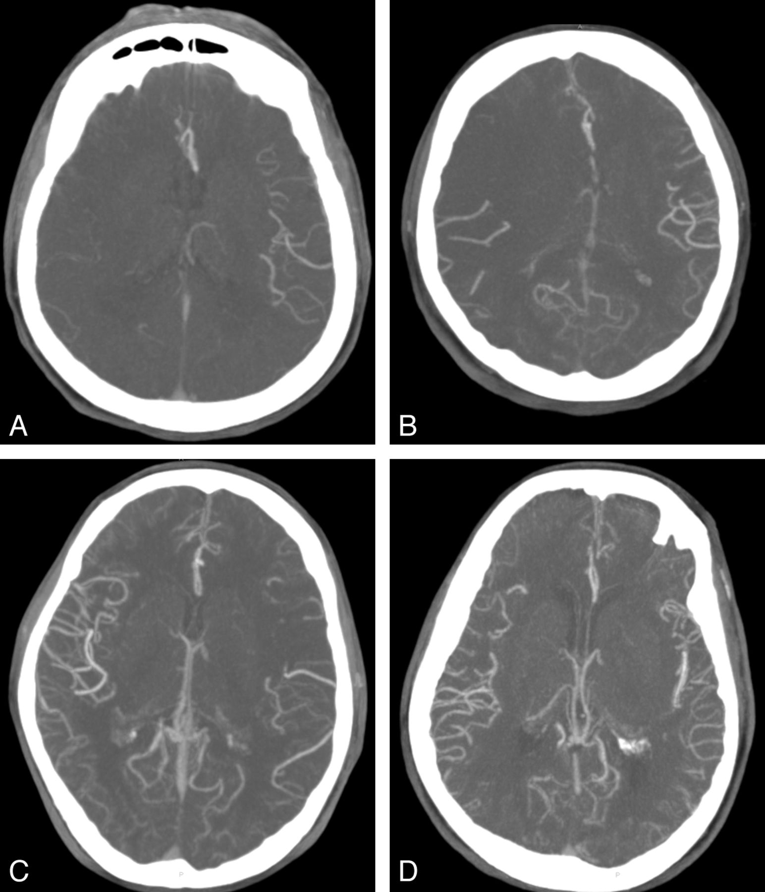
CTAs of all patients were reviewed by 3 neurointerventionists (B.Y., P.M., and R.D.), with consensus opinion reached on collateral supply. The reviewers were blinded to all clinical information during the consensus collateral grading. The CTA source images (20-mm, axial) were edited with MPR technique for assessment of leptomeningeal collaterals based on distribution of vessels at the Sylvian fissure and the leptomeningeal convexity. The MPR images were assessed with an MIP technique at 32 mm to evaluate the leptomeningeal collaterals. Collateral statuses were divided into 4 categories (Fig 1): score 0 = absence of contrast reaching the cortical surface of the affected hemisphere; score 1 = contrast reaching the cortical surface but not the Sylvian fissure; score 2 = contrast reaching the Sylvian fissure but opacifying <50% of hemisphere; score 3 = contrast reaching the Sylvian fissure and opacifying >50% of the hemisphere.

Fig 1.
  • Download figure
  • Open in new tab
  • Download powerpoint
Fig 1.

Leptomeningeal collateral CTA scores; A, score = 0; B, score = 1; C, score = 2; D, score = 3.

The Thrombolysis in Cerebral Infarction system was used to grade recanalization success.22 TICI grade 0 is no perfusion and no antegrade flow beyond the point of occlusion. TICI grade 1 is penetration with minimal perfusion. The contrast material passes beyond the area of obstruction but fails to opacify the entire cerebral bed distal to the obstruction for the duration of the angiographic run. TICI grade 2 is partial perfusion. The contrast material passes beyond the obstruction and opacifies the arterial bed distal to the obstruction. However, the rate of entry of contrast into the vessel distal to the obstruction or its rate of clearance from the distal bed or both are perceptibly slower than its entry into and/or clearance from comparable areas not perfused by the previously occluded vessel (eg, the opposite cerebral artery or the arterial bed proximal to the obstruction). In TICI grade 2a, only partial filling (less than two-thirds) of the entire vascular territory is visualized; in TICI grade 2b, complete filling of all of the expected vascular territory is visualized, but the filling is slower than normal. TICI grade 3 is complete perfusion; antegrade flow into the bed distal to the obstruction occurs as promptly as that into the obstruction and clearance.

Statistics

Statistical analysis was performed by using the STATA, Version 12 IC statistics package (StataCorp, College Station, Texas). We included the following baseline clinical variables: age, sex, comorbidities (history of hypertension, diabetes, atrial fibrillation, coronary artery disease, TIA, or stroke), baseline NIHSS score, IV-tPA, intra-arterial tissue plasminogen activator, onset time to CTA, and onset time to recanalization. Continuous variables were reported as median ± interquartile range (IQR). Categoric variables were reported as proportions. P values were reported by using the Fisher exact test for categoric variables and the Wilcoxon-Mann-Whitney rank sum test for continuous variables (age, NIHSS score, and time-to-event data). Effect sizes were reported as odds ratios for categoric variables and Hodges-Lehman median differences for continuous variables (age, NIHSS score, and time-to-event data). Receiver operating characteristic analysis curves were used to determine the collateral score threshold with functional outcome (3-month mRS score). Univariate and multivariate analyses were used to investigate the association between the dichotomized leptomeningeal collateral score with functional outcomes, mortality, and intracranial hemorrhages. Given the strong clinical association with clinical outcomes, we planned a priori to include the following independent variables in the model: age, baseline NIHSS score, TICI grade, and IV-tPA. A 2-sided P value <.05 was considered significant.

Results

Of the initial 104 patients with acute ischemic stroke with ICA and/or MCA occlusion who underwent IAT, CTA was not available for 14 patients before IAT. Three patients had poor image quality on CTA, and these patients were excluded. Eighty-seven patients with both adequate CTA and angiographic data with clinical outcome (mRS) at 90 days available were entered into the leptomeningeal collateral assessment. The baseline characteristics did not differ in a statistically significant manner between the patients with and without CTA (Table 1). In the CTA group, the median age was 66 years (IQR, 54–76 years). Fifty (57%) patients were men. The median NIHSS score at admission was 18 (IQR, 14–20). Forty (46%) subjects were treated with IV-tPA; 13 subjects (15%) received intra-arterial tPA. Sixty-five patients (75%) were treated with the Solitaire device (Covidien, Irvine, California), 10 (12%) patients were treated with the Merci retriever (Concentric Medical, Mountain View, California), and 3 (3%) were treated with the Penumbra System (Penumbra, Alameda, California).

View this table:
  • View inline
  • View popup
Table 1:

Baseline characteristics of the patient cohort with and without CTA

Leptomeningeal Collaterals

According to the leptomeningeal score, 34 subjects (39%) were graded as three, 30 subjects (35%) were graded as two, 12 subjects (14%) were graded as 1, and 11 subjects (13%) were graded as zero. Receiver operating characteristic analysis demonstrated the suitability of the collateral score ≥3 for determining good functional outcome of mRS ≤2 with an area under the curve of 0.6513 (sensitivity, 51.1%; specificity, 73.8%). The leptomeningeal score was dichotomized into good (score = 3) and poor (score = 0–2) on the basis of the receiver operating characteristic analysis. A higher percentage of IV-tPA was administered for a collateral score of 3 than for a collateral score of 0–2, (P = .008). The remainder of the baseline clinical variables did not differ in a statistically significant way between the 2 groups (Table 2).

View this table:
  • View inline
  • View popup
Table 2:

Comparison of patients for the dichotomized leptomeningeal collateral score

Outcome

Three-month outcome was favorable (mRS 0–2) in 47 subjects (54%), and the remaining 40 subjects (46%) had mRS scores of 3–6 (Fig 2). Patients with a good collateral score of 3 had higher odds of good functional outcome than patients with a collateral score of 0–2 (OR = 3.130; 95% CI, 1.252–7.825; P = .016). This finding remained significant after adjustment for administration of IV-tPA (OR = 2.998; 95% CI, 1.154–7.786; P = .024). In the multivariate analysis, patients with a collateral score of 3 had significantly higher odds of good outcome at 3-month follow-up after adjustment for age, baseline NIHSS score, IV-tPA, and TICI grades (adjusted OR = 2.985; 95% CI, 1.027–8.673; P = .045) (Table 3).

Fig 2.
  • Download figure
  • Open in new tab
  • Download powerpoint
Fig 2.

Distribution of mRS scores according to the leptomeningeal collateral scores.

View this table:
  • View inline
  • View popup
Table 3:

Univariate analysis of associations for outcomes for the dichotomized leptomeningeal score

Survival

Of the 13 deaths at 1 month, patients with poor collateral scores of 0–2 exhibited a trend toward a higher death rate than those with a collateral score of 3 (11 versus 2) (OR = 0.239; 95% CI, 0.049–1.153; P = .070), though no statistically significant differences were found.

Intracranial Hemorrhage

Although patients with a good collateral score of 3 had fewer episodes of intracranial hemorrhage than patients with a poor collateral score of 0–2 (4 versus 13), no statistically significant difference was detected (OR = 0.41; 95% CI, 0.122–1.385; P = .174).

Discussion

We found that a favorable pattern of leptomeningeal collaterals, as measured by CTA on admission, was associated with improved functional outcomes at 3 months in a cohort of patients with acute ischemic stroke with ICA or proximal MCA occlusion. In a multivariate model, a good leptomeningeal collateral score remained independently associated with good clinical outcome after adjustment for age, baseline NIHSS score, IV-tPA, and recanalization status.

Various studies have used CTA to score collaterals status14,17,19,20,23 with either CTA source images or by using MIP. Tan et al17 demonstrated that CTA-MIP was the best technique to quantify the degree of collateral circulation. In their retrospective analysis, they reported a good correlation between the CTA-based collateral score and the final infarct volume within 6 months.17 In another study, Miteff et al14 assessed retrograde filling of the MCA by 3 categories of collateral scoring and concluded that a good collateral status was one of the significant univariate predictors of favorable outcome.14 Menon et al21 included 138 patients (MCA-M1 and/or intracranial occlusion) in a retrospective single-center study and demonstrated good regional leptomeningeal anastomoses scores in 37.6% of patients, which correlated strongly with the size of the infarct core at baseline and were a strong independent predictor of final infarct and clinical outcome.

In a recent study, Souza et al23 found that CTA collaterals correlate with admission diffusion-weighted imaging infarct size and that a malignant collateral profile is highly specific for large admission DWI lesion size and poor functional outcome. In another study, Angermaier et al24 demonstrated that the CTA collateral grade was an independent predictor of final infarct volume in patients with stroke treated with endovascular therapy. On the other hand, Rosenthal et al18 reported that CTA collaterals had a positive impact on the outcomes of patients who did not achieve complete recanalization and had no impact in patients who were completely recanalized. Tan et al25 reported that good CTA collaterals correlated with improved outcomes in uni- but not in multivariate analyses. Our study reveals that a good CTA collateral score had a positive impact on the outcomes of patients in both uni- and multivariate analyses independent of recanalization status.

The system of leptomeningeal anastomoses is anastomotic connections between distal branches of the cerebral arteries on the surface of the brain that permit blood flow from the territory of an unobstructed artery into the territory of an occluded artery and constitute the secondary network of cerebral collateral circulation apart from the circle of Willis.26 Although the criterion standard for the assessment of leptomeningeal anastomoses is conventional DSA, the extent of leptomeningeal anastomoses formation seen on conventional DSA and with clinical outcome correlates well with collateral blood flow as assessed by CTA.15,27,28 CTA is quicker, simpler, and noninvasive and uses IV-administered contrast to visualize the extent of leptomeningeal anastomoses with intra- and extracranial vasculature.19,21

There is currently no consensus on what the presence or absence of collateral circulation means with respect to treatment choices for patients with acute ischemic stroke. Some authors believe a minority of patients with robust leptomeningeal collateral circulation will have minimal damage and experience excellent recovery without recanalization.29 On the other hand, some neurointerventionalists may want to try more aggressive IAT because there may be salvageable tissue in patients with good collateral vessel formation. Given the risks inherent in the therapeutic procedure and the clinical risk of futile recanalization,30 practitioners need a way to consistently select patients whom IAT is likely to benefit rather than harm. Given the significance of leptomeningeal collaterals, they should be taken into account with other validated scoring systems, namely the Houston Intra-Arterial Therapy 30 and the Totaled Heath Risks in Vascular Events score,31 in patient selection for IAT.

Our study has some limitations. First, selection bias is unavoidable in a retrospective single-center study. We sought to minimize reading bias by blinding the observers to clinical data and consensus achieved for the collateral grading. The CTA collateral scoring system was modified from Tan et al.17 Although our collateral grading derived from a consensus by 3 experienced neurointerventionists, the utility and value of the score requires further validation in a larger study before collateral scores are used as a primary means of selecting patients for revascularization therapy. Potential biases may have influenced the selection of cases for IAT on the basis of the CTA and clinical data.

Conclusions

Evaluation of the leptomeningeal collateral blood supply before IAT for anterior circulation intracranial arterial occlusion stroke (ICA and/or MCA) with a collateral score based on CTA-MIP reconstructions is independently associated with functional outcome at 3 months. We believe that leptomeningeal collateral assessment should be included in future trials investigating the clinical efficacy of IAT.

References

  1. 1.↵
    The National Institute of Neurological Disorders and Stroke rt-PA Stroke Study Group. Tissue plasminogen activator for acute ischemic stroke. N Engl J Med 1995;333:1581–87
    CrossRefPubMed
  2. 2.↵
    1. Wahlgren N,
    2. Ahmed N,
    3. Eriksson N,
    4. et al
    . Multivariable analysis of outcome predictors and adjustment of main outcome results to baseline data profile in randomized controlled trials Safe Implementation of Thrombolysis in Stroke-Monitoring Study (SITS-MOST). Stroke 2008;39:3316–22
    Abstract/FREE Full Text
  3. 3.↵
    1. Lees KR,
    2. Bluhmki E,
    3. von Kummer R,
    4. et al
    . Time to treatment with intravenous alteplase and outcome in stroke: an updated pooled analysis of ECASS, ATLANTIS, NINDS, and EPITHET trials. Lancet 2010;375:1695–703
    CrossRefPubMed
  4. 4.↵
    1. Bhatia R,
    2. Hill MD,
    3. Shobha N,
    4. et al
    . Low rates of acute recanalization with intravenous recombinant tissue plasminogen activator in ischemic stroke: real-world experience and a call for action. Stroke 2010;41:2254–58
    Abstract/FREE Full Text
  5. 5.↵
    1. Christou I,
    2. Felberg RA,
    3. Demchuk AM,
    4. et al
    . Intravenous tissue plasminogen activator and flow improvement in acute ischemic stroke patients with internal carotid artery occlusion. J Neuroimaging 2002;12:119–23
    CrossRefPubMed
  6. 6.↵
    1. Zangerle A,
    2. Kiechl S,
    3. Spiegel M,
    4. et al
    . Recanalization after thrombolysis in stroke patients: predictors and prognostic implications. Neurology 2007;68:39–44
    CrossRef
  7. 7.↵
    1. Meyers PM,
    2. Schumacher HC,
    3. Connolly ES Jr.,
    4. et al
    . Current status of endovascular stroke treatment. Circulation 2011;123:2591–601
    FREE Full Text
  8. 8.↵
    1. Smith WS,
    2. Sung G,
    3. Saver J,
    4. et al
    . Mechanical thrombectomy for acute ischemic stroke: final results of the Multi MERCI trial. Stroke 2008;39:1205–12
    Abstract/FREE Full Text
  9. 9.↵
    1. Clark W,
    2. Lutsep H,
    3. Barnwell S,
    4. et al
    . The Penumbra Pivotal Stroke trial: safety and effectiveness of a new generation of mechanical devices for clot removal in intracranial large vessel occlusive disease. Stroke 2009;40:2761–68
    Abstract/FREE Full Text
  10. 10.↵
    1. Broderick JP,
    2. Palesch YY,
    3. Demchuk AM,
    4. et al.
    , for the Interventional Management of Stroke (IMS) III Investigators. Endovascular therapy after intravenous t-PA versus t-PA alone for stroke. N Engl J Med 2013;368:893–903
    CrossRefPubMed
  11. 11.↵
    1. Ciccone A,
    2. Valvassori L,
    3. Nichelatti M,
    4. et al
    . Endovascular treatment for acute ischemic stroke. N Engl J Med 2013;368:904–13
    CrossRefPubMed
  12. 12.↵
    1. Chimowitz MI
    . Endovascular treatment for acute ischemic stroke: still unproven. N Engl J Med 2013;368:954–55
  13. 13.↵
    1. Dalkara T,
    2. Arsava EM
    . Can restoring incomplete microcirculatory reperfusion improve stroke outcome after thrombolysis? J Cereb Blood Flow Metab 2012;32:2091–209
    CrossRefPubMed
  14. 14.↵
    1. Miteff F,
    2. Levi CR,
    3. Bateman GA,
    4. et al
    . The independent predictive utility of computed tomography angiographic collateral status in acute ischaemic stroke. Brain 2009;132(pt 8):2231–2238
    Abstract/FREE Full Text
  15. 15.↵
    1. Bang OY,
    2. Saver JL,
    3. Buck BH,
    4. et al.
    , for the UCLA Collateral Investigators. Impact of collateral flow on tissue fate in acute ischaemic stroke. J Neurol Neurosurg Psychiatry 2007;79:625–29
    CrossRefPubMed
  16. 16.↵
    1. Bang OY,
    2. Saver JL,
    3. Kim SJ,
    4. et al
    . Collateral flow predicts response to endovascular therapy for acute ischemic stroke. Stroke 2011;42:693–99
    Abstract/FREE Full Text
  17. 17.↵
    1. Tan JC,
    2. Dillon WP,
    3. Liu S,
    4. et al
    . Systematic comparison of perfusion-CT and CT-angiography in acute stroke patients. Ann Neurol 2007;61:533–43
    CrossRefPubMed
  18. 18.↵
    1. Rosenthal ES,
    2. Schwamm LH,
    3. Roccatagliata L,
    4. et al
    . Role of recanalization in acute stroke outcome: rationale for a CT angiogram-based “benefit of recanalization” model. AJNR Am J Neuroradiol 2008;29:1471–75
    Abstract/FREE Full Text
  19. 19.↵
    1. Maas MB,
    2. Lev MH,
    3. Ay H,
    4. et al
    . Collateral vessels on CT angiography predict outcome in acute ischemic stroke. Stroke 2009;40:3001–05
    Abstract/FREE Full Text
  20. 20.↵
    1. Lima FO,
    2. Furie KL,
    3. Silva GS,
    4. et al
    . The pattern of leptomeningeal collaterals on CT angiography is a strong predictor of long-term functional outcome in stroke patients with large vessel intracranial occlusion. Stroke 2010;41:2316–22
    Abstract/FREE Full Text
  21. 21.↵
    1. Menon BK,
    2. Smith EE,
    3. Modi J,
    4. et al
    . Regional leptomeningeal score on CT angiography predicts clinical and imaging outcomes in patients with acute anterior circulation occlusions. AJNR Am J Neuroradiol 2011;32:1640–45
    Abstract/FREE Full Text
  22. 22.↵
    1. Higashida RT,
    2. Anthony J Furlan,
    3. et al
    . Trial design and reporting standards for intraarterial cerebral thrombolysis for acute ischemic stroke. J Vasc Interv Radiol 2003;14(9 Pt 2):S493–94
    CrossRefPubMed
  23. 23.↵
    1. Souza LC,
    2. Yoo AJ,
    3. Chaudhry ZA,
    4. et al
    . Malignant CTA collateral profile is highly specific for large admission DWI infarct core and poor outcome in acute stroke. AJNR Am J Neuroradiol 2012;33:1331–36
    Abstract/FREE Full Text
  24. 24.↵
    1. Angermaier A,
    2. Langner S,
    3. Kirsch M,
    4. et al
    . CT-angiographic collateralization predicts final infarct volume after intra-arterial thrombolysis for acute anterior circulation ischemic stroke. Cerebrovasc Dis 2011;31:177–84
    CrossRefPubMed
  25. 25.↵
    1. Tan IY,
    2. Demchuk AM,
    3. Hopyan J,
    4. et al
    . CT angiography clot burden score and collateral score: correlation with clinical and radiologic outcomes in acute middle cerebral artery infarct. AJNR Am J Neuroradiol 2009;30:525–31
    Abstract/FREE Full Text
  26. 26.↵
    1. Liebeskind DS
    . Collaterals in acute stroke: beyond the clot. Neuroimaging Clin N Am 2005;15:553–573, x
    CrossRefPubMed
  27. 27.↵
    1. Wildermuth S,
    2. Knauth M,
    3. Brandt T,
    4. et al
    . Role of CT angiography in patient selection for thrombolytic therapy in acute hemispheric stroke. Stroke 1998;29:935–38
    Abstract/FREE Full Text
  28. 28.↵
    1. Liebeskind DS
    . Collateral circulation. Stroke 2003;34:2279–84
    Abstract/FREE Full Text
  29. 29.↵
    1. Tomsick TA,
    2. Khatri P,
    3. Jovin T,
    4. et al
    . Equipoise among recanalization strategies. Neurology 2010;74:1069–76
    CrossRef
  30. 30.↵
    1. Hallevi Hen,
    2. Barreto AD,
    3. Liebeskind DS,
    4. et al
    . Identifying patients at high risk for poor outcome after intra-arterial therapy for acute ischemic stroke. Stroke 2009;40:1780–85
    Abstract/FREE Full Text
  31. 31.↵
    1. Flint AC,
    2. Kamel H,
    3. Rao VA,
    4. et al
    . Validation of the Totaled Heath Risks In Vascular Events (THRIVE) score for outcome prediction in endovascular stroke treatment. Int J Stroke 2014;9:32–39
    CrossRefPubMed
  • Received July 25, 2013.
  • Accepted after revision August 26, 2013.
  • © 2014 by American Journal of Neuroradiology
View Abstract
PreviousNext
Back to top

In this issue

American Journal of Neuroradiology: 35 (4)
American Journal of Neuroradiology
Vol. 35, Issue 4
1 Apr 2014
  • Table of Contents
  • Index by author
  • Complete Issue (PDF)
Advertisement
Print
Download PDF
Email Article

Thank you for your interest in spreading the word on American Journal of Neuroradiology.

NOTE: We only request your email address so that the person you are recommending the page to knows that you wanted them to see it, and that it is not junk mail. We do not capture any email address.

Enter multiple addresses on separate lines or separate them with commas.
The Impact of Arterial Collateralization on Outcome after Intra-Arterial Therapy for Acute Ischemic Stroke
(Your Name) has sent you a message from American Journal of Neuroradiology
(Your Name) thought you would like to see the American Journal of Neuroradiology web site.
CAPTCHA
This question is for testing whether or not you are a human visitor and to prevent automated spam submissions.
Cite this article
S. Seeta Ramaiah, L. Churilov, P. Mitchell, R. Dowling, B. Yan
The Impact of Arterial Collateralization on Outcome after Intra-Arterial Therapy for Acute Ischemic Stroke
American Journal of Neuroradiology Apr 2014, 35 (4) 667-672; DOI: 10.3174/ajnr.A3862

Citation Manager Formats

  • BibTeX
  • Bookends
  • EasyBib
  • EndNote (tagged)
  • EndNote 8 (xml)
  • Medlars
  • Mendeley
  • Papers
  • RefWorks Tagged
  • Ref Manager
  • RIS
  • Zotero
0 Responses
Respond to this article
Share
Bookmark this article
The Impact of Arterial Collateralization on Outcome after Intra-Arterial Therapy for Acute Ischemic Stroke
S. Seeta Ramaiah, L. Churilov, P. Mitchell, R. Dowling, B. Yan
American Journal of Neuroradiology Apr 2014, 35 (4) 667-672; DOI: 10.3174/ajnr.A3862
del.icio.us logo Twitter logo Facebook logo Mendeley logo
  • Tweet Widget
  • Facebook Like
  • Google Plus One
Purchase

Jump to section

  • Article
    • Abstract
    • ABBREVIATIONS:
    • Materials and Methods
    • Results
    • Discussion
    • Conclusions
    • References
  • Figures & Data
  • Info & Metrics
  • Responses
  • References
  • PDF

Related Articles

  • No related articles found.
  • PubMed
  • Google Scholar

Cited By...

  • Circle of Willis variation and outcome after intra-arterial treatment
  • Impact of single phase CT angiography collateral status on functional outcome over time: results from the MR CLEAN Registry
  • Interrogating the Functional Correlates of Collateralization in Patients with Intracranial Stenosis Using Multimodal Hemodynamic Imaging
  • Impact of collaterals on the efficacy and safety of endovascular treatment in acute ischaemic stroke: a systematic review and meta-analysis
  • Collateral flow as causative of good outcomes in endovascular stroke therapy
  • Crossref (23)
  • Google Scholar

This article has been cited by the following articles in journals that are participating in Crossref Cited-by Linking.

  • Impact of collaterals on the efficacy and safety of endovascular treatment in acute ischaemic stroke: a systematic review and meta-analysis
    Xinyi Leng, Hui Fang, Thomas W H Leung, Chen Mao, Zhongrong Miao, Liping Liu, Ka Sing Wong, David S Liebeskind
    Journal of Neurology, Neurosurgery & Psychiatry 2016 87 5
  • The cerebral collateral circulation: Relevance to pathophysiology and treatment of stroke
    Myron D. Ginsberg
    Neuropharmacology 2018 134
  • Collateral flow as causative of good outcomes in endovascular stroke therapy
    Sunil A Sheth, Nerses Sanossian, Qing Hao, Sidney Starkman, Latisha K Ali, Doojin Kim, Nestor R Gonzalez, Satoshi Tateshima, Reza Jahan, Gary R Duckwiler, Jeffrey L Saver, Fernando Vinuela, David S Liebeskind
    Journal of NeuroInterventional Surgery 2016 8 1
  • Expanding the concept of neuroprotection for acute ischemic stroke: The pivotal roles of reperfusion and the collateral circulation
    Myron D. Ginsberg
    Progress in Neurobiology 2016 145-146
  • Determinants of leptomeningeal collateral flow in stroke patients with a middle cerebral artery occlusion
    Tom van Seeters, Geert Jan Biessels, L. Jaap Kappelle, Yolanda van der Graaf, Birgitta K. Velthuis
    Neuroradiology 2016 58 10
  • Impact of single phase CT angiography collateral status on functional outcome over time: results from the MR CLEAN Registry
    Ivo GH Jansen, Maxim JHL Mulder, Robert-Jan B Goldhoorn, Anna MM Boers, Adriaan CGM van Es, Lonneke SF Yo, Jeannette Hofmeijer, Jasper M Martens, Marianne AA van Walderveen, Bas FW van der Kallen, Sjoerd FM Jenniskens, Kilian M Treurniet, Henk A Marquering, Marieke ES Sprengers, Wouter J Schonewille, Joost CJ Bot, Geert J Lycklama a Nijeholt, Hester F Lingsma, David S Liebeskind, Jelis Boiten, Jan Albert Vos, Yvo BWEM Roos, Robert J van Oostenbrugge, Aad van der Lugt, Wim H van Zwam, Diederik WJ Dippel, Ido R van den Wijngaard, Charles BLM Majoie
    Journal of NeuroInterventional Surgery 2019 11 9
  • A meta‐analysis of collateral status and outcomes of mechanical thrombectomy
    Jiacheng Qian, Lu Fan, Weiqing Zhang, Jian Wang, Jianting Qiu, Yujie Wang
    Acta Neurologica Scandinavica 2020 142 3
  • Collateral state and the effect of endovascular reperfusion therapy on clinical outcome in ischemic stroke patients
    Johannes C. Gerber, Marketa Petrova, Pawel Krukowski, Matthias Kuhn, Andrij Abramyuk, Ulf Bodechtel, Imanuel Dzialowski, Kay Engellandt, Hagen Kitzler, Lars‐Peder Pallesen, Hauke Schneider, Ruediger von Kummer, Volker Puetz, Jennifer Linn
    Brain and Behavior 2016 6 9
  • Transcranial Doppler and Transcranial Color Duplex in Defining Collateral Cerebral Blood Flow
    Maher Saqqur, Khurshid Khan, Carol Derksen, Andrei Alexandrov, Ashfaq Shuaib
    Journal of Neuroimaging 2018 28 5
  • Prediction of favorable outcome by percent improvement in patients with acute ischemic stroke treated with endovascular stent thrombectomy
    Yuezhou Cao, Shuiping Wang, Wen Sun, Qiliang Dai, Wei Li, Jin Cai, Xinying Fan, Wusheng Zhu, Yunyun Xiong, Yunfei Han, Wenjie Zi, Shiquan Yang, Jia Chen, Xinfeng Liu
    Journal of Clinical Neuroscience 2017 38

More in this TOC Section

  • Usefulness of Quantitative Susceptibility Mapping for the Diagnosis of Parkinson Disease
  • Evaluating the Effects of White Matter Multiple Sclerosis Lesions on the Volume Estimation of 6 Brain Tissue Segmentation Methods
  • White Matter Alterations in the Brains of Patients with Active, Remitted, and Cured Cushing Syndrome: A DTI Study
Show more Brain

Similar Articles

Advertisement

Indexed Content

  • Current Issue
  • Accepted Manuscripts
  • Article Preview
  • Past Issues
  • Editorials
  • Editors Choice
  • Fellow Journal Club
  • Letters to the Editor

Cases

  • Case Collection
  • Archive - Case of the Week
  • Archive - Case of the Month
  • Archive - Classic Case

Special Collections

  • Special Collections

Resources

  • News and Updates
  • Turn around Times
  • Submit a Manuscript
  • Author Policies
  • Manuscript Submission Guidelines
  • Evidence-Based Medicine Level Guide
  • Publishing Checklists
  • Graphical Abstract Preparation
  • Imaging Protocol Submission
  • Submit a Case
  • Become a Reviewer/Academy of Reviewers
  • Get Peer Review Credit from Publons

Multimedia

  • AJNR Podcast
  • AJNR SCANtastic
  • Video Articles

About Us

  • About AJNR
  • Editorial Board
  • Not an AJNR Subscriber? Join Now
  • Alerts
  • Feedback
  • Advertise with us
  • Librarian Resources
  • Permissions
  • Terms and Conditions

American Society of Neuroradiology

  • Not an ASNR Member? Join Now

© 2025 by the American Society of Neuroradiology All rights, including for text and data mining, AI training, and similar technologies, are reserved.
Print ISSN: 0195-6108 Online ISSN: 1936-959X

Powered by HighWire