
Article Figures & Data
Figures
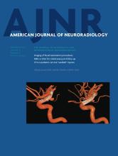
- Fig 1.
Hyperperfusion in the right basal ganglia is demonstrated by increased cerebral blood flow (arrows, A) and increased cerebral blood volume (arrows, B). Also seen is a large region of right middle cerebral artery territory ischemia as evidenced by reduced CBF (A) with relative preservation of CBV (B). Lateral digital subtraction angiogram from a right carotid artery injection demonstrates early opacification of the basal vein of Rosenthal (solid arrow, C) and the internal cerebral vein (dashed arrow, C). Follow-up MR imaging 12 hours later shows restricted diffusion indicating infarction of the right basal ganglia, along with patchy cortical infarctions in the right MCA territory (D).
- Fig 2.
Hyperperfusion of the left basal ganglia is shown by increased cerebral blood flow (arrow, A) and increased cerebral blood volume (arrow, B) on CT perfusion. Follow-up FLAIR MR imaging 3.5 days later shows regions of T2 hyperintensity indicating infarction of this same region (C and D).
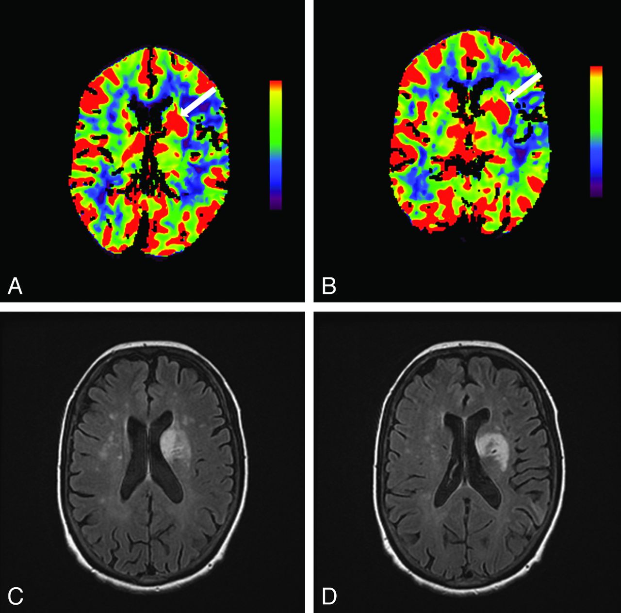
- Fig 3.
Increased cerebral blood flow in the right basal ganglia (arrow, A) in addition to a large region of reduced CBF in the right middle cerebral artery territory is shown on the CT perfusion scan at presentation. Noncontrast head CT 1 day later demonstrates right MCA infarction but apparent sparing of the right basal ganglia (B). Follow-up CT at day 6 shows hypoattenuation involving right basal ganglia (arrow, C) in addition to evolution of a large right MCA infarction with mass effect.


Tables
Baseline patient characteristics
Age/Sex HTN CAD DM A. fib Initial BP, (mm Hg) NIHSS MCA Territory Time to CTP (mins)a TICI Grade ICH 1 79/F Y N N N 125/63 16 R 343 2a Y 2 61/M N Y N Y 116/70 21 L 187 2b Y 3 61/M N N N N 114/72 17 R 125 2b N 4 66/M Y N N Y 155/99 21 L 190 2b N 5 61/F Y N N N 140/80 20 R 230 0 N 6 65/F Y N N N 146/92 16 L 210 NAb N Note:—HTN indicates hypertension; CAD, coronary artery disease; DM, diabetes mellitus; A. fib, atrial fibrillation; BP, blood pressure; ICH, intracranial hemorrhage; R, right; L, left; Y, yes; N, no; NA, not applicable.
↵a TICI 0 indicates no perfusion; 2a, perfusion of ≤50% of MCA distribution; 2b, perfusion of >50% of MCA distribution; 3, full perfusion.
↵b Endovascular therapy was not attempted because of large core on CTP.


