
Article Figures & Data
Figures
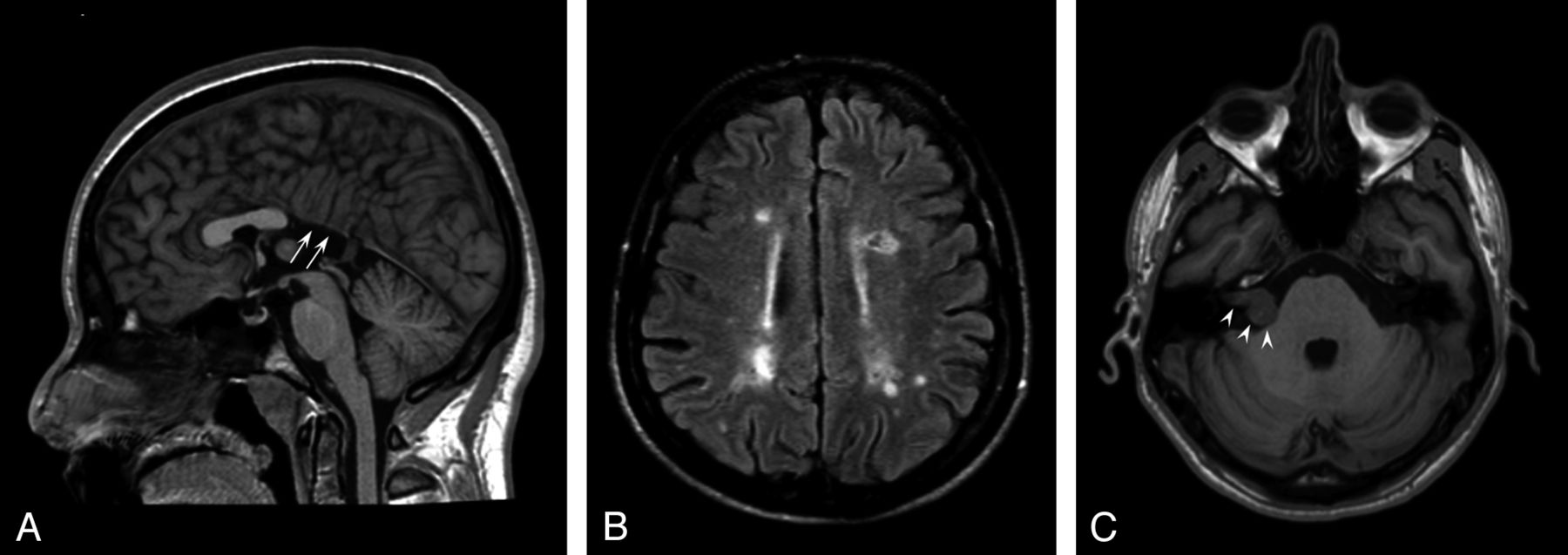
- Fig 1.
Conventional brain MR imaging findings in 45 patients with Kallmann syndrome. A, Midsagittal T1-weighted image of a patient with agenesia of the posterior portion of the corpus callosum (white arrows). B, Axial FLAIR image of the patient disclosing several multiple sclerosis–like white matter signal abnormalities in the centrum semiovale bilaterally. C, Non-enhanced axial T1-weighted image at the level of the internal acoustic meatus showing a right intra/extrameatal dumbell-shaped mass consistent with an acoustic schwannoma.
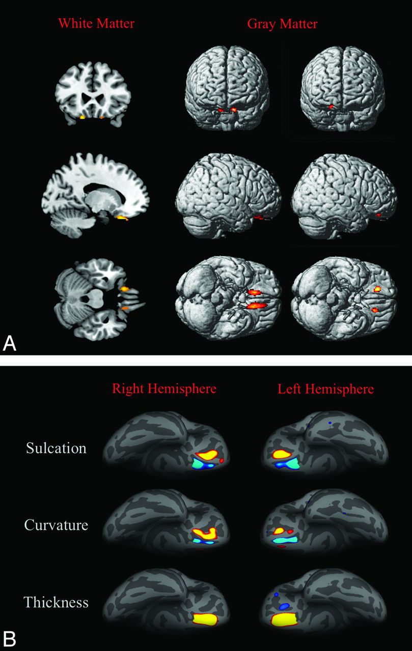
- Fig 2.
3D-T1-based whole-brain analyses on 42 patients with Kallmann syndrome versus 23 controls. A, Voxel-based morphometry findings. Clusters of significantly decreased white matter volume (colored areas in the multiplanar reconstructions in the first column) were detected exclusively and symmetrically in the posterior portion of the medial orbital-frontal gyrus close to the olfactory sulcus; no regions of increased white matter volume were detected in our sample. Clusters of significantly decreased (second column) and increased (third column) gray matter volume are shown as colored cortical areas in the volume-rendering technique images within or close to the olfactory sulci. B, Sulcation, curvature, and thickness findings. Colored areas represent increased (yellow-red) and decreased (blue) values in patients with KS. Almost all differences are clustered within the olfactory sulci and the neighboring cortex of the rectus and medial orbital-frontal gyri.


Tables
Cortical areas >40 mm2 with significant differences between patients with Kallmann syndrome and controls by sulcation, curvature, and cortical thickness analyses
Hemisphere Size (mm2) Local Maximum, Talairach FDR Threshold Cerebral Region X Y Z Sulcation Right 394.05 ↓ 11.2 13.8 −14.9 3.3429 Olfactory sulcus and contiguous cortex of the rectus gyrus Right 325.72 ↑ 16.4 28.7 −23.7 3.3429 Medial orbital-frontal gyrus Left 518.07 ↓ −9.8 16.0 −15.4 3.3159 Olfactory sulcus and contiguous cortex of the rectus gyrus Left 252.41 ↑ −19.6 33.2 −17.3 3.3159 Medial orbital-frontal gyrus Curvature Right 372.85 ↑ 14.4 32.3 −25.3 3.4704 Medial orbital-frontal gyrus Right 77.61 ↓ 11.7 16.6 −14.2 3.4704 Olfactory sulcus Right 44.71 ↓ 11.5 37.9 −19.1 3.4704 Olfactory sulcus Left 196.94 ↓ −11.6 38.7 −20.0 3.4704 Olfactory sulcus Left 114.79 ↑ −19.6 32.7 −14.9 3.4704 Medial orbital-frontal gyrus Thickness Right 568.97 ↑ −14.1 30.7 −19.2 3.3834 Olfactory sulcus and contiguous cortex of the rectus and medial orbital-frontal gyri Left 686.38 ↑ 13.7 33.4 −23.3 3.2803 Olfactory sulcus and contiguous cortex of the rectus and medial orbital-frontal gyri Left 78.39 ↓ 12.7 48.6 33.5 3.2803 Medial orbital-frontal sulcus Left 40.54 ↓ 45.2 −62.3 36.9 3.2803 Lateral orbital-frontal gyrus Note:—↑ indicates that patients with KS showed increased sulcation, curvature, and cortical thickness; ↓, patients with KS showed decreased sulcation, curvature, and cortical thickness; FDR, false discovery rate.


