Advertisement

Main menu

  • Home
  • Content
    • Current Issue
    • Accepted Manuscripts
    • Article Preview
    • Past Issue Archive
    • AJNR Case Collection
    • Case of the Week Archive
    • Classic Case Archive
    • Case of the Month Archive
  • Special Collections
    • Spinal CSF Leak Articles (Jan 2020-June 2024)
    • 2024 AJNR Journal Awards
    • Most Impactful AJNR Articles
  • Multimedia
    • AJNR Podcast
    • AJNR Scantastics
    • Video Articles
  • For Authors
    • Submit a Manuscript
    • Author Policies
    • Fast publishing of Accepted Manuscripts
    • Graphical Abstract Preparation
    • Manuscript Submission Guidelines
    • Imaging Protocol Submission
    • Submit a Case for the Case Collection
  • About Us
    • About AJNR
    • Editorial Board
  • More
    • Become a Reviewer/Academy of Reviewers
    • Subscribers
    • Permissions
    • Alerts
    • Feedback
    • Advertisers
    • ASNR Home
  • Other Publications
    • ajnr

User menu

  • Alerts
  • Log in

Search

  • Advanced search
American Journal of Neuroradiology

American Journal of Neuroradiology

ASHNR American Society of Functional Neuroradiology ASHNR American Society of Pediatric Neuroradiology ASSR
  • Alerts
  • Log in

Advanced Search

  • Home
  • Content
    • Current Issue
    • Accepted Manuscripts
    • Article Preview
    • Past Issue Archive
    • AJNR Case Collection
    • Case of the Week Archive
    • Classic Case Archive
    • Case of the Month Archive
  • Special Collections
    • Spinal CSF Leak Articles (Jan 2020-June 2024)
    • 2024 AJNR Journal Awards
    • Most Impactful AJNR Articles
  • Multimedia
    • AJNR Podcast
    • AJNR Scantastics
    • Video Articles
  • For Authors
    • Submit a Manuscript
    • Author Policies
    • Fast publishing of Accepted Manuscripts
    • Graphical Abstract Preparation
    • Manuscript Submission Guidelines
    • Imaging Protocol Submission
    • Submit a Case for the Case Collection
  • About Us
    • About AJNR
    • Editorial Board
  • More
    • Become a Reviewer/Academy of Reviewers
    • Subscribers
    • Permissions
    • Alerts
    • Feedback
    • Advertisers
    • ASNR Home
  • Follow AJNR on Twitter
  • Visit AJNR on Facebook
  • Follow AJNR on Instagram
  • Join AJNR on LinkedIn
  • RSS Feeds

Welcome to the new AJNR, Updated Hall of Fame, and more. Read the full announcements.


AJNR is seeking candidates for the position of Associate Section Editor, AJNR Case Collection. Read the full announcement.


Getting new auth cookie, if you see this message a lot, tell someone!
Research ArticleSpine

Double Inversion Recovery Sequence of the Cervical Spinal Cord in Multiple Sclerosis and Related Inflammatory Diseases

I. Riederer, D.C. Karampinos, M. Settles, C. Preibisch, J.S. Bauer, J.F. Kleine, M. Mühlau and C. Zimmer
American Journal of Neuroradiology January 2015, 36 (1) 219-225; DOI: https://doi.org/10.3174/ajnr.A4093
I. Riederer
aFrom the Departments of Diagnostic and Interventional Neuroradiology (I.R., C.P., J.S.B., J.F.K., C.Z.)
  • Find this author on Google Scholar
  • Find this author on PubMed
  • Search for this author on this site
D.C. Karampinos
bDiagnostic and Interventional Radiology (D.C.K., M.S.)
  • Find this author on Google Scholar
  • Find this author on PubMed
  • Search for this author on this site
M. Settles
bDiagnostic and Interventional Radiology (D.C.K., M.S.)
  • Find this author on Google Scholar
  • Find this author on PubMed
  • Search for this author on this site
C. Preibisch
aFrom the Departments of Diagnostic and Interventional Neuroradiology (I.R., C.P., J.S.B., J.F.K., C.Z.)
cNeuroimaging Center at the Technische Universität München (C.P., M.M.)
  • Find this author on Google Scholar
  • Find this author on PubMed
  • Search for this author on this site
J.S. Bauer
aFrom the Departments of Diagnostic and Interventional Neuroradiology (I.R., C.P., J.S.B., J.F.K., C.Z.)
  • Find this author on Google Scholar
  • Find this author on PubMed
  • Search for this author on this site
J.F. Kleine
aFrom the Departments of Diagnostic and Interventional Neuroradiology (I.R., C.P., J.S.B., J.F.K., C.Z.)
  • Find this author on Google Scholar
  • Find this author on PubMed
  • Search for this author on this site
M. Mühlau
cNeuroimaging Center at the Technische Universität München (C.P., M.M.)
dDepartment of Neurology (M.M.), Klinikum rechts der Isar, Technische Universität München, Munich, Germany.
  • Find this author on Google Scholar
  • Find this author on PubMed
  • Search for this author on this site
C. Zimmer
aFrom the Departments of Diagnostic and Interventional Neuroradiology (I.R., C.P., J.S.B., J.F.K., C.Z.)
  • Find this author on Google Scholar
  • Find this author on PubMed
  • Search for this author on this site
  • Article
  • Figures & Data
  • Info & Metrics
  • Responses
  • References
  • PDF
Loading

Article Figures & Data

Figures

  • Tables
  • Fig 1.
    • Download figure
    • Open in new tab
    • Download powerpoint
    Fig 1.

    Bland-Altman analysis of the lesion counts of reader 1 and 2 for the DIR sequence (upper row) and the T2WI TSE sequence (lower row). There was an almost identical, nonsignificant bias in both techniques, with reader 2 counting on average 0.33 more lesions on DIR images and 0.37 more lesions on T2 images than reader 1 (95% CI, −3.2–2.6; correlation coefficient, r = 0.97; and −2.8–2.1, r = 0.94, respectively).
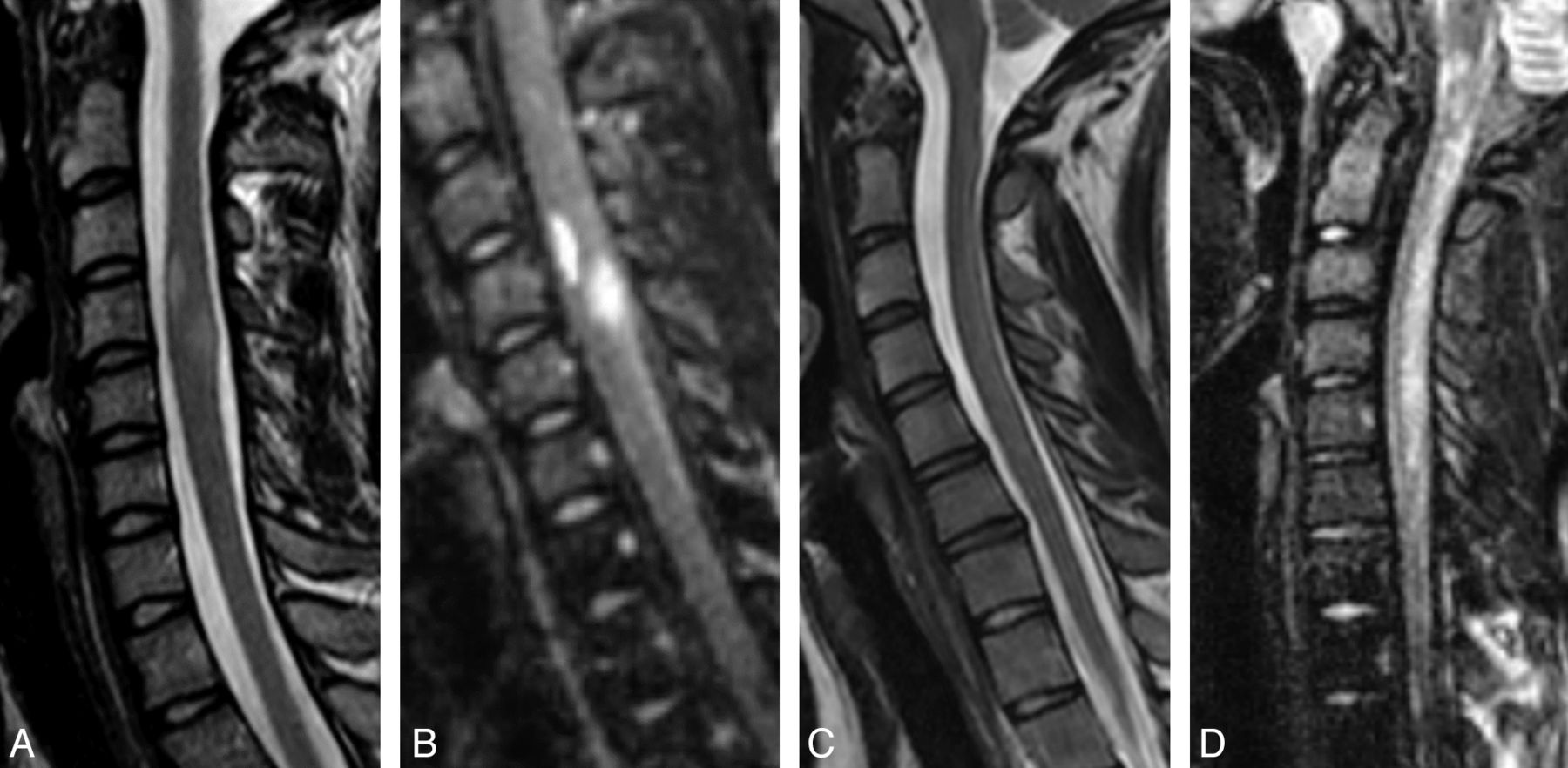
  • Fig 2.
    • Download figure
    • Open in new tab
    • Download powerpoint
    Fig 2.

    Examples of 2 patients with lesions visible only in the DIR images and not in the T2WI TSE images. Sagittal (A and F) and axial (D and H) T2WI TSE images; sagittal (B and G), coronal (C), and axial (E and I) reconstructions of the 3D DIR sequence of the spinal cord. The group of images on the left (I, A–E) shows the cervical spinal cord of a 52-year-old female patient. Note the lesion in the spinal cord at the C4 vertebral body level, which is only visible in the DIR sequence. This patient indicated pain in her left shoulder, weakness of her left arm, and tingling in her left palm. In T2WI TSE images, 1 lesion was visible in the cervical spinal cord at the C2/C3 level with discrete contrast enhancement (not shown in the image). At first, the differential diagnosis included neoplasm and inflammation. Due to the cervical 3D DIR sequence, another small lesion was detected at the C4 lateral level on the right (arrow), favoring the diagnosis of cervical myelitis. The group of images on the right (II, F–I) shows the spinal cord of a 49-year-old patient with clinically isolated syndrome. Note the lesion in the spinal cord at the C7 vertebral body level, which is only visible in the DIR sequence.
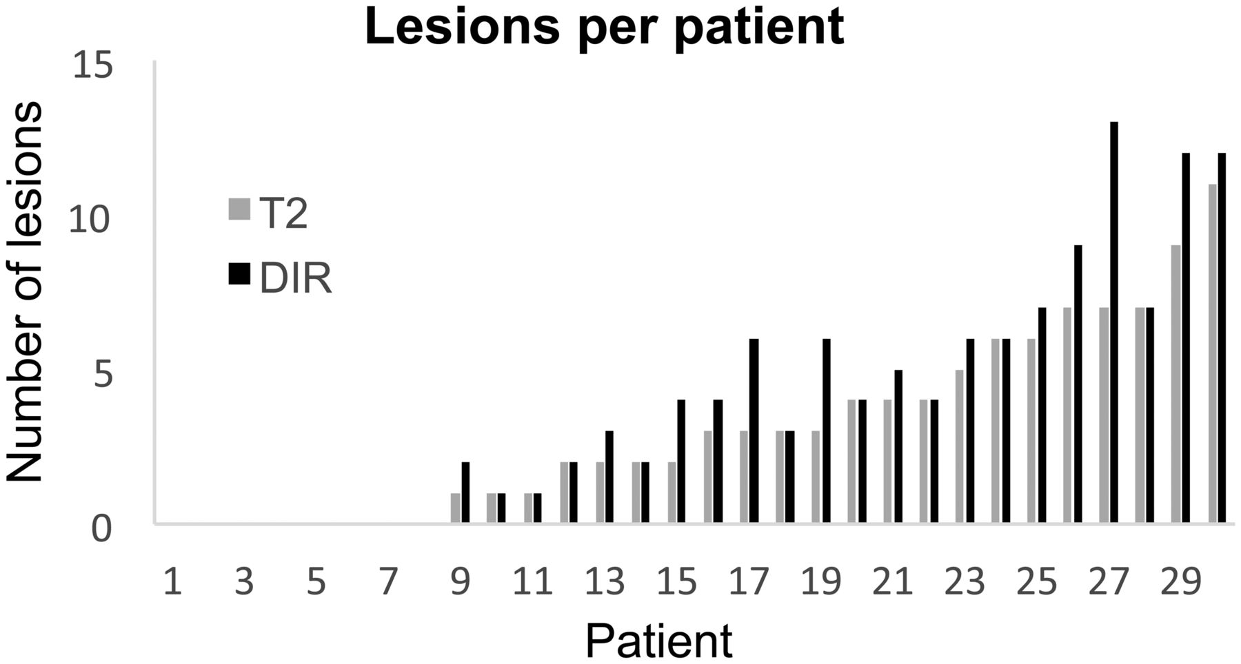
  • Fig 3.
    • Download figure
    • Open in new tab
    • Download powerpoint
    Fig 3.

    The number of lesions per patient according to the consensus reading of both radiologists in the T2WI TSE and DIR sequences. Volunteers are not included. Range, 0–13 (DIR), 0–11 (T2WI TSE); mean, 3.97 ± 3.85 (DIR), 3.10 ± 3.00 (T2WI TSE); P < .01.

  • Fig 4.
    • Download figure
    • Open in new tab
    • Download powerpoint
    Fig 4.

    Sagittal T2WI TSE (A and C) and DIR (B and D) images of the cervical spinal cord of 2 patients. A and B, Two lesions in the spinal cord at the C4 vertebral body level of a 22-year-old female patient with clinically isolated syndrome. C and D, A diffuse myelitis of an 18-year-old female patient with MS. The lesion-to-background contrast sCNR in DIR images is remarkably higher.

  • Fig 5.
    • Download figure
    • Open in new tab
    • Download powerpoint
    Fig 5.

    Sagittal (A), coronal (B), and axial (C) reconstructions of 3D DIR images of the cervical spinal cord of a 42-year-old female patient with primary-progressive MS. Elongated lesions in the lateral spinal cord are visible on both sides (arrow).

  • Fig 6.
    • Download figure
    • Open in new tab
    • Download powerpoint
    Fig 6.

    Sagittal T2WI TSE (A) and DIR (B) sequence of the thoracic spinal cord of a 35-year-old female patient with clinically isolated syndrome. On both sequences, a hyperintense lesion is visible in the spinal cord at the T7 level (arrow).

Tables

  • Figures
  • MRI acquisition parameters of the 3D DIR and axial and sagittal T2WI TSE sequences

    Sequence3D DIR2D T2WI TSE
    Imaging planeSagittalSagittalAxial
    Acquisition matrix208 × 208 × 300212 × 233308 × 207
    Acquisition voxel size (mm3)1.2 × 1.2 × 1.30.94 × 1.18 × 20.65 × 0.88 × 4
    TR (ms)550030714238
    TE (ms)287100100
    TSE factor1732925
    IR delays (ms)2550/450
    Flip/refocusing angleT2prep with TE = 125 ms and 4 refocusing pulses90°/120°90°/120°
    Acquisition time7 min 36 sec3 min 47 sec3 min 15 sec
    Sections3001528
    • Note:—IR indicates inversion recovery; T2prep, preparation pulse to ensure T2 weighting.

PreviousNext
Back to top

In this issue

American Journal of Neuroradiology: 36 (1)
American Journal of Neuroradiology
Vol. 36, Issue 1
1 Jan 2015
  • Table of Contents
  • Index by author
  • Complete Issue (PDF)
Advertisement
Print
Download PDF
Email Article

Thank you for your interest in spreading the word on American Journal of Neuroradiology.

NOTE: We only request your email address so that the person you are recommending the page to knows that you wanted them to see it, and that it is not junk mail. We do not capture any email address.

Enter multiple addresses on separate lines or separate them with commas.
Double Inversion Recovery Sequence of the Cervical Spinal Cord in Multiple Sclerosis and Related Inflammatory Diseases
(Your Name) has sent you a message from American Journal of Neuroradiology
(Your Name) thought you would like to see the American Journal of Neuroradiology web site.
CAPTCHA
This question is for testing whether or not you are a human visitor and to prevent automated spam submissions.
Cite this article
I. Riederer, D.C. Karampinos, M. Settles, C. Preibisch, J.S. Bauer, J.F. Kleine, M. Mühlau, C. Zimmer
Double Inversion Recovery Sequence of the Cervical Spinal Cord in Multiple Sclerosis and Related Inflammatory Diseases
American Journal of Neuroradiology Jan 2015, 36 (1) 219-225; DOI: 10.3174/ajnr.A4093

Citation Manager Formats

  • BibTeX
  • Bookends
  • EasyBib
  • EndNote (tagged)
  • EndNote 8 (xml)
  • Medlars
  • Mendeley
  • Papers
  • RefWorks Tagged
  • Ref Manager
  • RIS
  • Zotero
0 Responses
Respond to this article
Share
Bookmark this article
Double Inversion Recovery Sequence of the Cervical Spinal Cord in Multiple Sclerosis and Related Inflammatory Diseases
I. Riederer, D.C. Karampinos, M. Settles, C. Preibisch, J.S. Bauer, J.F. Kleine, M. Mühlau, C. Zimmer
American Journal of Neuroradiology Jan 2015, 36 (1) 219-225; DOI: 10.3174/ajnr.A4093
del.icio.us logo Twitter logo Facebook logo Mendeley logo
  • Tweet Widget
  • Facebook Like
  • Google Plus One
Purchase

Jump to section

  • Article
    • Abstract
    • ABBREVIATIONS:
    • Materials and Methods
    • Results
    • Discussion
    • Conclusions
    • Acknowledgments
    • Footnotes
    • References
  • Figures & Data
  • Info & Metrics
  • Responses
  • References
  • PDF

Related Articles

  • No related articles found.
  • PubMed
  • Google Scholar

Cited By...

  • Enhancing Lesion Detection in Inflammatory Myelopathies: A Deep Learning-Reconstructed Double Inversion Recovery MRI Approach
  • Author Response: Asymptomatic Optic Nerve Lesions: An Underestimated Cause of Silent Retinal Atrophy in MS
  • Gadolinium-Enhanced 3D T1-Weighted Black-Blood MR Imaging for the Detection of Acute Optic Neuritis
  • Improved Cervical Cord Lesion Detection with 3D-MP2RAGE Sequence in Patients with Multiple Sclerosis
  • A 3T Phase-Sensitive Inversion Recovery MRI Sequence Improves Detection of Cervical Spinal Cord Lesions and Shows Active Lesions in Patients with Multiple Sclerosis
  • Pre- and Postcontrast 3D Double Inversion Recovery Sequence in Multiple Sclerosis: A Simple and Effective MR Imaging Protocol
  • Evaluation of Focal Cervical Spinal Cord Lesions in Multiple Sclerosis: Comparison of White Matter-Suppressed T1 Inversion Recovery Sequence versus Conventional STIR and Proton Density-Weighted Turbo Spin-Echo Sequences
  • Comparison of Sagittal FSE T2, STIR, and T1-Weighted Phase-Sensitive Inversion Recovery in the Detection of Spinal Cord Lesions in MS at 3T
  • Improved Lesion Detection by Using Axial T2-Weighted MRI with Full Spinal Cord Coverage in Multiple Sclerosis
  • Crossref (36)
  • Google Scholar

This article has been cited by the following articles in journals that are participating in Crossref Cited-by Linking.

  • Spinal cord MRI in multiple sclerosis—diagnostic, prognostic and clinical value
    Hugh Kearney, David H. Miller, Olga Ciccarelli
    Nature Reviews Neurology 2015 11 6
  • Advances in spinal cord imaging in multiple sclerosis
    Marcello Moccia, Serena Ruggieri, Antonio Ianniello, Ahmed Toosy, Carlo Pozzilli, Olga Ciccarelli
    Therapeutic Advances in Neurological Disorders 2019 12
  • Comparison of Sagittal FSE T2, STIR, and T1-Weighted Phase-Sensitive Inversion Recovery in the Detection of Spinal Cord Lesions in MS at 3T
    P. Alcaide-Leon, A. Pauranik, L. Alshafai, S. Rawal, J. Oh, W. Montanera, G. Leung, A. Bharatha
    American Journal of Neuroradiology 2016 37 5
  • Magnetic Resonance Imaging and Clinical Features in Acute and Subacute Myelopathies
    Stefan Weidauer, Marlies Wagner, Michael Nichtweiß
    Clinical Neuroradiology 2017 27 4
  • Chondrocalcinosis is associated with increased knee joint degeneration over 4 years: data from the Osteoarthritis Initiative
    S.C. Foreman, A.S. Gersing, C.E. von Schacky, G.B. Joseph, J. Neumann, N.E. Lane, C.E. McCulloch, M.C. Nevitt, T.M. Link
    Osteoarthritis and Cartilage 2020 28 2
  • Improved Lesion Detection by Using Axial T2-Weighted MRI with Full Spinal Cord Coverage in Multiple Sclerosis
    S. Galler, J.- P. Stellmann, K. L. Young, D. Kutzner, C. Heesen, J. Fiehler, S. Siemonsen
    American Journal of Neuroradiology 2016 37 5
  • The Role of Advanced Magnetic Resonance Imaging Techniques in Multiple Sclerosis Clinical Trials
    Kedar R. Mahajan, Daniel Ontaneda
    Neurotherapeutics 2017 14 4
  • Detecting optic nerve lesions in clinically isolated syndrome and multiple sclerosis: double-inversion recovery magnetic resonance imaging in comparison with visually evoked potentials
    Isabelle Riederer, Mark Mühlau, Muna-Miriam Hoshi, Claus Zimmer, Justus F. Kleine
    Journal of Neurology 2019 266 1
  • 3D PSIR MRI at 3 Tesla improves detection of spinal cord lesions in multiple sclerosis
    S. Mirafzal, A. Goujon, R. Deschamps, K. Zuber, J. C. Sadik, O. Gout, Augustin Lecler, J. Savatovsky
    Journal of Neurology 2020 267 2
  • Evaluation of Chondrocalcinosis and Associated Knee Joint Degeneration Using MR Imaging: Data from the Osteoarthritis Initiative
    Alexandra S. Gersing, Benedikt J. Schwaiger, Ursula Heilmeier, Gabby B. Joseph, Luca Facchetti, Martin Kretzschmar, John A. Lynch, Charles E. McCulloch, Michael C. Nevitt, Lynne S. Steinbach, Thomas M. Link
    European Radiology 2017 27 6

More in this TOC Section

  • MP2RAGE 7T in MS Lesions of the Cervical Spine
  • Bern Score Validity for SIH
  • Deep Learning for STIR Spine MRI Quality
Show more Spine

Similar Articles

Advertisement

Indexed Content

  • Current Issue
  • Accepted Manuscripts
  • Article Preview
  • Past Issues
  • Editorials
  • Editors Choice
  • Fellow Journal Club
  • Letters to the Editor

Cases

  • Case Collection
  • Archive - Case of the Week
  • Archive - Case of the Month
  • Archive - Classic Case

Special Collections

  • Special Collections

Resources

  • News and Updates
  • Turn around Times
  • Submit a Manuscript
  • Author Policies
  • Manuscript Submission Guidelines
  • Evidence-Based Medicine Level Guide
  • Publishing Checklists
  • Graphical Abstract Preparation
  • Imaging Protocol Submission
  • Submit a Case
  • Become a Reviewer/Academy of Reviewers
  • Get Peer Review Credit from Publons

Multimedia

  • AJNR Podcast
  • AJNR SCANtastic
  • Video Articles

About Us

  • About AJNR
  • Editorial Board
  • Not an AJNR Subscriber? Join Now
  • Alerts
  • Feedback
  • Advertise with us
  • Librarian Resources
  • Permissions
  • Terms and Conditions

American Society of Neuroradiology

  • Not an ASNR Member? Join Now

© 2025 by the American Society of Neuroradiology All rights, including for text and data mining, AI training, and similar technologies, are reserved.
Print ISSN: 0195-6108 Online ISSN: 1936-959X

Powered by HighWire