Advertisement

Main menu

  • Home
  • Content
    • Current Issue
    • Accepted Manuscripts
    • Article Preview
    • Past Issue Archive
    • AJNR Case Collection
    • Case of the Week Archive
    • Classic Case Archive
    • Case of the Month Archive
  • Special Collections
    • Spinal CSF Leak Articles (Jan 2020-June 2024)
    • 2024 AJNR Journal Awards
    • Most Impactful AJNR Articles
  • Multimedia
    • AJNR Podcast
    • AJNR Scantastics
    • Video Articles
  • For Authors
    • Submit a Manuscript
    • Author Policies
    • Fast publishing of Accepted Manuscripts
    • Graphical Abstract Preparation
    • Manuscript Submission Guidelines
    • Imaging Protocol Submission
    • Submit a Case for the Case Collection
  • About Us
    • About AJNR
    • Editorial Board
  • More
    • Become a Reviewer/Academy of Reviewers
    • Subscribers
    • Permissions
    • Alerts
    • Feedback
    • Advertisers
    • ASNR Home
  • Other Publications
    • ajnr

User menu

  • Alerts
  • Log in

Search

  • Advanced search
American Journal of Neuroradiology

American Journal of Neuroradiology

ASHNR American Society of Functional Neuroradiology ASHNR American Society of Pediatric Neuroradiology ASSR
  • Alerts
  • Log in

Advanced Search

  • Home
  • Content
    • Current Issue
    • Accepted Manuscripts
    • Article Preview
    • Past Issue Archive
    • AJNR Case Collection
    • Case of the Week Archive
    • Classic Case Archive
    • Case of the Month Archive
  • Special Collections
    • Spinal CSF Leak Articles (Jan 2020-June 2024)
    • 2024 AJNR Journal Awards
    • Most Impactful AJNR Articles
  • Multimedia
    • AJNR Podcast
    • AJNR Scantastics
    • Video Articles
  • For Authors
    • Submit a Manuscript
    • Author Policies
    • Fast publishing of Accepted Manuscripts
    • Graphical Abstract Preparation
    • Manuscript Submission Guidelines
    • Imaging Protocol Submission
    • Submit a Case for the Case Collection
  • About Us
    • About AJNR
    • Editorial Board
  • More
    • Become a Reviewer/Academy of Reviewers
    • Subscribers
    • Permissions
    • Alerts
    • Feedback
    • Advertisers
    • ASNR Home
  • Follow AJNR on Twitter
  • Visit AJNR on Facebook
  • Follow AJNR on Instagram
  • Join AJNR on LinkedIn
  • RSS Feeds

Welcome to the new AJNR, Updated Hall of Fame, and more. Read the full announcements.


AJNR is seeking candidates for the position of Associate Section Editor, AJNR Case Collection. Read the full announcement.


Research ArticleBrain

Diagnostic Yield of Emergency Department Arch-to-Vertex CT Angiography in Patients with Suspected Acute Stroke

A.R. Deipolyi, L.M. Hamberg, R.G. Gonzaléz, J.A. Hirsch and G.J. Hunter
American Journal of Neuroradiology February 2015, 36 (2) 265-268; DOI: https://doi.org/10.3174/ajnr.A4112
A.R. Deipolyi
aFrom the Department of Radiology (A.R.D., R.G.G., J.A.H., G.J.H.), Massachusetts General Hospital, Boston, Massachusetts
  • Find this author on Google Scholar
  • Find this author on PubMed
  • Search for this author on this site
L.M. Hamberg
bDepartment of Radiology (L.M.H.), Brigham and Women's Hospital, Boston, Massachusetts.
  • Find this author on Google Scholar
  • Find this author on PubMed
  • Search for this author on this site
R.G. Gonzaléz
aFrom the Department of Radiology (A.R.D., R.G.G., J.A.H., G.J.H.), Massachusetts General Hospital, Boston, Massachusetts
  • Find this author on Google Scholar
  • Find this author on PubMed
  • Search for this author on this site
J.A. Hirsch
aFrom the Department of Radiology (A.R.D., R.G.G., J.A.H., G.J.H.), Massachusetts General Hospital, Boston, Massachusetts
  • Find this author on Google Scholar
  • Find this author on PubMed
  • Search for this author on this site
G.J. Hunter
aFrom the Department of Radiology (A.R.D., R.G.G., J.A.H., G.J.H.), Massachusetts General Hospital, Boston, Massachusetts
  • Find this author on Google Scholar
  • Find this author on PubMed
  • Search for this author on this site
  • Article
  • Figures & Data
  • Info & Metrics
  • Responses
  • References
  • PDF
Loading

Abstract

BACKGROUND AND PURPOSE: Our aim was to investigate how often relevant diagnostic findings in an arch-to-vertex CTA scan, obtained specifically as part of the acute stroke CT protocol, are located in the head, neck, and upper chest regions.

MATERIALS AND METHODS: Radiology reports were reviewed in 302 consecutive patients (170 men, 132 women; median ages, 66 and 73 years, respectively) who underwent emergency department investigation of suspected acute stroke between January and July 2010. Diagnostic CTA findings relevant to patient management were recorded for the head, neck, and chest regions individually. Additionally, the contributions to the total CTA scan effective dose were estimated from each of the 3 anatomic regions by using the ImPACT CT Dose Calculator.

RESULTS: Of the 302 patients, 161 (54%) had relevant diagnostic findings in the head; 94 (31%), in the neck; and 4 (1%), in the chest. The estimated contributions to the total CTA scan dose from each body region, head, neck, and upper chest, were 14 ± 2%, 33 ± 5%, and 53 ± 6%, respectively.

CONCLUSIONS: Most clinically relevant findings are in the head and neck, supporting inclusion of these regions in arch-to-vertex CTA performed specifically in patients with acute stroke in the emergency department. Further studies are required to investigate extending the scan to the upper chest because only 1% of patients in our study had clinically relevant findings in the mediastinum, yet half the CTA effective dose was due to scanning in this region.

In the evaluation of patients presenting to the emergency department within 4.5 hours of the onset of acute stroke or strokelike symptoms, emphasis is placed on the immediate identification of inclusion and exclusion criteria for the administration of intravenous thrombolytic therapy.1⇓⇓⇓⇓–6 Subsequent investigation of risk factors for stroke is an elective process and typically does not begin until after the acute event has passed.7⇓–9

With improvement in CT technology, the limitations on scan coverage of neurovascular CTA have relaxed, and single-pass imaging from the aortic arch to the skull vertex has become commonplace.10⇓⇓⇓⇓–15 Nevertheless, we have found no studies that evaluated benefit to the patient from this practice. Furthermore, current guidelines for the emergency management of patients with acute stroke remain focused on noncontrast head CT and CTA evaluation only of the intracranial circulation.1,16–17

Our primary purpose was to investigate the incidence of diagnostically relevant findings reported in the head, neck, and chest components of an arch-to-vertex CTA performed in the emergency department as part of the immediate evaluation and triage of acute stroke. A secondary aim was to estimate the individual contributions to the total effective dose in these regions.

Materials and Methods

Patient Population and Clinical Findings

This retrospective study was approved by our institutional review body, waiving consent in accordance with the Health Insurance Portability and Accountability Act. Included in the study were adult patients with signs and symptoms of acute stroke who presented to the emergency department between January 1 and July 31, 2010, and who underwent acute stroke CT imaging as part of their emergency diagnostic evaluation. There were no exclusion criteria. From the electronic medical record, age, weight, sex, NIHSS score at the time of presentation in the emergency department, and the treatment received were recorded for each patient.

All patients were included in the determination of the diagnostic information from individual regions of the CTA. The radiology reports were reviewed, and the presence and number of relevant diagnostic findings in the head, neck, and chest portions of the CTA scan were identified. Relevant diagnostic findings were defined as follows: 1) in the head, any vascular pathology affecting the intracranial circulation, for example, >50% vessel stenosis, occlusion or partial occlusion of major vessels, loss of gray-white differentiation in a vascular territory, aneurysm, tumor, or hemorrhage; 2) in the neck, >50% stenosis of the common or internal carotid arteries, >70% stenosis of the vertebral arteries, or dissection of any vessel; and 3) in the chest, thrombus, dissection, or aneurysm of the aorta >5 cm. If there was uncertainty about the meaning of a report, joint review of both the report and the images was undertaken to resolve it. For the purposes of stenosis categorization, NASCET criteria were used.18

CTA Imaging

CTA scanning was performed as part of the acute stroke CT protocol on either a 64- or 16-section multidetector row CT scanner (LightSpeed VCT and LightSpeed Pro16; GE Healthcare, Milwaukee, Wisconsin). For the CTA, a single axial scan was acquired through the aortic root and a region of interest was placed in the ascending aorta for use with SmartPrep software (GE Healthcare), a manufacturer-specific bolus-tracking method that allows consistent timing of CTA imaging with respect to an individual patient's contrast hemodynamics. Contrast material (iopamidol, Isovue 370; Bracco Diagnostics, Princeton, New Jersey) was injected in 2 phases: first, 60 mL at 4 mL/s, immediately followed by 15 mL at 2 mL/s, followed by a 40-mL saline chase. The change in Hounsfield unit number in the aortic region of interest was monitored during injection; CTA imaging was triggered 10 seconds after this change exceeded 75 Hounsfield units. On the 64-section scanner, the scanning parameters for CTA were the following: 120 kV(peak) x-ray tube voltage, automatic tube-current modulation, 32-cm scan FOV, 64 × 0.625 mm detector configuration, 0.5-second gantry rotation time, a 0.52 pitch factor, and 40-mm beam width. On the 16-section scanner, the CTA scanning parameters were the following: either 120 or 140 kVp x-ray tube voltage, automatic tube current modulation, 25-cm scan FOV, 16 × 0.625 mm detector configuration, 0.5-second gantry rotation time, 0.94 pitch factor, and 10-mm beam width. For all protocols, 1.25-mm images were reconstructed at 0.625-mm intervals by using a 512 × 512 image matrix and the manufacturer's standard reconstruction kernel.

Effective Dose Estimation

Effective dose contributions for each protocol were estimated by using the ImPACT CT Patient Dosimetry Calculator (ImPACT, London, United Kingdom).19–20 This software uses a mathematic, anthropomorphic phantom to represent an average-sized, 70-kg reference adult. If used for effective dose estimation in smaller patients, the software underestimates the effective dose, and in larger patients, it overestimates it. Thus, only those patients in our cohort with weights within 10 kg of the reference adult (60–80 kg) were included in the effective dose analysis.

Because all 3 CTA protocols had the tube current modulation option turned on, an average milliampere value used for the head, neck, and upper chest region was determined for the patients studied. This was facilitated by writing custom software (Visual Studio 2010; Microsoft, Redmond, Washington) that obtained the milliampere values for each section within a CTA series, together with their corresponding section location coordinates from the DICOM headers. From these data, a milliampere value was individually determined for each body region for all patients.

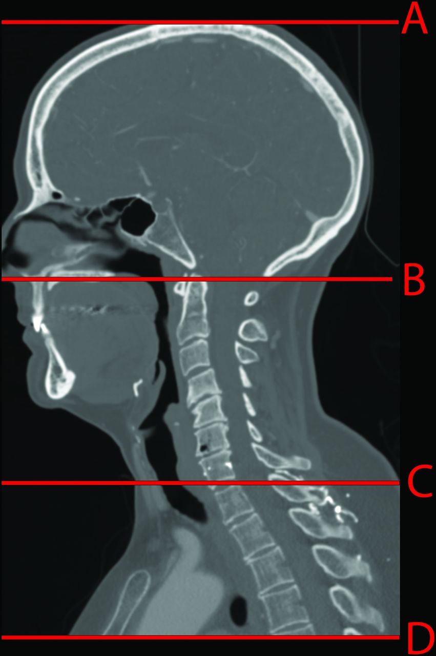
The body regions were determined by using anatomic landmarks. The head region was counted from the skull vertex to the top of the C1 ring; the neck region was taken from the top of the dens to the bottom of the C7 vertebra; and the chest region, from the bottom of C7 to the lowest level scanned (Figure). The start and end locations for each body region were recorded on our institutional PACS system (Impax 5.3; Agfa-Gevaert, Mortsel, Belgium) as the section location coordinates marking the relevant body region boundaries.

Figure.
  • Download figure
  • Open in new tab
  • Download powerpoint
Figure.

Template for the anatomic landmarks used to determine the head, neck, and chest regions. The red horizontal lines on the sagittal reformat demonstrate the location of the 4 landmarks used: the skull vertex (A), the top of the C1 arch (B), the bottom of the C7 vertebra (C), and the most inferior section scanned (D). The head region was defined as between A and B; the neck region, between B and C; and the chest region, between C and D.

Descriptive and comparative statistics and significance testing (unpaired Student t test) were performed by using Excel 2010 (Microsoft).

Results

Of the 302 patients identified in the study period, there were 170 men (mean age, 65 years; median age, 66 years; range, 19–90 years) and 132 women (mean age, 69 years; median age, 73 years; range, 23–100 years). Weight measurements were available in 267 patients (88%, 267/302) with a mean of 82 ± 19 kg. There were 111 patients weighing 60–80 kg (42%, 111/267), with a mean of 71 ± 6 kg. Data for these patients were used for estimation of effective dose contributions.

In all patients, the average NIHSS score at the time of presentation to the emergency department was 7.7 (range, 0–26). Ninety-three patients (31%, 93/302) were eligible for IV-tPA. Of these, 63% (59/93) received IV-tPA; 37% did not because they fell outside the time window for treatment (<4.5 hours), because their lesion was too large (more than one-third of the MCA territory), or there were medical contraindications such as recent trauma, systemic bleeding, or surgery; 6.6% of patients (20/302) proceeded to intra-arterial therapy. The mean NIHSS score in the intra-arterially treated group was 12.4, which was significantly higher than that in the remaining patients who had an average scale value of 7.3 (P < .01).

In the head portion of the CTA, 53% of patients (161/302) had significant vascular-related pathology. In 78% of these patients (129/161), the reported findings were directly relevant to the patient's ischemic stroke symptoms, with vessel cutoff in the territory of the patient's infarct.

In the neck portion of the CTA, relevant vascular pathology was found in 31% of patients (94/302). Of these, 64% (60/94) had at least moderate internal carotid artery stenosis, 30% (28/94) had significant vertebral artery stenosis, 6% (6/94) had both vertebral and internal carotid arterial stenosis, and 4% (4/94) had internal carotid or vertebral artery dissections.

In the upper chest, relevant vascular pathology was found in 1.3% of patients (4/302); this consisted of extensive aortic plaque associated with moderate or severe stenosis of a common carotid origin in all 4 cases.

The subset of 111 average-sized patients, for whom the body region contributions to the total effective dose were estimated, demonstrated a 14 ± 2%, 33 ± 5%, and 53 ± 6% contribution to the total effective dose from the head, neck, and chest regions, respectively.

Discussion

We have found that a very small minority of findings on neurovascular CTA, performed expressly for the purposes of triage of patients with acute stroke symptoms, are in the chest. While some neurointerventional experts argue for the routine inclusion of the upper chest in an emergency department CTA, it is unclear how often 3D road maps from the scan are actually available before intra-arterial intervention. Increasing public awareness of potential radiation risks from medical x-rays has resulted in renewed scrutiny of the radiation dose burden from CT scanning.21 We found that approximately half of the effective dose from arch-to-vertex CTA is due to the chest portion of the scan because of the high radiosensitivity of the lungs.22 On the basis of our observations, the chest portion of the CTA does not appear to contribute meaningfully to the management or triage of patients with acute stroke; thus, an approximately 50% reduction in effective dose could be achieved simply by limiting CTA to the vasculature above the shoulders, without compromising patient care.

The present study is limited by its retrospective nature because only the radiology report was used to determine the presence of relevant findings in the 3 regions of the CTA. In the acute setting, interpretation of the images by a neuroradiologist, as they appear on the scanner console, happens in real- or near-real-time, meeting the recommendations of the American Heart Association that stipulate interpretation of imaging studies within 45 minutes of patient arrival in the emergency department.1 Discussion among neuroradiology, neurointerventional, and neurology personnel at that time will likely have a substantial impact on the chosen management pathway, yet it may not have been fully represented in the final radiology report and thus may have been unavailable for the present analysis. Thus, we may under-represent the actual findings used at the time of treatment. Addressing this issue and the issues of road-mapping before intra-arterial therapy and the management value of the intrathoracic component of the CTA would require prospective trials with appropriate power to answer these questions definitively. Another potential limitation is how well our study cohort represents the larger population of patients with acute stroke. Given the low number of relevant positive findings in the chest in 302 patients, we believe it is unlikely that substantially different results would be found in a larger study group.

Conclusions

In summary, we found that CT angiography of the head, neck, and upper chest performed in the emergency department, specifically to manage patients with acute stroke symptoms, provided diagnostically useful, potentially management-altering information only from the head and neck components of the scan. No information relevant to acute management of such patients was forthcoming from the chest portion of the CTA, yet this component accounted for approximately 50% of the effective dose of the CTA scan. Further evidence-based studies are needed to more fully evaluate these findings.

Footnotes

  • Disclosures: Joshua A. Hirsch—UNRELATED: Consultancy: CareFusion, Comments: Received fees related to vertebral augmentation; Stock/Stock Options: stroke development company.

REFERENCES

  1. 1.↵
    1. Jauch EC,
    2. Saver JL,
    3. Adams HP, et al
    . Guidelines for the early management of adults with ischemic stroke: a guideline for healthcare professionals from the American Heart Association/American Stroke Association. Stroke 2013;44:870–947
    Abstract/FREE Full Text
  2. 2.↵
    1. Del Zoppo GJ,
    2. Saver JL,
    3. Jauch EC, et al
    . Expansion of the time window for treatment of acute ischemic stroke with intravenous tissue plasminogen activator: a science advisory from the American Heart Association/American Stroke Association. Stroke 2009;40:2945–48
    FREE Full Text
  3. 3.↵
    1. Wechsler LR
    . Intravenous thrombolytic therapy for acute ischemic stroke. N Engl J Med 2011;364:2138–46
    CrossRefPubMed
  4. 4.↵
    1. Wardlaw JM,
    2. Murray V,
    3. Berge E, et al
    . Recombinant tissue plasminogen activator for acute ischaemic stroke: an updated systematic review and meta-analysis. Lancet 2012;379:2364–72
    CrossRefPubMed
  5. 5.↵
    1. Adair G,
    2. Grant J,
    3. Pandhi N, et al
    . Clinical inquires: when should patients with stroke receive thrombolytics? J Fam Pract 2004;53:656, 658–59
    PubMed
  6. 6.↵
    1. Hacke W,
    2. Kaste M,
    3. Bluhmki E, et al
    . Thrombolysis with alteplase 3 to 4.5 hours after acute ischemic stroke. N Engl J Med 2008;359:1317–29
    CrossRefPubMed
  7. 7.↵
    1. Morris JG,
    2. Duffis EJ,
    3. Fisher M
    . Cardiac workup of ischemic stroke: can we improve our diagnostic yield? Stroke 2009;40:2893–98
    Abstract/FREE Full Text
  8. 8.↵
    1. Chung JH,
    2. Mitsumori LM,
    3. Ordovas KG, et al
    . Heart as a source of stroke: imaging evaluation with computed tomography. J Thorac Imaging 2012;27:W52–60
    CrossRefPubMed
  9. 9.↵
    1. Boussel L,
    2. Cakmak S,
    3. Wintermark M, et al
    . Ischemic stroke: etiologic work-up with multidetector CT of the heart and extra- and intracranial arteries. Radiology 2011;258:206–12
    CrossRefPubMed
  10. 10.↵
    1. Shrier DA,
    2. Tanaka H,
    3. Numaguchi Y, et al
    . CT angiography in the evaluation of acute stroke. AJNR Am J Neuroradiol 1997;18:1011–20
    Abstract
  11. 11.↵
    1. Knauth M,
    2. von Kummer R,
    3. Jansen O, et al
    . Potential of CT angiography in acute ischemic stroke. AJNR Am J Neuroradiol 1997;18:1001–10
    Abstract
  12. 12.↵
    1. Hunter GJ,
    2. Hamberg LM,
    3. Ponzo JA, et al
    . Assessment of cerebral perfusion and arterial anatomy in hyperacute stroke with three-dimensional functional CT: early clinical results. AJNR Am J Neuroradiol 1998;19:29–37
    Abstract
  13. 13.↵
    1. Smith WS,
    2. Roberts HC,
    3. Chuang NA, et al
    . Safety and feasibility of a CT protocol for acute stroke: combined CT, CT angiography, and CT perfusion imaging in 53 consecutive patients. AJNR Am J Neuroradiol 2003;24:688–90
    Abstract/FREE Full Text
  14. 14.↵
    1. Latchaw RE,
    2. Alberts MJ,
    3. Lev MH, et al
    . Recommendations for imaging of acute ischemic stroke: a scientific statement from the American Heart Association. Stroke 2009;40:3646–78
    FREE Full Text
  15. 15.↵
    1. Leiva-Salinas C,
    2. Wintermark M
    . Imaging of acute ischemic stroke. Neuroimaging Clin N Am 2010;20:455–68
    CrossRefPubMed
  16. 16.↵
    1. Gentile NT,
    2. Cernetich J,
    3. Kanamalla US, et al
    . Expedited computed tomography perfusion and angiography in acute ischemic stroke: a feasibility study. J Emerg Med 2012;43:308–15
    CrossRefPubMed
  17. 17.↵
    1. Kidwell CS,
    2. Wintermark M
    . The role of CT and MRI in the emergency evaluation of persons with suspected stroke. Curr Neurol Neurosci Rep 2010;10:21–28
    CrossRefPubMed
  18. 18.↵
    1. Chang YJ,
    2. Golby AJ,
    3. Albers GW
    . Detection of carotid stenosis: from NASCET results to clinical practice. Stroke 1995;26:1325–28
    Abstract/FREE Full Text
  19. 19.↵
    1. Jones DG,
    2. Shrimpton PC
    . Survey of CT Practice in the UK. Part 3: Normalized Organ Doses Calculated using Monte Carlo Techniques. National Radiological Protection Board-250. London: Her Majesty's Stationery Office, 1991
  20. 20.↵
    1. Shrimpton PC,
    2. Jones DG,
    3. Hillier MC, et al
    . Survey of CT Practice in the UK. Part 2: Dosimetric Aspects. National Radiological Protection Board-R249. London: Her Majesty's Stationery Office; 1991
  21. 21.↵
    1. Boone JM,
    2. Hendee WR,
    3. McNitt-Gray MF, et al
    . Radiation exposure from CT scans: how to close our knowledge gaps, monitor and safeguard exposure: proceedings and recommendations of the Radiation Dose Summit, sponsored by National Radiological Protection Board, February 24–25, 2011. Radiology 2012;265:544–54
    CrossRefPubMed
  22. 22.↵
    International Commission on Radiological Protection. The 2007 Recommendations of the International Commission on Radiological Protection. Stockholm: International Commission on Radiological Protection. ICRP Publication 103, Ann. ICRP 37 (2–4), 2007
  • Received April 23, 2014.
  • Accepted after revision August 5, 2014.
  • © 2015 by American Journal of Neuroradiology
View Abstract
PreviousNext
Back to top

In this issue

American Journal of Neuroradiology: 36 (2)
American Journal of Neuroradiology
Vol. 36, Issue 2
1 Feb 2015
  • Table of Contents
  • Index by author
  • Complete Issue (PDF)
Advertisement
Print
Download PDF
Email Article

Thank you for your interest in spreading the word on American Journal of Neuroradiology.

NOTE: We only request your email address so that the person you are recommending the page to knows that you wanted them to see it, and that it is not junk mail. We do not capture any email address.

Enter multiple addresses on separate lines or separate them with commas.
Diagnostic Yield of Emergency Department Arch-to-Vertex CT Angiography in Patients with Suspected Acute Stroke
(Your Name) has sent you a message from American Journal of Neuroradiology
(Your Name) thought you would like to see the American Journal of Neuroradiology web site.
CAPTCHA
This question is for testing whether or not you are a human visitor and to prevent automated spam submissions.
Cite this article
A.R. Deipolyi, L.M. Hamberg, R.G. Gonzaléz, J.A. Hirsch, G.J. Hunter
Diagnostic Yield of Emergency Department Arch-to-Vertex CT Angiography in Patients with Suspected Acute Stroke
American Journal of Neuroradiology Feb 2015, 36 (2) 265-268; DOI: 10.3174/ajnr.A4112

Citation Manager Formats

  • BibTeX
  • Bookends
  • EasyBib
  • EndNote (tagged)
  • EndNote 8 (xml)
  • Medlars
  • Mendeley
  • Papers
  • RefWorks Tagged
  • Ref Manager
  • RIS
  • Zotero
0 Responses
Respond to this article
Share
Bookmark this article
Diagnostic Yield of Emergency Department Arch-to-Vertex CT Angiography in Patients with Suspected Acute Stroke
A.R. Deipolyi, L.M. Hamberg, R.G. Gonzaléz, J.A. Hirsch, G.J. Hunter
American Journal of Neuroradiology Feb 2015, 36 (2) 265-268; DOI: 10.3174/ajnr.A4112
del.icio.us logo Twitter logo Facebook logo Mendeley logo
  • Tweet Widget
  • Facebook Like
  • Google Plus One
Purchase

Jump to section

  • Article
    • Abstract
    • Materials and Methods
    • Results
    • Discussion
    • Conclusions
    • Footnotes
    • REFERENCES
  • Figures & Data
  • Info & Metrics
  • Responses
  • References
  • PDF

Related Articles

  • No related articles found.
  • PubMed
  • Google Scholar

Cited By...

  • Site and Rate of Occlusive Disease in Cervicocerebral Arteries: A CT Angiography Study of 2209 Patients with Acute Ischemic Stroke
  • Initial hospital management of patients with emergent large vessel occlusion (ELVO): report of the standards and guidelines committee of the Society of NeuroInterventional Surgery
  • Crossref (16)
  • Google Scholar

This article has been cited by the following articles in journals that are participating in Crossref Cited-by Linking.

  • Initial hospital management of patients with emergent large vessel occlusion (ELVO): report of the standards and guidelines committee of the Society of NeuroInterventional Surgery
    Ryan A McTaggart, Sameer A Ansari, Mayank Goyal, Todd A Abruzzo, Barb Albani, Adam J Arthur, Michael J Alexander, Felipe C Albuquerque, Blaise Baxter, Ketan R Bulsara, Michael Chen, Josser E Delgado Almandoz, Justin F Fraser, Donald Frei, Chirag D Gandhi, Don V Heck, Steven W Hetts, M Shazam Hussain, Michael Kelly, Richard Klucznik, Seon-Kyu Lee, Thabele Leslie-Mawzi, Philip M Meyers, Charles J Prestigiacomo, G Lee Pride, Athos Patsalides, Robert M Starke, Peter Sunenshine, Peter A Rasmussen, Mahesh V Jayaraman
    Journal of NeuroInterventional Surgery 2017 9 3
  • Detecting Large Vessel Occlusion at Multiphase CT Angiography by Using a Deep Convolutional Neural Network
    Matthew T. Stib, Justin Vasquez, Mary P. Dong, Yun Ho Kim, Sumera S. Subzwari, Harold J. Triedman, Amy Wang, Hsin-Lei Charlene Wang, Anthony D. Yao, Mahesh Jayaraman, Jerrold L. Boxerman, Carsten Eickhoff, Ugur Cetintemel, Grayson L. Baird, Ryan A. McTaggart
    Radiology 2020 297 3
  • Detection of emergent large vessel occlusion stroke with CT angiography is high across all levels of radiology training and grayscale viewing methods
    Chelsea A. Boyd, Mahesh V. Jayaraman, Grayson L. Baird, William S. Einhorn, Matthew T. Stib, Michael K. Atalay, Jerrold L. Boxerman, Ana P. Lourenco, Gaurav Jindal, Douglas T. Hidlay, Eleanor L. DiBiasio, Ryan A. McTaggart
    European Radiology 2020 30 8
  • Leaving No Large Vessel Occlusion Stroke Behind
    Ryan A. McTaggart, Jessalyn K. Holodinsky, Johanna M. Ospel, Andrew K. Cheung, Nathan W. Manning, Jason D. Wenderoth, Thanh G. Phan, Richard Beare, Kendall Lane, Richard A. Haas, Noreen Kamal, Mayank Goyal, Mahesh V. Jayaraman
    Stroke 2020 51 7
  • Site and Rate of Occlusive Disease in Cervicocerebral Arteries: A CT Angiography Study of 2209 Patients with Acute Ischemic Stroke
    D.C. Rotzinger, P.J. Mosimann, R.A. Meuli, P. Maeder, P. Michel
    American Journal of Neuroradiology 2017 38 5
  • Acute ischemic stroke imaging: a practical approach for diagnosis and triage
    Joseph Yeen Young, Pamela Whitney Schaefer
    The International Journal of Cardiovascular Imaging 2016 32 1
  • Interrater variability for CT angiography evaluation between neurologists and neuroradiologist in acute stroke patients
    Michal Bar, Jiri Kral, Tomas Jonszta, Vaclav Marcian, Martin Kuliha, Robert Mikulik
    The British Journal of Radiology 2017 90 1071
  • Radiology workload in clinical implementation of thrombectomy for acute ischemic stroke: experience from The Netherlands
    Bram A. C. M. Fasen, Roeland J. J. Heijboer, Frans-Jan H. Hulsmans, Robert M. Kwee
    Neuroradiology 2020 62 7
  • Unresolved Issues in Thrombectomy
    Mahesh V. Jayaraman, Ryan A. McTaggart, Mayank Goyal
    Current Neurology and Neuroscience Reports 2017 17 9
  • CT cervico-cerebral angiography in acute stroke. Can we justify aortic arch imaging?
    Gavin Sugrue, Michael K. O’Reilly, Danielle Byrne, Matthew T. Crockett, Sean Murphy, Eoin C. Kavanagh
    Irish Journal of Medical Science (1971 -) 2019 188 2

More in this TOC Section

  • Optimal MRI Sequence for Identifying Occlusion Location in Acute Stroke: Which Value of Time-Resolved Contrast-Enhanced MRA?
  • SWI or T2*: Which MRI Sequence to Use in the Detection of Cerebral Microbleeds? The Karolinska Imaging Dementia Study
  • Progression of Microstructural Damage in Spinocerebellar Ataxia Type 2: A Longitudinal DTI Study
Show more Brain

Similar Articles

Advertisement

Indexed Content

  • Current Issue
  • Accepted Manuscripts
  • Article Preview
  • Past Issues
  • Editorials
  • Editors Choice
  • Fellow Journal Club
  • Letters to the Editor

Cases

  • Case Collection
  • Archive - Case of the Week
  • Archive - Case of the Month
  • Archive - Classic Case

Special Collections

  • Special Collections

Resources

  • News and Updates
  • Turn around Times
  • Submit a Manuscript
  • Author Policies
  • Manuscript Submission Guidelines
  • Evidence-Based Medicine Level Guide
  • Publishing Checklists
  • Graphical Abstract Preparation
  • Imaging Protocol Submission
  • Submit a Case
  • Become a Reviewer/Academy of Reviewers
  • Get Peer Review Credit from Publons

Multimedia

  • AJNR Podcast
  • AJNR SCANtastic
  • Video Articles

About Us

  • About AJNR
  • Editorial Board
  • Not an AJNR Subscriber? Join Now
  • Alerts
  • Feedback
  • Advertise with us
  • Librarian Resources
  • Permissions
  • Terms and Conditions

American Society of Neuroradiology

  • Not an ASNR Member? Join Now

© 2025 by the American Society of Neuroradiology All rights, including for text and data mining, AI training, and similar technologies, are reserved.
Print ISSN: 0195-6108 Online ISSN: 1936-959X

Powered by HighWire