
Article Figures & Data
Figures
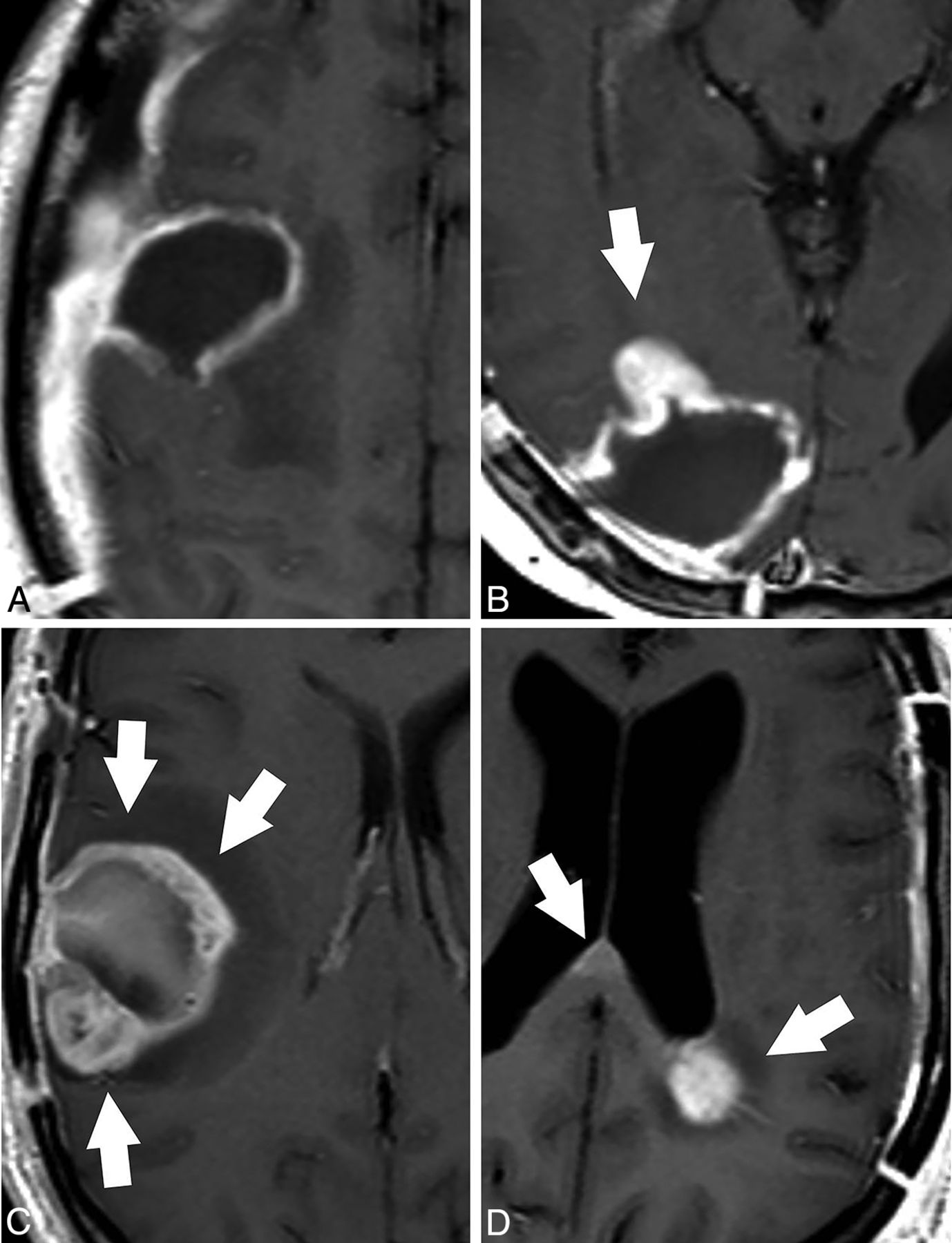
- Fig 1.
Patterns of tumor growth in the PRMR examination in patients with glioblastoma. Axial contrast-enhanced T1-weighted images. A, No growth. The MR examination shows a thin homogeneous enhancement in the wall of the surgical cavity that is considered normal evolution after surgery. B, Focal growth. A focal-enhancing nodule is found at the anterior margin of the surgical cavity in this study (arrow). C, Global growth. Thick irregular enhancement in the margins of the surgical cavity involving more than half of the surgical cavity (arrows). Note the presence of some hyperintense postsurgical material in the surgical cavity. D, Distant growth. Focal contrast enhancement is found in the juxtaventricular parietal lobe and in the splenium of the corpus callosum (arrows), distant to the surgical cavity (not shown), after resection of a left temporal lobe glioblastoma.
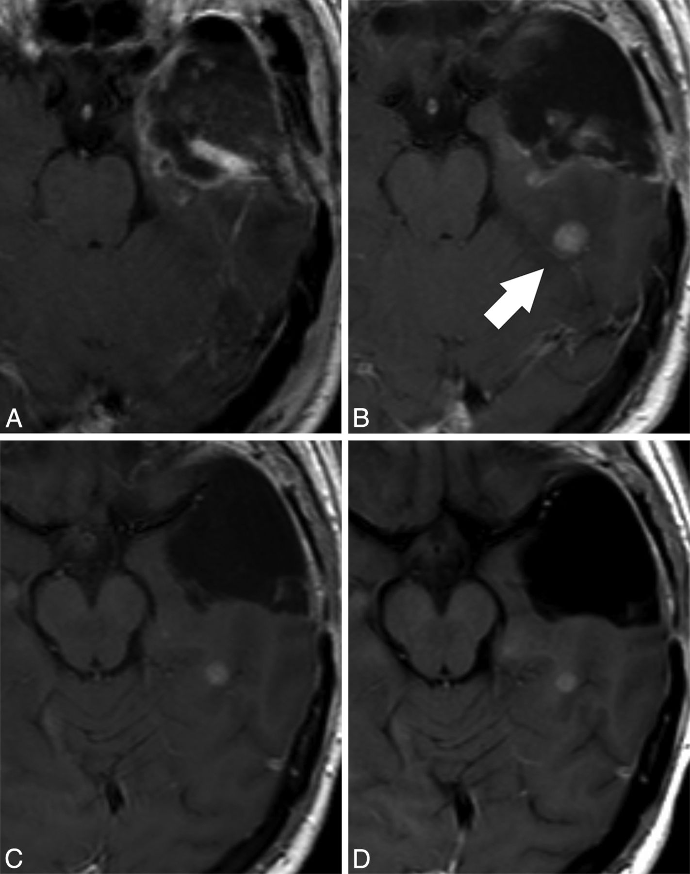
- Fig 2.
Representative case in which the response assessment is progressive disease (pseudoprogression) in reference to the EPMR examination and stable disease in reference to the PRMR examination. Contrast-enhanced T1-weighted images performed at A, 24 hours after surgery (EPMR), B, 3 days before radiation therapy (PRMR), C, 27 days after radiation therapy, and D, 135 days after radiation therapy. A contrast-enhancing nodule appears in the PRMR (arrow, B), distant to the surgical cavity. As a relevant consequence, the radiation therapy field design was modified to include the nodule in the target volume. The postradiation examination (C) shows a small decrease in the size of the nodule, which remains stable in an MR examination performed 4 months after radiation therapy (D). In this particular case, the PRMR provides evidence that the nodule appeared before treatment and remained stable after the treatment. The response assessment would be progressive disease compared with the EPMR (pseudoprogression because the nodule remained stable in the next follow-up MR examination performed 4 months after radiation therapy) and stable disease compared with the PRMR.
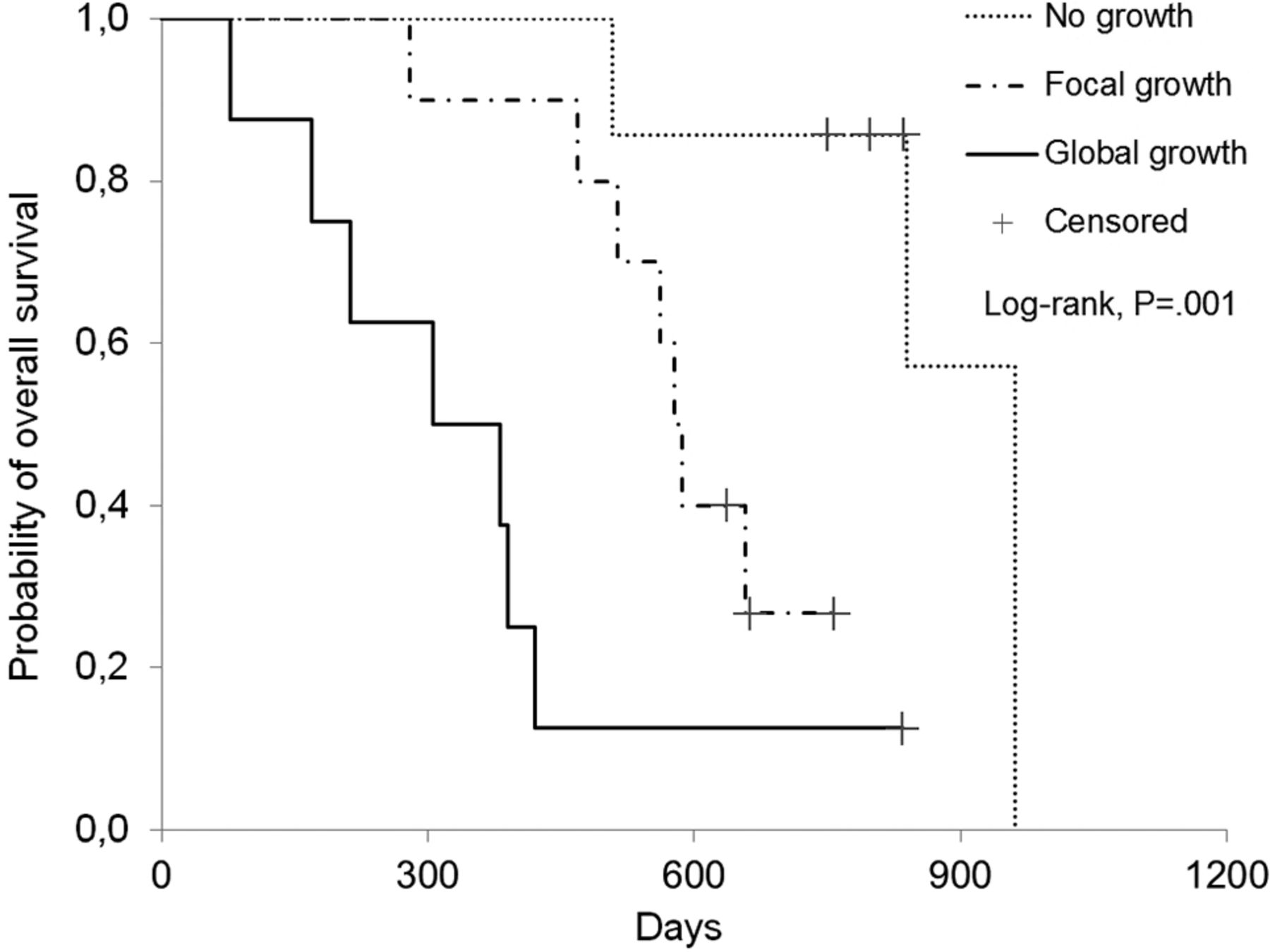
- Fig 3.
Survival curves of patients stratified according to the pattern of tumor growth in the PRMR. Patients with “no growth” in the PRMR showed significantly improved survival (median survival, 837 days) compared with patients with “focal growth” (median survival, 582 days) and “global growth” (median survival, 344 days; P = .001).


Tables
Characteristics Age (yr) (mean [range]) 57 [26–73] Sex (No. of patients) Men 19 Women 9 Karnofsky Performance Status (No. of patients) 70–80 9 90–100 19 Time between surgery and EPMR (No. of patients) <24 h 2 24–48 h 15 48–72 h 11 Time between surgery and radiotherapy (d) (mean [range]) 39 [27–64] Time between PRMR and radiotherapy (No. of patients) 1 d 8 2–3 d 9 4–5 d 9 6–7 d 2 Time between radiotherapy and follow-up with MRI (d) (mean [range]) 28 [16–39] - Table 2:
Concordance among tumor response assessment performed 2–6 weeks after completion of radiotherapy with both EPMR and PRMR as baseline examsa
PRMR as Baseline EPMR as Baseline True Progressionb Pseudoprogressionb Stable Disease Partial Response Total True Progressionb 9 0 0 0 9 Pseudoprogressionb 0 3 0 0 3 Stable Disease 0 3 8 1 12 Partial Response 0 1 1 2 4 Total 9 7 9 3 28 - Table 3:
Overall survival of patients stratified by the extent of resection evaluated on the EPMR and by the pattern of tumor growth on the PRMR
Characteristic Patients Survival (d) (median; range) Hazard Ratio (95% CI) P Value Total Alive Extent of resection evaluated on the EPMR .002 Total 7 4 835; 169–961 0.099 (0.022–0.445) Between 95% and 100% 11 4 637; 213–1115 0.188 (0.060–0.591) Below 95% 10 0 386; 78–577 1b Pattern of growth on the PRMR .001 No growth 7 4 837; 508–961 0.047 (0.006–0.393) Focal 10 3 582; 279–758 0.294 (0.098–0.885) Global 8 1 344; 78–835 1b Distanta 3 0 334; 286–1115 NE


Jump to section
Related Articles
Cited By...
- No citing articles found.