Advertisement

Main menu

  • Home
  • Content
    • Current Issue
    • Accepted Manuscripts
    • Article Preview
    • Past Issue Archive
    • AJNR Case Collection
    • Case of the Week Archive
    • Classic Case Archive
    • Case of the Month Archive
  • Special Collections
    • Spinal CSF Leak Articles (Jan 2020-June 2024)
    • 2024 AJNR Journal Awards
    • Most Impactful AJNR Articles
  • Multimedia
    • AJNR Podcast
    • AJNR Scantastics
    • Video Articles
  • For Authors
    • Submit a Manuscript
    • Author Policies
    • Fast publishing of Accepted Manuscripts
    • Graphical Abstract Preparation
    • Manuscript Submission Guidelines
    • Imaging Protocol Submission
    • Submit a Case for the Case Collection
  • About Us
    • About AJNR
    • Editorial Board
  • More
    • Become a Reviewer/Academy of Reviewers
    • Subscribers
    • Permissions
    • Alerts
    • Feedback
    • Advertisers
    • ASNR Home
  • Other Publications
    • ajnr

User menu

  • Alerts
  • Log in

Search

  • Advanced search
American Journal of Neuroradiology

American Journal of Neuroradiology

ASHNR American Society of Functional Neuroradiology ASHNR American Society of Pediatric Neuroradiology ASSR
  • Alerts
  • Log in

Advanced Search

  • Home
  • Content
    • Current Issue
    • Accepted Manuscripts
    • Article Preview
    • Past Issue Archive
    • AJNR Case Collection
    • Case of the Week Archive
    • Classic Case Archive
    • Case of the Month Archive
  • Special Collections
    • Spinal CSF Leak Articles (Jan 2020-June 2024)
    • 2024 AJNR Journal Awards
    • Most Impactful AJNR Articles
  • Multimedia
    • AJNR Podcast
    • AJNR Scantastics
    • Video Articles
  • For Authors
    • Submit a Manuscript
    • Author Policies
    • Fast publishing of Accepted Manuscripts
    • Graphical Abstract Preparation
    • Manuscript Submission Guidelines
    • Imaging Protocol Submission
    • Submit a Case for the Case Collection
  • About Us
    • About AJNR
    • Editorial Board
  • More
    • Become a Reviewer/Academy of Reviewers
    • Subscribers
    • Permissions
    • Alerts
    • Feedback
    • Advertisers
    • ASNR Home
  • Follow AJNR on Twitter
  • Visit AJNR on Facebook
  • Follow AJNR on Instagram
  • Join AJNR on LinkedIn
  • RSS Feeds

Welcome to the new AJNR, Updated Hall of Fame, and more. Read the full announcements.


AJNR is seeking candidates for the position of Associate Section Editor, AJNR Case Collection. Read the full announcement.


Research ArticleSpine

Transforaminal Insertion of a Thermocouple on the Posterior Vertebral Wall Combined with Hydrodissection during Lumbar Spinal Radiofrequency Ablation

R. Lecigne, J. Garnon, R.L. Cazzato, P. Auloge, D. Dalili, G. Koch and A. Gangi
American Journal of Neuroradiology October 2019, 40 (10) 1786-1790; DOI: https://doi.org/10.3174/ajnr.A6233
R. Lecigne
aFrom the Department of Radiology (R.L.), Pôle d'imagerie, Centre Hospitalier Universitaire, Cedex, France
  • Find this author on Google Scholar
  • Find this author on PubMed
  • Search for this author on this site
  • ORCID record for R. Lecigne
J. Garnon
bDepartment of Interventional Radiology (J.G., R.L.C., P.A., G.K., A.G.), Hopitaux Universitaires de Strasbourg, Strasbourg, France
  • Find this author on Google Scholar
  • Find this author on PubMed
  • Search for this author on this site
  • ORCID record for J. Garnon
R.L. Cazzato
bDepartment of Interventional Radiology (J.G., R.L.C., P.A., G.K., A.G.), Hopitaux Universitaires de Strasbourg, Strasbourg, France
  • Find this author on Google Scholar
  • Find this author on PubMed
  • Search for this author on this site
  • ORCID record for R.L. Cazzato
P. Auloge
bDepartment of Interventional Radiology (J.G., R.L.C., P.A., G.K., A.G.), Hopitaux Universitaires de Strasbourg, Strasbourg, France
  • Find this author on Google Scholar
  • Find this author on PubMed
  • Search for this author on this site
  • ORCID record for P. Auloge
D. Dalili
cDepartment of Radiology and Radiological Science (D.D.), Johns Hopkins University School of Medicine, Baltimore, Maryland.
  • Find this author on Google Scholar
  • Find this author on PubMed
  • Search for this author on this site
  • ORCID record for D. Dalili
G. Koch
bDepartment of Interventional Radiology (J.G., R.L.C., P.A., G.K., A.G.), Hopitaux Universitaires de Strasbourg, Strasbourg, France
  • Find this author on Google Scholar
  • Find this author on PubMed
  • Search for this author on this site
  • ORCID record for G. Koch
A. Gangi
bDepartment of Interventional Radiology (J.G., R.L.C., P.A., G.K., A.G.), Hopitaux Universitaires de Strasbourg, Strasbourg, France
  • Find this author on Google Scholar
  • Find this author on PubMed
  • Search for this author on this site
  • ORCID record for A. Gangi
  • Article
  • Figures & Data
  • Info & Metrics
  • Responses
  • References
  • PDF
Loading

Abstract

SUMMARY: The purpose of the present article is to describe the technique of transforaminal insertion of an ultrathin thermosensor in the anterior epidural space in 13 patients treated by radiofrequency ablation. The mean time taken to position the thermosensor was 10.6 minutes (range, 5–38 minutes). Technical success was 93% (correct positioning in 13/14 levels). Additional hydrodissection was performed through the same access in 11 cases. No postoperative neural deficit was elicited in any of the cases.

ABBREVIATION:

RFA
radiofrequency ablation

Inadvertent thermal ablation of neural structures located in the lumbar canal is a rare but potentially debilitating complication following percutaneous thermal ablation of lesions located in the vertebral body, if the expected ablation zone abuts the posterior wall of the vertebra.1⇓–3 Using a thermosensor, one can monitor local temperatures to ensure that threshold temperatures are not breached in a critical area.4,5 However, precise positioning of the thermometer at an ideal interface between the lumbar spine and the dural sac can be challenging. We herein aim to describe the technique of transforaminal insertion of an ultrathin thermocouple into the anterior epidural space while performing radiofrequency ablation of spinal metastases to monitor the temperature at the interface between the posterior part of the vertebral body and the dura.

MATERIALS AND METHODS

This is a single-center retrospective study. All patients gave informed consent for the procedure. Institutional review board approval was waived.

Study Population and Procedures

Between April 2015 and August 2018, thirteen patients (7 women, 6 men; mean age, 60.8 years) with 14 lumbar tumors were treated in our institution using radiofrequency ablation (RFA) and associated planned local temperature monitoring using the transforaminal approach. Patient, lesion, and RFA characteristics are listed in Table 1.

View this table:
  • View inline
  • View popup
Table 1:

Patients, lesions, and RFA characteristics

All procedures were performed with the patient under general anesthesia in the prone position, with either conebeam CT guidance (n = 7 patients) or combined CT and fluoroscopic guidance (n = 1), depending on room availability at the time of treatment. Thermal ablation was conducted according to the manufacturer's recommendations. If the temperature exceeded 45° and was not controllable with active hydrodissection, RFA was stopped. After completion of the ablation, additional cementoplasty was performed to ensure bone consolidation and reduce the risk of secondary postablation fracture.

Technique of the Double-Oblique Transforaminal Approach

Optimal insertion of the thermometer requires a double-oblique approach; the ideal trajectory is from cranial to caudal and from lateral to medial (Figs 1 and 2). First, an 18-ga spinal needle is advanced in the very medial part of the foramen. Subsequently, a 20-cm-long, 28-ga reusable monopolar nitinol radiofrequency electrode (MultGen; Stryker, Kalamazoo, Michigan) is inserted coaxially through the spinal needle and gently advanced. Although little resistance can be felt, the electrode should be advanced until it reaches the posterior vertebral wall/posterior part of the tumor. The electrode is used only for its capacity to display the temperature (and is therefore referred to as a thermosensor). The various steps of the double-oblique transforaminal approach are presented in Fig 3.

Fig 1.
  • Download figure
  • Open in new tab
  • Download powerpoint
Fig 1.

Double-oblique transforaminal approach on fluoroscopy (patient 12). Anterior-posterior (A) and lateral (B) fluoroscopic projections demonstrate the tip of the thermosensor (white arrow) just posterior to the vertebral body, at its mid portion (B), and in the midline (A), thanks to an oblique approach in both anterior-posterior and lateral views.

Fig 2.
  • Download figure
  • Open in new tab
  • Download powerpoint
Fig 2.

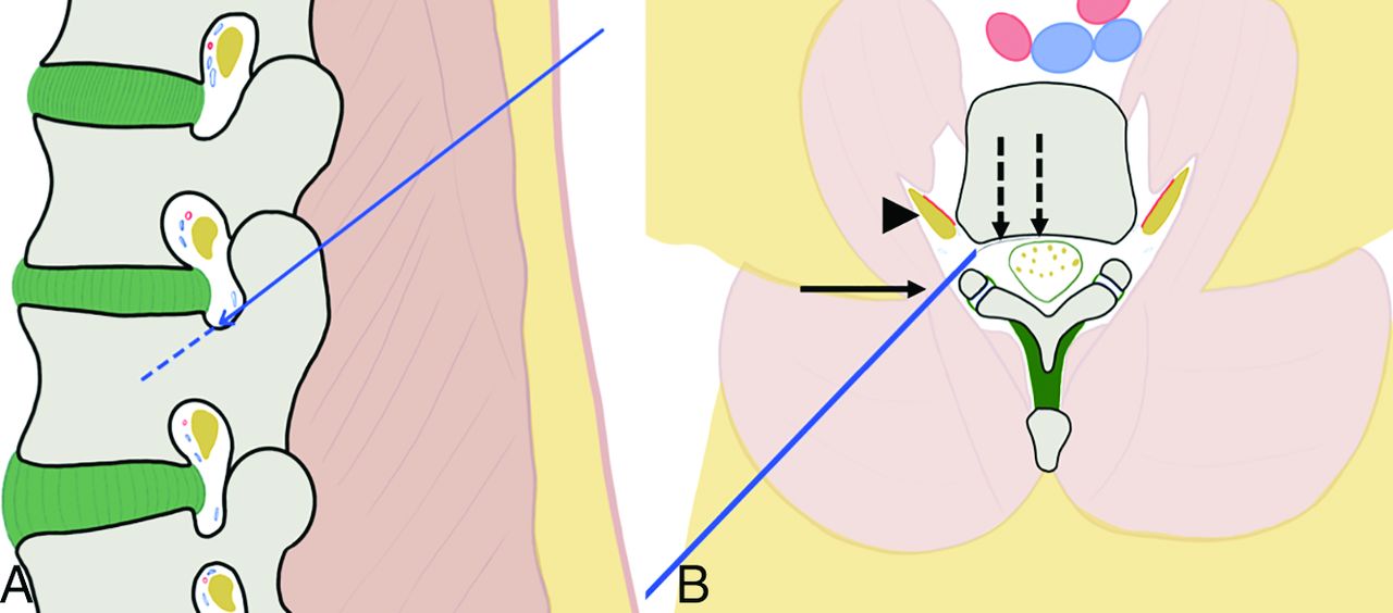
Representation and principle of the double-oblique approach. A, Drawing from a sagittal perspective. The craniocaudal approach in the sagittal plane enables going through the posterior and inferior parts of the foramen, away from the radicular nerve and vessels. B, Drawing from an oblique axial view (in the axis of the sagittal angulation). The lateromedial approach allows slipping along the facet joint with the 18-ga needle (arrow), away from the nerve and vessels (arrowhead). The 28-ga thermosensor (dotted arrows) can then be advanced into the anterior epidural space toward the posterior wall of the vertebral body.

Fig 3.
  • Download figure
  • Open in new tab
  • Download powerpoint
Fig 3.

Fluoroscopic details of the technique of the transforaminal approach. A, The craniocaudal angulation in the sagittal plane is estimated on the lateral projection to point the 18-ga needle (white arrow) used as a landmark on the skin of the patient toward the posterior and inferior parts of the foramen. B, The 35° oblique view (from the anterior-posterior view) is then used to define the distance of the entry point laterally. The 18-ga spinal needle (arrow) should be pointed toward the lateral part of the facet joint (dotted line). C, The needle is advanced in the oblique view toward the foramen. D, Once in the vicinity of the foramen, the lateral view confirms that the needle enters it at its posterior and inferior parts. E, Satisfactory localization of the needle tip (arrow) inside the foramen is confirmed on anterior-posterior projection. F, The 28-ga thermosensor (dotted arrow) is gently advanced into the canal until it reaches the midline (G), where resistance is felt. H, At this point, the tip of the thermometer (dotted arrow) should be located at the middle portion of the vertebral body on the lateral view.

Additionally, hydrodissection using dextrose mixed with contrast (50:50 ratio for optimal visualization) can be combined with temperature monitoring. Here, before the insertion of the thermosensor, a Y-valve is connected to the hub of the 18-ga needle, and once the thermosensor has been introduced through the distal port of the valve, fluid may be injected through the lateral port (Fig 4).

Fig 4.
  • Download figure
  • Open in new tab
  • Download powerpoint
Fig 4.

Thermal monitoring combined with hydrodissection. A, Lateral fluoroscopic view demonstrates the 18-ga needle in the foramen (arrow) and the thermosensor in contact with the posterior wall (dotted arrow). B, Conebeam CT acquisition with reconstruction in the axis of the needle and thermometer confirms the findings of fluoroscopy with the 18-ga needle (arrow) and the thermosensor (dotted arrow). C, Lateral view after injection of dextrose mixed with contrast shows satisfactory diffusion of the fluid into the anterior epidural space (white asterisks) separating the dural sac from the vertebral body. D, This is again outlined on the conebeam CT acquisition, which demonstrates the hydrodissection (black asterisk) between the posterior wall and the dural sac.

RESULTS

Detailed results are listed in Table 2. The mean time to optimally position the thermosensor was 10.6 minutes (range, 5–38 minutes). There was a single case requiring >12 minutes for thermosensor insertion (a case requiring conebeam CT acquisition). Optimal positioning was achieved in 13 cases with a technical success rate of 93%. In the case considered unsuccessful, the thermometer was in the epidural space but 4 mm cranial and 5 mm lateral to the optimum, desired end point. Additional hydrodissection was performed through the same access in 11 cases and was effective in all cases except 2 (both cases with epidural extension). The mean maximal temperature displayed by the thermosensor was 42° (range, 39°–45°). None of the cases had premature stoppage of ablation. In a single case (performed with monopolar RFA), continuous irrigation of the anterior epidural space had to be performed to maintain the temperature at 45°. No postoperative neural deficits were encountered. MR imaging was performed in the early postoperative period in all patients within a mean of 13 days (range, 1–30 days) with no evidence of epidural hematoma, and fluid had completely reabsorbed in all patients who had benefited from additional hydrodissection.

View this table:
  • View inline
  • View popup
Table 2:

Results of transforaminal approach

DISCUSSION

As demonstrated in this case series, the transforaminal insertion of a small thermosensor into the anterior epidural space at the midlevel of a vertebral body is technically feasible, can be performed with stand-alone fluoroscopy, and adds little time to the procedure. Double-oblique access is mandatory to access the midlevel of the vertebral body wall because it allows lining up of the thermosensor with the ideal trajectory. Because the thermocouple is very thin, a more direct approach (similar to a discal puncture) cannot achieve such positioning because the thermometer will not “take the curve” along the posterior part of the vertebral body. The craniocaudal approach should always be favored to avoid transgressing the danger zone located at the anterior and superior parts of the foramina, which contains the radicular nerve and adjacent vessels.6 The double-oblique approach allows the safest passage (posterior and inferior) through the foramen.7,8 In the aging spine, the anatomy of the foramen can, however, be modified by a bulging disc and/or osteophytes. Careful examination of the planning CT is therefore recommended before thermosensor insertion.

Although more challenging than a posterior epidural or a straight transforaminal approach, the technique is the only method to access the anterior epidural space at exactly the level of the RFA probes where fluctuations in local temperature are believed to be the most critical because of the shape and extent of the expected ablation zone. Moreover, it can easily be combined with hydrodissection, which allows separation of the dural sac from the ablation area if the tumor does not extend into the anterior epidural fat. The major challenge of the technique is to precisely navigate through the foramen under fluoroscopy with a double-oblique approach. Any deviation from the ideal trajectory might lead to either a suboptimal position or the impossibility of advancing the thermosensor because of bony interposition.

In this series, we had a single case in which active fluid injection was required to maintain the local temperature at a maximum threshold of 45°. This excellent safety profile of ablation might be explained by the preferential use of dedicated bone bipolar RFA devices that are known not to diffuse through an intact osseous cortex and form smaller, precise, and more predictable ablation zones than other ablation techniques (cryoablation, microwave, monopolar RFA).2,9 Nevertheless, local thermal monitoring gives extra confidence and reassurance to the operating physician who can follow the real-time evolution of local temperatures, thereby increasing the safety profile of the procedure, even though the present study is not powered enough to statistically prove it. This technique is particularly useful if the procedure is being performed with the patient under general anesthesia without access to intraprocedural neurophysiologic testing to monitor neural conduction.10 It can theoretically be used in combination with all heat- or cold-based ablation modalities (RFA, microwave, laser, cryoablation) to avoid breaching above 45° or below 0°, which are known to be the neurotoxic threshold temperatures.11 Although favorable and with many potential uses, this technique is unfortunately not transferable to the dorsal spine because of the interposition of the ribs, which preclude a required steeper sagittal approach. The present study is limited by the small cohort of patients, which does not allow definitive conclusions regarding efficacy and safety. Moreover, all the procedures were performed by interventional radiologists trained in the spinal procedure, likely representing a bias in terms of reproducibility.

Footnotes

  • Disclosures: Julien Garnon—UNRELATED: Consultancy: BTG-Galil Medical; Payment for Lectures Including Service on Speakers Bureaus: Canon Medical, Medtronic. Afshin Gangi—UNRELATED: Payment for Lectures Including Service on Speakers Bureaus: Canon Medical; Patents (Planned, Pending or Issued): AprioMed.

References

  1. 1.↵
    1. Khan MA,
    2. Deib G,
    3. Deldar B, et al
    . Efficacy and safety of percutaneous microwave ablation and cementoplasty in the treatment of painful spinal metastases and myeloma. AJNR Am J Neuroradiol 2018;39:1376–83 doi:10.3174/ajnr.A5680 pmid:29794238
    Abstract/FREE Full Text
  2. 2.↵
    1. Wallace AN,
    2. Hillen TJ,
    3. Friedman MV, et al
    . Percutaneous spinal ablation in a sheep model: protective capacity of an intact cortex, correlation of ablation parameters with ablation zone size, and correlation of postablation MRI and pathologic findings. AJNR Am J Neuroradiol 2017;38:1653–59 doi:10.3174/ajnr.A5228 pmid:28572150
    Abstract/FREE Full Text
  3. 3.↵
    1. Westbroek EM,
    2. Goodwin ML,
    3. Hui F, et al
    . Thermal injury to the spinal cord, a rare complication of percutaneous microwave spine tumor ablation: case report. J Clin Neurosci 2019;64:50–54 doi:10.1016/j.jocn.2019.03.018 pmid:30922532
    CrossRefPubMed
  4. 4.↵
    1. Tsoumakidou G,
    2. Buy X,
    3. Garnon J, et al
    . Percutaneous thermal ablation: how to protect the surrounding organs. Tech Vasc Interv Radiol 2011;14:170–76 doi:10.1053/j.tvir.2011.02.009 pmid:21767784
    CrossRefPubMed
  5. 5.↵
    1. Kastler A,
    2. Krainik A,
    3. Sakhri L, et al
    . Feasibility of real-time intraprocedural temperature control during bone metastasis thermal microwave ablation: a bicentric retrospective study. J Vasc Interv Radiol 2017;28:366–71 doi:10.1016/j.jvir.2016.09.030 pmid:28007329
    CrossRefPubMed
  6. 6.↵
    1. Hoshide R,
    2. Feldman E,
    3. Taylor W
    . Cadaveric analysis of the Kambin's triangle. Cureus 2016;8:e475 doi:10.7759/cureus.475 pmid:27004152
    CrossRefPubMed
  7. 7.↵
    1. Mandell JC,
    2. Czuczman GJ,
    3. Gaviola GC, et al
    . The lumbar neural foramen and transforaminal epidural steroid injections: an anatomic review with key safety considerations in planning the percutaneous approach. AJR Am J Roentgenol 2017;209:W26–35 doi:10.2214/AJR.16.17471 pmid:28504548
    CrossRefPubMed
  8. 8.↵
    1. Kennedy DJ,
    2. Dreyfuss P,
    3. Aprill CN, et al
    . Paraplegia following image-guided transforaminal lumbar spine epidural steroid injection: two case reports. Pain Med 2009;10:1389–94 doi:10.1111/j.1526-4637.2009.00728.x pmid:19863744
    CrossRefPubMed
  9. 9.↵
    1. Cazzato RL,
    2. Garnon J,
    3. Caudrelier J, et al
    . Low-power bipolar radiofrequency ablation and vertebral augmentation for the palliative treatment of spinal malignancies. Int J Hyperthermia 2018;34:1282–88 doi:10.1080/02656736.2017.1422557 pmid:29347855
    CrossRefPubMed
  10. 10.↵
    1. Kurup AN,
    2. Schmit GD,
    3. Morris JM, et al
    . Avoiding complications in bone and soft-tissue ablation. Cardiovasc Intervent Radiol 2017;40:166–76 doi:10.1007/s00270-016-1487-y pmid:27826789
    CrossRefPubMed
  11. 11.↵
    1. Tsoumakidou G,
    2. Koch G,
    3. Caudrelier J, et al
    . Image-guided spinal ablation: a review. Cardiovasc Intervent Radiol 2016;39:1229–38 doi:10.1007/s00270-016-1402-6 pmid:27329231
    CrossRefPubMed
  • Received May 31, 2019.
  • Accepted after revision August 1, 2019.
  • © 2019 by American Journal of Neuroradiology
View Abstract
PreviousNext
Back to top

In this issue

American Journal of Neuroradiology: 40 (10)
American Journal of Neuroradiology
Vol. 40, Issue 10
1 Oct 2019
  • Table of Contents
  • Index by author
  • Complete Issue (PDF)
Advertisement
Print
Download PDF
Email Article

Thank you for your interest in spreading the word on American Journal of Neuroradiology.

NOTE: We only request your email address so that the person you are recommending the page to knows that you wanted them to see it, and that it is not junk mail. We do not capture any email address.

Enter multiple addresses on separate lines or separate them with commas.
Transforaminal Insertion of a Thermocouple on the Posterior Vertebral Wall Combined with Hydrodissection during Lumbar Spinal Radiofrequency Ablation
(Your Name) has sent you a message from American Journal of Neuroradiology
(Your Name) thought you would like to see the American Journal of Neuroradiology web site.
CAPTCHA
This question is for testing whether or not you are a human visitor and to prevent automated spam submissions.
Cite this article
R. Lecigne, J. Garnon, R.L. Cazzato, P. Auloge, D. Dalili, G. Koch, A. Gangi
Transforaminal Insertion of a Thermocouple on the Posterior Vertebral Wall Combined with Hydrodissection during Lumbar Spinal Radiofrequency Ablation
American Journal of Neuroradiology Oct 2019, 40 (10) 1786-1790; DOI: 10.3174/ajnr.A6233

Citation Manager Formats

  • BibTeX
  • Bookends
  • EasyBib
  • EndNote (tagged)
  • EndNote 8 (xml)
  • Medlars
  • Mendeley
  • Papers
  • RefWorks Tagged
  • Ref Manager
  • RIS
  • Zotero
0 Responses
Respond to this article
Share
Bookmark this article
Transforaminal Insertion of a Thermocouple on the Posterior Vertebral Wall Combined with Hydrodissection during Lumbar Spinal Radiofrequency Ablation
R. Lecigne, J. Garnon, R.L. Cazzato, P. Auloge, D. Dalili, G. Koch, A. Gangi
American Journal of Neuroradiology Oct 2019, 40 (10) 1786-1790; DOI: 10.3174/ajnr.A6233
del.icio.us logo Twitter logo Facebook logo Mendeley logo
  • Tweet Widget
  • Facebook Like
  • Google Plus One
Purchase

Jump to section

  • Article
    • Abstract
    • ABBREVIATION:
    • MATERIALS AND METHODS
    • RESULTS
    • DISCUSSION
    • Footnotes
    • References
  • Figures & Data
  • Info & Metrics
  • Responses
  • References
  • PDF

Related Articles

  • No related articles found.
  • PubMed
  • Google Scholar

Cited By...

  • No citing articles found.
  • Crossref (3)
  • Google Scholar

This article has been cited by the following articles in journals that are participating in Crossref Cited-by Linking.

  • Temperature Assessment During Radio Frequency Ablation in Ex Vivo Long Bone by Fiber Bragg Grating Sensors
    Francesca De Tommasi, Emiliano Schena, Massimiliano Carassiti, Paul Jutte, Venkatasubramanian Kalpathy Venkiteswaran, Sarthak Misra
    IEEE Sensors Journal 2024 24 4
  • Safety of Thermosensor Insertion in the Midline of the Spinal Canal Anterior to the Dura: A Cadaveric Study
    Julien Garnon, Irène Olivier, Romain Lecigne, Melissa Fesselier, Danoob Dalili, Pierre Auloge, Roberto Luigi Cazzato, Jack Jennings, Guillaume Koch, Afshin Gangi
    CardioVascular and Interventional Radiology 2021 44 12
  • Fiber-Optic Temperature Field Monitoring Based on Deep Learning Network for Tumor Thermal Ablation
    Weikang Liu, Zheyu Zhang, Guofeng Yan
    Journal of Lightwave Technology 2024 42 17

More in this TOC Section

Spine

  • Bern Score Validity for SIH
  • MP2RAGE 7T in MS Lesions of the Cervical Spine
  • Resisted Inspiration for CSF-Venous Fistula
Show more Spine

Interventional

  • SAVE vs. Solumbra Techniques for Thrombectomy
  • CT Perfusion&Reperfusion in Acute Ischemic Stroke
  • Delayed Reperfusion Post-Thrombectomy&Thrombolysis
Show more Interventional

Similar Articles

Advertisement

Indexed Content

  • Current Issue
  • Accepted Manuscripts
  • Article Preview
  • Past Issues
  • Editorials
  • Editors Choice
  • Fellow Journal Club
  • Letters to the Editor

Cases

  • Case Collection
  • Archive - Case of the Week
  • Archive - Case of the Month
  • Archive - Classic Case

Special Collections

  • Special Collections

Resources

  • News and Updates
  • Turn around Times
  • Submit a Manuscript
  • Author Policies
  • Manuscript Submission Guidelines
  • Evidence-Based Medicine Level Guide
  • Publishing Checklists
  • Graphical Abstract Preparation
  • Imaging Protocol Submission
  • Submit a Case
  • Become a Reviewer/Academy of Reviewers
  • Get Peer Review Credit from Publons

Multimedia

  • AJNR Podcast
  • AJNR SCANtastic
  • Video Articles

About Us

  • About AJNR
  • Editorial Board
  • Not an AJNR Subscriber? Join Now
  • Alerts
  • Feedback
  • Advertise with us
  • Librarian Resources
  • Permissions
  • Terms and Conditions

American Society of Neuroradiology

  • Not an ASNR Member? Join Now

© 2025 by the American Society of Neuroradiology All rights, including for text and data mining, AI training, and similar technologies, are reserved.
Print ISSN: 0195-6108 Online ISSN: 1936-959X

Powered by HighWire