
Article Figures & Data
Figures
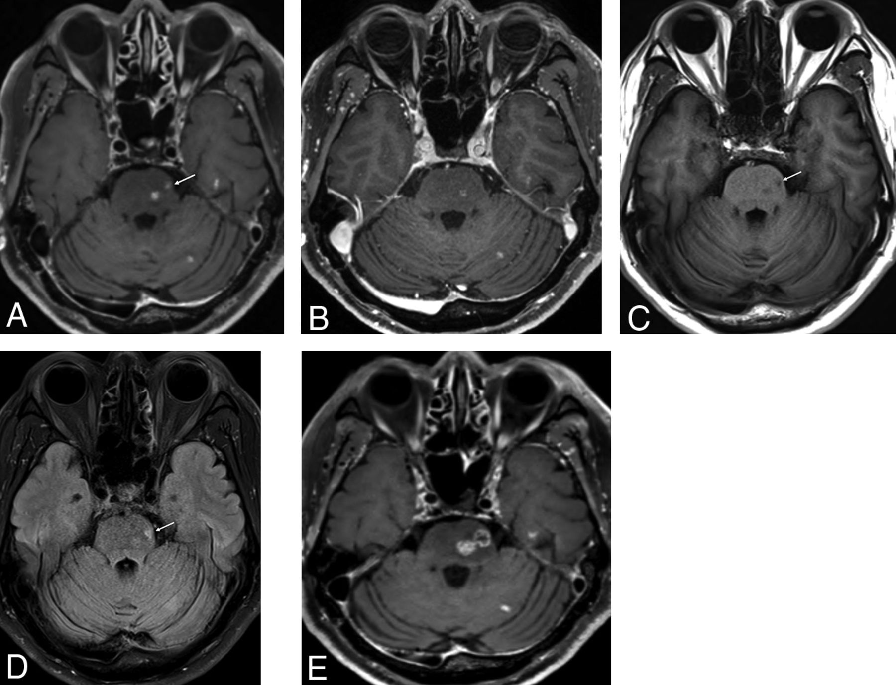
- Fig 1.
MR images of a 62-year-old male patient with lung cancer. Contrast-enhanced SPACE with DANTE (A) clearly shows a focal enhancing lesion in the left pons (arrow), but this is barely visible in MPRAGE (B). However, this lesion was accompanied by signal changes in the precontrast 2D T1-weighted (C) and FLAIR images (D) and an increase in size on follow-up MR imaging after 3 months (E).
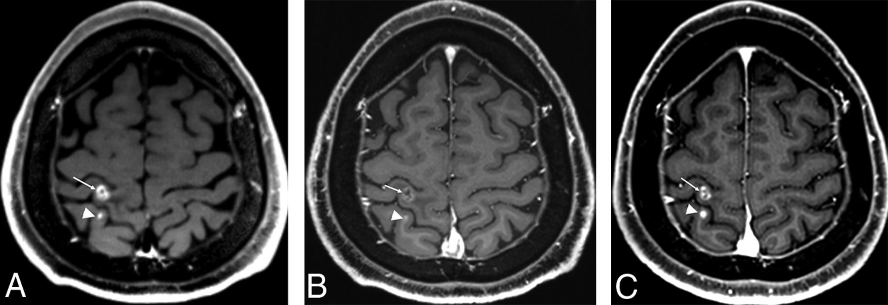
- Fig 2.
MR images of a 54-year-old male patient with lung cancer. Contrast-enhanced SPACE with DANTE (A) clearly shows two enhancing lesions in the right parietal lobe (arrow and arrowhead). One enhancing lesion is clearly visible (arrow), but the other enhancing lesion at the posterior aspect (arrowhead) is poorly visible on MPRAGE (B). However, this lesion has increased (arrowhead) in size and shows increased enhancement on the follow-up MR imaging 3 months later (C).
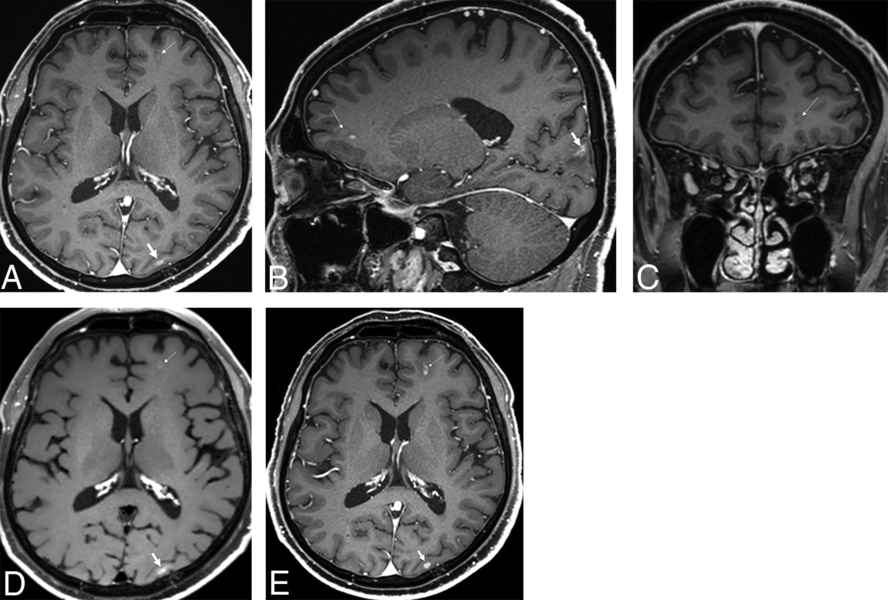
- Fig 3.
Minute and faint enhancing lesions at the left frontal and occipital lobes were missed by observer 2 (A). A left frontal lesion also shows faint enhancement on sagittal and coronal reconstructed MPRAGE images (B and C). However, these enhancing lesions show more prominent enhancement on SPACE with DANTE (D) and increased size on the follow-up MR imaging (E) 4 months later.
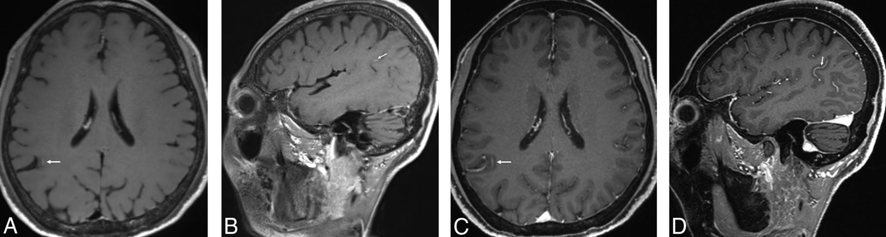
- Fig 4.
Contrast-enhanced SPACE with DANTE (A and B) shows a focal, linear enhancing lesion in the right parietal lobe. However, we could easily recognize this as a false-positive finding caused by incomplete vessel suppression on MPRAGE images (C and D).
- Fig 5.
The CNRlesion/parenchyma of SPACE with DANTE (8.62) (A) was higher than that of MPRAGE (3.49) (B).


Tables
SPACE with DANTE MPRAGE P Value Lesion diameter ≤5 mm Observer 1 No. of lesionsa 328 (4.39 ± 7.54) 175 (2.96 ± 5.13) .0006 Sensitivity (%)b 87.4 49.4 PPV (%)b 98.1 95.6 Observer 2 No. of lesionsa 324 (4.50 ± 7.52) 150 (2.76 ± 5.20) <.0001 Sensitivity (%)b 86.86 51.14 PPV (%)b 97.8 98.4 ICCc 0.99 0.98 Lesion diameter >5 mm Observer 1 No. of lesionsa 186 (2.62 ± 4.51) 168 (2.37 ± 3.52) .0978 Sensitivity (%)b 94.74 85.79 PPV (%)b 96.77 97.02 Observer 2 No. of lesionsa 188 (2.65 ± 4.37) 169 (2.38 ± 3.92) .0531 Sensitivity (%)b 95.26 87.37 PPV (%)b 96.28 98.22 ICCc 0.99 0.98 SPACE with DANTE MPRAGE P Value Lesion diameter ≤5 mm Observer 1 0.904 0.698 Observer 2 0.861 0.702 Mean ± SD 0.882 ± 0.023 0.700 ± 0.038 .0017 Lesion diameter >5 mm Observer 1 0.957 0.921 Observer 2 0.943 0.928 Mean ± SD 0.950 ± 0.013 0.925 ± 0.014 .1762 ↵a Data for observers 1 and 2 are mean value of FOM data compared between SPACE with DANTE and MPRAGE.
- Table 3:
CNRlesion/parenchyma and CNRwhite matter/gray matter of SPACE with DANTE and MPRAGEa
SPACE with DANTE MPRAGE P Value CNR (n = 51) Lesion/parenchyma 52.3 ± 43.1 17.5 ± 19.3 <.0001 White matter/gray matter −0.65 ± 1.39 3.08 ± 1.39 <.0001 ↵a Data are presented as means. Values were calculated using paired t tests.


Jump to section
Related Articles
Cited By...
- Diagnostic Confidence of Contrast-Enhanced T1-Weighted MRI for the Detection of Brain Metastases: 3D FSE versus 3D GRE-Based Sequences
- Compressed Sensitivity Encoding Artificial Intelligence Accelerates Brain Metastasis Imaging by Optimizing Image Quality and Reducing Scan Time
- Usefulness of Wave-CAIPI for Postcontrast 3D T1-SPACE in the Evaluation of Brain Metastases
- Utility of Contrast-Enhanced T2 FLAIR for Imaging Brain Metastases Using a Half-dose High-Relaxivity Contrast Agent