
Article Figures & Data
Figures
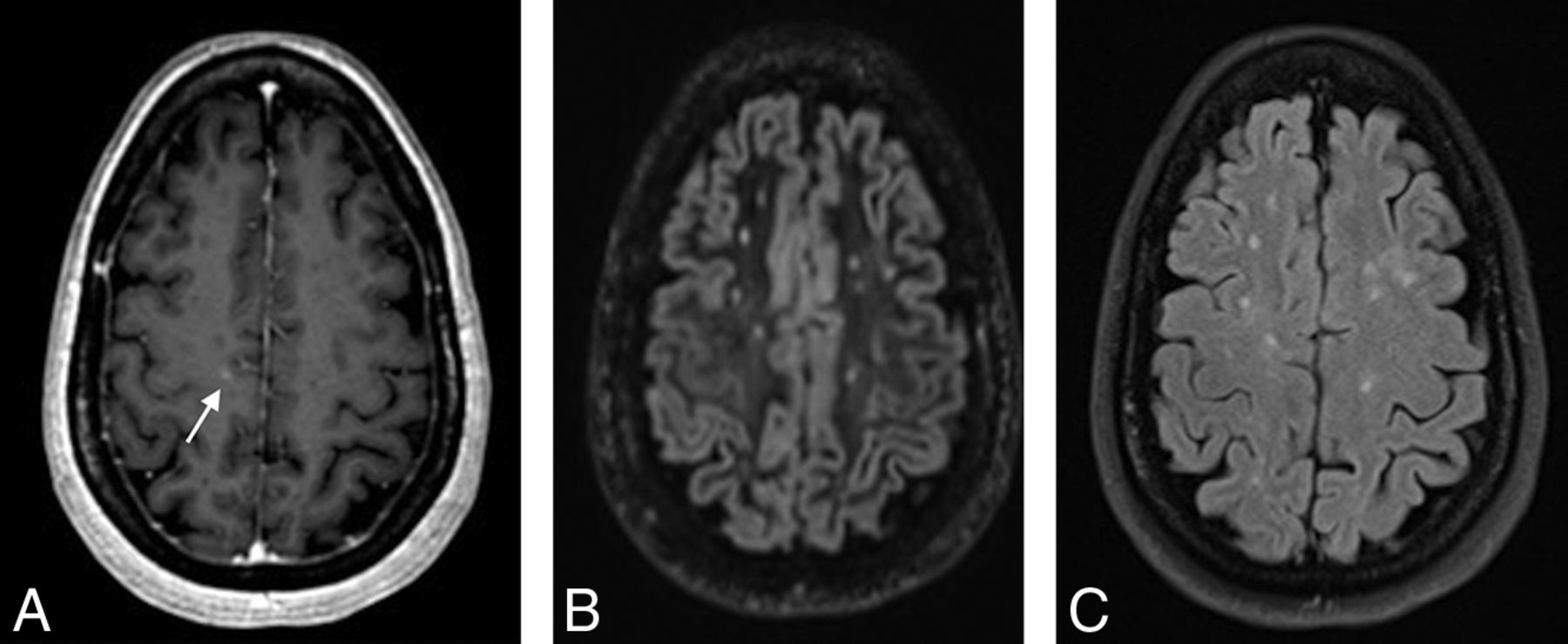
- Fig 1.
3D contrast-enhanced T1 MPRAGE (A) demonstrating a faint right frontal subcortical lesion (arrow) not visible on 3D DIR (B) and 2D FLAIR (C).
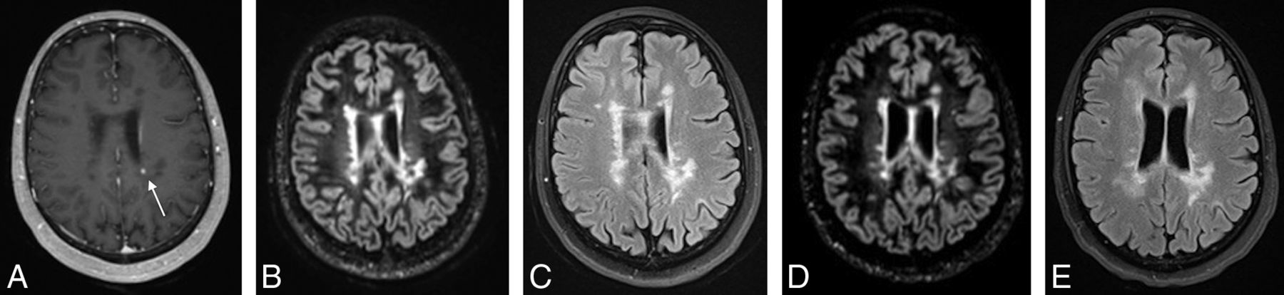
- Fig 2.
3D contrast-enhanced T1 MPRAGE (A) demonstrating a left periventricular enhancing lesion (arrow) in a region of confluent white matter lesions that is not detectable as new between the more recent 3D-DIR (B) and 2D-FLAIR (C) and prior 3D-DIR (D) and 2D-FLAIR (E).


Tables
- Table 1:
The number of examinations categorized by T2 lesion burden and number of enhancing lesions
T2 Lesion Burden No. of MR Imaging Examinations No. of MR Imaging Enhancing Lesions At Least 1 0 1 2 3 4 Mild (<10 lesions) 47/252 (19%) 44 3 3/252 (6%) Moderate (11–20 lesions) 89/252 (35%) 78 7 3 1 11/252 (12%) Severe (>20 lesions) 116/252 (46%) 99 10 1 4 2 17/252 (15%) Total 252 221 20 4 4 3 34/252 (13%) - Table 2:
Relationship between new signal abnormality and enhancement in patients with MS with prior comparison MR imaging examinations available
No Enhancing Lesion Enhancing Lesion Present Total New DIR/FLAIR 10 (13%) 6 (8%) 16 (21%) Stable DIR/FLAIR 59 (78%) 1 (1%) 60 (79%) Total 69 (91%) 7 (9%) 76


