
Article Figures & Data
Figures
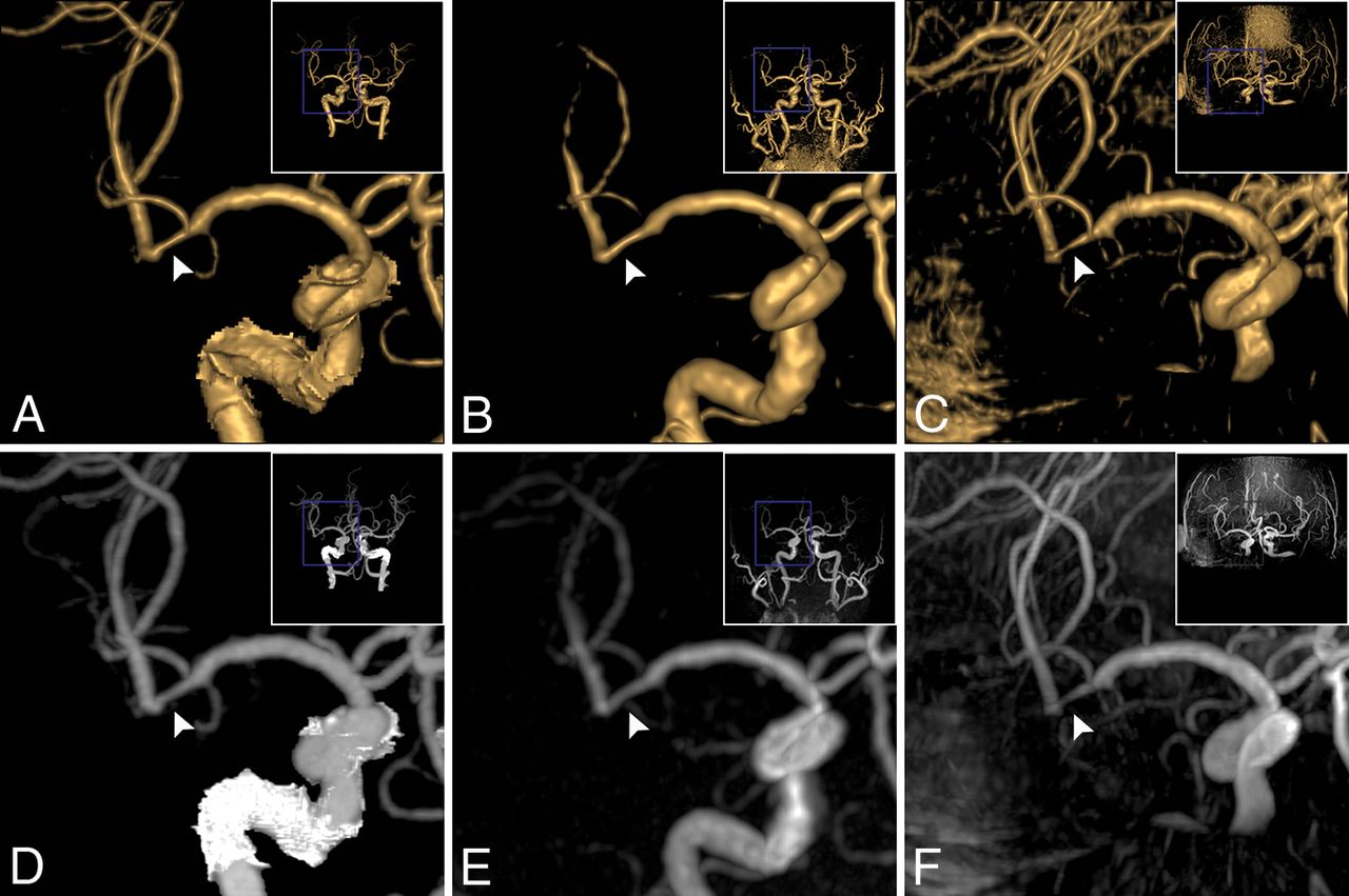
- Fig 1.
Coronal projection of stenosis in the right MCA M2 segment (a 74-year-old man). A stenosis (34%, grade 2) was observed on VR of CTA (A, white arrowhead) and on VR of zTE-MRA (B, white arrowhead); the stenosis (32%, grade 2) was equal to that on CTA. The stenosis on VR of TOF (C, white arrowhead) was overestimated (72%, grade 3). In correspondence with MIP of CTA (D, white arrowhead), flow signal in the stenosis lesion was homogeneous on MIP of zTE-MRA (E, white arrowhead, score 4) and was heterogeneous on TOF-MRA (F, white arrowhead, score 3).
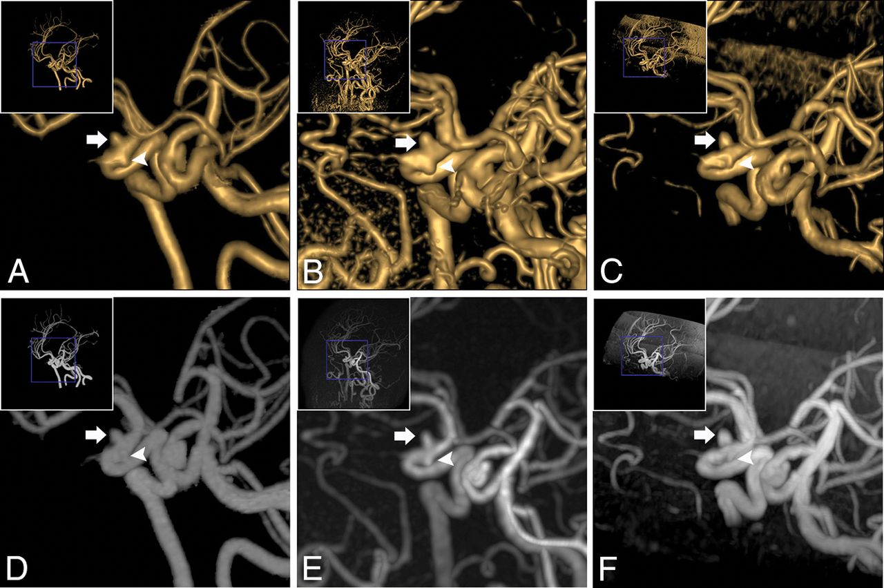
- Fig 2.
Oblique projection of aneurysms in the right ICA siphon segment (a 63-year-old woman). Two aneurysms were found on VR of CTA (A): The diameters were 3.0 × 3.2 mm (white arrow) and 2.4 × 2.1 mm (white arrowhead), respectively. Equal findings were observed on VR of zTE (B, 3.3 × 3.5 mm, white arrow; 2.× 2.4 mm, white arrowhead). On VR of TOF-MRA (C), the large one (3.1 × 3.4 mm, white arrow) was equal to the one on CTA. However, the tiny one was not evident (1.5 × 1.4 mm, white arrowhead). Concerning MIP of CTA (D, white arrow and white arrowhead), the same results were observed on MIP of zTE-MRA (E, white arrow and white arrowhead) and TOF-MRA (F, white arrow and white arrowhead).
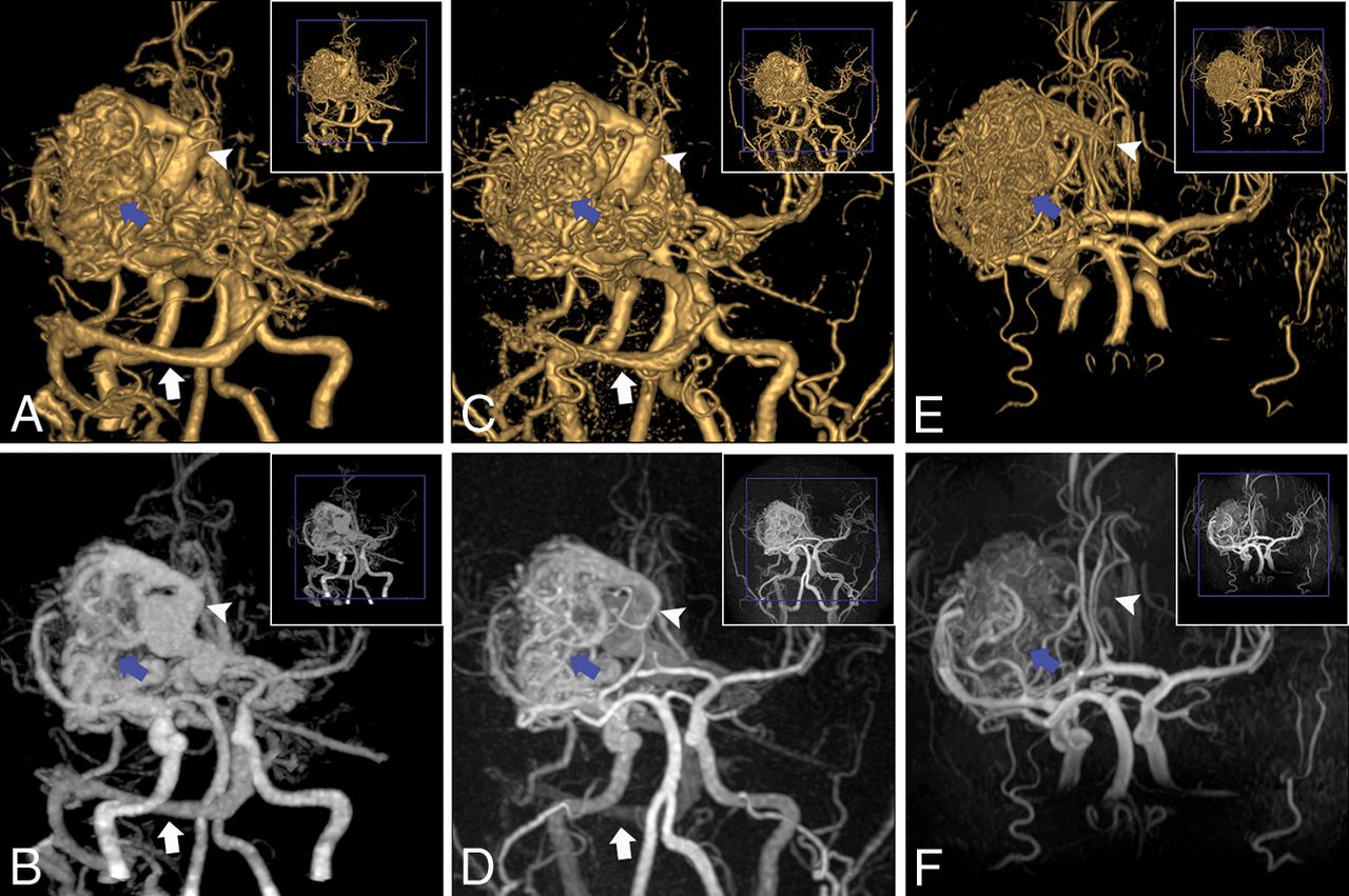
- Fig 3.
An AVM in the left MCA M1 segment (a 27-year-old man). For the AVM, the nidus (blue arrow), draining vein (white arrowhead), and venous sinus (transverse sinus and sigmoid sinus, white arrow) were clearly depicted on VR and MIP of CTA (A and B) and zTE-MRA (C and D), whereas they were not well-defined (nidus, blue arrow; draining vein, white arrowhead) and were missed (venous sinus) on VR and MIP of TOF-MRA (E and F).
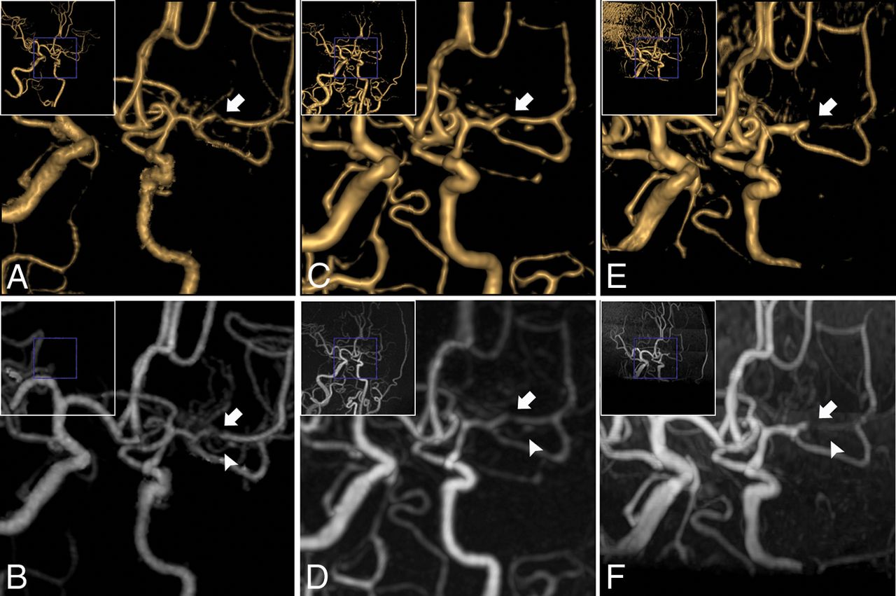
- Fig 4.
Oblique projection of Moyamoya disease in the left MCA M1 segment (a 43-year-old woman). The stenosed MCA (white arrowhead) and developed collateral vessels (white arrow) were scanned on VR and MIP of CTA (A and B) and zTE-MRA (C and D). A false occlusion in MCA (white arrowhead) and ill-defined collateral vessels (white arrow) were seen on VR and MIP of TOF-MRA (E and F).


Tables
TR/TE (ms) Flip Angle FOV (cm) Matrix Thickness (mm) Slices NEX Bandwidth (kHz) Slabs Label Duration (sec) Coverage Time (min:sec) zTE-MRA 862/0.016 3° 15 × 15 166 × 166 1.2 320 1 31.25 – 2 Calvarium-mandible 5:48 TOF-MRA 25/3.4 15° 30 × 24 320 × 256 1.4 256 1 41.67 3 – Cingulate cortex–mesencephalon 5:08 Note:— –indicates no data available; TR, repetition time; TE, echo time; FOV, field of view; NEX, number of excitation.
Collimation Pitch FOV (cm) Gantry Rotation Time (ms) Thickness (mm) Slices Tube Voltage (kV[peak]) Tube Current (mAs) Dose-Length Product (mGy × cm) Coverage CTA 128 × 0.625 0.758 17 × 17 400 0.6 715 100–120 100–450 220–608 Aortic arch–vertex zTE-MRA A Grade zTE-MRA B Grade 0 1 2 3 4 Total 0 1 0 0 0 0 1 (2.27%) 1 0 7 3 0 0 10 (22.73%) 2 0 0 14 0 0 14 (31.82%) 3 0 0 4 8 0 12 (27.27%) 4 0 0 0 1 6 7 (15.91%) Total 1 (2.27%) 7 (15.91%) 21 (47.73%) 9 (20.45%) 6 (13.64%) 44 ↵a Grading criterion: NASCET. Data represent the number of cases. A and B are observers A and B.
TOF-MRA A Grade TOF-MRA B Grade 0 1 2 3 4 Total 0 0 0 0 0 0 0 (0.00%) 1 1 9 0 0 0 10 (22.73%) 2 0 1 8 4 0 13(29.55%) 3 0 0 4 7 0 11 (25.00%) 4 0 0 0 2 8 10 (22.73%) Total 1 (2.27%) 10 (22.73%) 12 (27.27%) 13 (29.55%) 8 (18.18%) 44 ↵a Grading criterion: NASCET. Data represent the number of cases. A and B are observers A and B.
- Table 6:
Classification of stenosis grade from observers for zTE-MRA, TOF-MRA, and CTA (n = 44)a
Stenosis Grade zTE-MRA TOF-MRA CTA 0 1 (2.27%) 0 (0.00%) 1 (2.27%) 1 11 (25.00%) 10 (22.73%) 11 (25.00%) 2 12 (27.27%) 12 (27.27%) 13 (29.55%) 3 11 (25.00%) 11 (25.00%) 10 (22.73%) 4 9 (20.45%) 11 (25.00%) 9 (20.45%) ↵a Grading criterion: NASCET. Data represent the number of cases.
Group MRA Sum ra Intercept (95% CI)b Slope (95% CI)b ICC Group tinyc zTE 15 0.84 −0.5084–1.1794 −0.4952–0.2093 0.83; 95% CI, 0.57–0.94 TOF 15 0.74 0.7000–2.6133 −1.0344 to −0.1923 0.64; 95% CI, 0.21–0.86 Group larged zTE 23 0.98 −0.6059–0.2130 −0.02625–0.1200 0.98, 95% CI, 0.97–0.99 TOF 23 0.95 −1.1571–0.3062 −0.01380–0.2520 0.95, 95% CI, 0.89–0.98


Jump to section
Related Articles
Cited By...
- No citing articles found.