Advertisement

Main menu

  • Home
  • Content
    • Current Issue
    • Accepted Manuscripts
    • Article Preview
    • Past Issue Archive
    • AJNR Case Collection
    • Case of the Week Archive
    • Classic Case Archive
    • Case of the Month Archive
  • Special Collections
    • Spinal CSF Leak Articles (Jan 2020-June 2024)
    • 2024 AJNR Journal Awards
    • Most Impactful AJNR Articles
  • Multimedia
    • AJNR Podcast
    • AJNR Scantastics
    • Video Articles
  • For Authors
    • Submit a Manuscript
    • Author Policies
    • Fast publishing of Accepted Manuscripts
    • Graphical Abstract Preparation
    • Manuscript Submission Guidelines
    • Imaging Protocol Submission
    • Submit a Case for the Case Collection
  • About Us
    • About AJNR
    • Editorial Board
  • More
    • Become a Reviewer/Academy of Reviewers
    • Subscribers
    • Permissions
    • Alerts
    • Feedback
    • Advertisers
    • ASNR Home
  • Other Publications
    • ajnr

User menu

  • Alerts
  • Log in

Search

  • Advanced search
American Journal of Neuroradiology

American Journal of Neuroradiology

ASHNR American Society of Functional Neuroradiology ASHNR American Society of Pediatric Neuroradiology ASSR
  • Alerts
  • Log in

Advanced Search

  • Home
  • Content
    • Current Issue
    • Accepted Manuscripts
    • Article Preview
    • Past Issue Archive
    • AJNR Case Collection
    • Case of the Week Archive
    • Classic Case Archive
    • Case of the Month Archive
  • Special Collections
    • Spinal CSF Leak Articles (Jan 2020-June 2024)
    • 2024 AJNR Journal Awards
    • Most Impactful AJNR Articles
  • Multimedia
    • AJNR Podcast
    • AJNR Scantastics
    • Video Articles
  • For Authors
    • Submit a Manuscript
    • Author Policies
    • Fast publishing of Accepted Manuscripts
    • Graphical Abstract Preparation
    • Manuscript Submission Guidelines
    • Imaging Protocol Submission
    • Submit a Case for the Case Collection
  • About Us
    • About AJNR
    • Editorial Board
  • More
    • Become a Reviewer/Academy of Reviewers
    • Subscribers
    • Permissions
    • Alerts
    • Feedback
    • Advertisers
    • ASNR Home
  • Follow AJNR on Twitter
  • Visit AJNR on Facebook
  • Follow AJNR on Instagram
  • Join AJNR on LinkedIn
  • RSS Feeds

Welcome to the new AJNR, Updated Hall of Fame, and more. Read the full announcements.


AJNR is seeking candidates for the position of Associate Section Editor, AJNR Case Collection. Read the full announcement.


Research ArticleAdult Brain

Detailed Arterial Anatomy and Its Anastomoses of the Sphenoid Ridge and Olfactory Groove Meningiomas with Special Reference to the Recurrent Branches from the Ophthalmic Artery

M. Hiramatsu, K. Sugiu, T. Hishikawa, J. Haruma, Y. Takahashi, S. Murai, K. Nishi, Y. Yamaoka, Y. Shimazu, K. Fujii, M. Kameda, K. Kurozumi and I. Date
American Journal of Neuroradiology November 2020, 41 (11) 2082-2087; DOI: https://doi.org/10.3174/ajnr.A6790
M. Hiramatsu
aFrom the Department of Neurological Surgery (M.H., K.S., T.H., J.H., Y.T., S.M., K.N., Y.Y., Y.S., K.F., M.K., K.K., I.D.), Okayama University Graduate School of Medicine, Dentistry and Pharmaceutical Sciences, Okayama, Japan
  • Find this author on Google Scholar
  • Find this author on PubMed
  • Search for this author on this site
  • ORCID record for M. Hiramatsu
K. Sugiu
aFrom the Department of Neurological Surgery (M.H., K.S., T.H., J.H., Y.T., S.M., K.N., Y.Y., Y.S., K.F., M.K., K.K., I.D.), Okayama University Graduate School of Medicine, Dentistry and Pharmaceutical Sciences, Okayama, Japan
  • Find this author on Google Scholar
  • Find this author on PubMed
  • Search for this author on this site
  • ORCID record for K. Sugiu
T. Hishikawa
aFrom the Department of Neurological Surgery (M.H., K.S., T.H., J.H., Y.T., S.M., K.N., Y.Y., Y.S., K.F., M.K., K.K., I.D.), Okayama University Graduate School of Medicine, Dentistry and Pharmaceutical Sciences, Okayama, Japan
  • Find this author on Google Scholar
  • Find this author on PubMed
  • Search for this author on this site
  • ORCID record for T. Hishikawa
J. Haruma
aFrom the Department of Neurological Surgery (M.H., K.S., T.H., J.H., Y.T., S.M., K.N., Y.Y., Y.S., K.F., M.K., K.K., I.D.), Okayama University Graduate School of Medicine, Dentistry and Pharmaceutical Sciences, Okayama, Japan
  • Find this author on Google Scholar
  • Find this author on PubMed
  • Search for this author on this site
  • ORCID record for J. Haruma
Y. Takahashi
aFrom the Department of Neurological Surgery (M.H., K.S., T.H., J.H., Y.T., S.M., K.N., Y.Y., Y.S., K.F., M.K., K.K., I.D.), Okayama University Graduate School of Medicine, Dentistry and Pharmaceutical Sciences, Okayama, Japan
  • Find this author on Google Scholar
  • Find this author on PubMed
  • Search for this author on this site
  • ORCID record for Y. Takahashi
S. Murai
aFrom the Department of Neurological Surgery (M.H., K.S., T.H., J.H., Y.T., S.M., K.N., Y.Y., Y.S., K.F., M.K., K.K., I.D.), Okayama University Graduate School of Medicine, Dentistry and Pharmaceutical Sciences, Okayama, Japan
  • Find this author on Google Scholar
  • Find this author on PubMed
  • Search for this author on this site
  • ORCID record for S. Murai
K. Nishi
aFrom the Department of Neurological Surgery (M.H., K.S., T.H., J.H., Y.T., S.M., K.N., Y.Y., Y.S., K.F., M.K., K.K., I.D.), Okayama University Graduate School of Medicine, Dentistry and Pharmaceutical Sciences, Okayama, Japan
  • Find this author on Google Scholar
  • Find this author on PubMed
  • Search for this author on this site
  • ORCID record for K. Nishi
Y. Yamaoka
aFrom the Department of Neurological Surgery (M.H., K.S., T.H., J.H., Y.T., S.M., K.N., Y.Y., Y.S., K.F., M.K., K.K., I.D.), Okayama University Graduate School of Medicine, Dentistry and Pharmaceutical Sciences, Okayama, Japan
  • Find this author on Google Scholar
  • Find this author on PubMed
  • Search for this author on this site
  • ORCID record for Y. Yamaoka
Y. Shimazu
aFrom the Department of Neurological Surgery (M.H., K.S., T.H., J.H., Y.T., S.M., K.N., Y.Y., Y.S., K.F., M.K., K.K., I.D.), Okayama University Graduate School of Medicine, Dentistry and Pharmaceutical Sciences, Okayama, Japan
  • Find this author on Google Scholar
  • Find this author on PubMed
  • Search for this author on this site
  • ORCID record for Y. Shimazu
K. Fujii
aFrom the Department of Neurological Surgery (M.H., K.S., T.H., J.H., Y.T., S.M., K.N., Y.Y., Y.S., K.F., M.K., K.K., I.D.), Okayama University Graduate School of Medicine, Dentistry and Pharmaceutical Sciences, Okayama, Japan
  • Find this author on Google Scholar
  • Find this author on PubMed
  • Search for this author on this site
  • ORCID record for K. Fujii
M. Kameda
aFrom the Department of Neurological Surgery (M.H., K.S., T.H., J.H., Y.T., S.M., K.N., Y.Y., Y.S., K.F., M.K., K.K., I.D.), Okayama University Graduate School of Medicine, Dentistry and Pharmaceutical Sciences, Okayama, Japan
  • Find this author on Google Scholar
  • Find this author on PubMed
  • Search for this author on this site
  • ORCID record for M. Kameda
K. Kurozumi
aFrom the Department of Neurological Surgery (M.H., K.S., T.H., J.H., Y.T., S.M., K.N., Y.Y., Y.S., K.F., M.K., K.K., I.D.), Okayama University Graduate School of Medicine, Dentistry and Pharmaceutical Sciences, Okayama, Japan
bDepartment of Neurosurgery (K.K.), Hamamatsu University School of Medicine, Hamamatsu, Shizuoka, Japan.
  • Find this author on Google Scholar
  • Find this author on PubMed
  • Search for this author on this site
  • ORCID record for K. Kurozumi
I. Date
aFrom the Department of Neurological Surgery (M.H., K.S., T.H., J.H., Y.T., S.M., K.N., Y.Y., Y.S., K.F., M.K., K.K., I.D.), Okayama University Graduate School of Medicine, Dentistry and Pharmaceutical Sciences, Okayama, Japan
  • Find this author on Google Scholar
  • Find this author on PubMed
  • Search for this author on this site
  • ORCID record for I. Date
  • Article
  • Figures & Data
  • Supplemental
  • Info & Metrics
  • Responses
  • References
  • PDF
Loading

Abstract

BACKGROUND AND PURPOSE: Detailed arterial anatomy of the sphenoid ridge and olfactory groove meningiomas is complicated due to the fine angioarchitecture and anastomoses between each feeder. Herein, we present details of the arterial anatomy and the relationships of feeders in these lesions.

MATERIALS AND METHODS: This study included 20 patients admitted to our department between April 2015 and March 2020. Conditions of subjects consisted of 16 sphenoid ridge meningiomas and 4 olfactory groove meningiomas. We mainly analyzed arterial anatomy using 3D rotational angiography and slab MIP images of these lesions. We also analyzed the anastomoses of each feeder.

RESULTS: We found that 19 (95%), 15 (75%), and 15 (75%) lesions had feeders from the ophthalmic, internal carotid, and external carotid arteries, respectively. As feeders from the ophthalmic artery, recurrent meningeal arteries were involved in 18 lesions (90%). Fifteen lesions (75%) had anastomoses between each feeder.

CONCLUSIONS: Most of the meningiomas in the sphenoid ridge and olfactory groove had feeders from the ophthalmic and internal carotid arteries. There were various anastomoses between each feeder. This is the first report to demonstrate the detailed arterial anatomy and frequency of recurrent branches from the ophthalmic artery and their anastomoses using detailed imaging techniques.

ABBREVIATIONS:

ECA
external carotid artery
EtA
ethmoidal artery
ILT
inferolateral trunk
LSOF
lateral part of the superior orbital fissure
MHT
meningohypophyseal trunk
MMA
middle meningeal artery
MSOF
medial part of the superior orbital fissure
OC
optic canal
OphA
ophthalmic artery
RMA
recurrent meningeal artery
SOF
superior orbital fissure
TOB
transosseous branch
WHO
World Health Organization

The dural arterial anatomy of the sphenoid ridge and anterior skull base is very complicated because there are many fine arteries from not only the external carotid artery (ECA) but also the ICA and ophthalmic artery (OphA).1 Moreover, there are various anastomoses between each artery.2⇓⇓-5 In the setting of hypervascular diseases related to the dura, such as meningioma or dural arteriovenous fistula in these regions, the angioarchitecture can be visualized more clearly. In the present study, we investigated the details of the arterial anatomy of sphenoid ridge and olfactory groove meningiomas with special reference to the recurrent branches of the OphA and anastomoses between each feeder.

MATERIALS AND METHODS

All procedures performed in studies involving human participants were carried out in accordance with the ethical standards of Okayama University Graduate School of Medicine, Dentistry and Pharmaceutical Sciences’s institutional research committee (1911–023). Written informed consent was obtained from all patients before DSA and treatment, but written informed consent for this study was not required because of the retrospective and noninvasive study design. Conditions of subjects consisted of 16 sphenoid ridge meningiomas and 4 olfactory groove meningiomas between April 2015 and March 2020. In this study period, we treated 29 patients with meningiomas in these regions, and we excluded 9 cases without 3D rotational DSA data because we could not analyze the details of the angioarchitecture in these cases.

We examined the meningioma location, tumor invasion, tumor size, histopathologic diagnosis according to World Health Organization (WHO) grade, and arterial anatomy of the feeders. Feeders were divided into branches of the OphA, ICA, and ECA (Fig 1). Branches of the OphA are the recurrent meningeal arteries (RMAs) described below, the ethmoidal artery (EtA), and the transosseous branch (TOB). Branches of the ICA are the inferolateral trunk (ILT) and the meningohypophyseal trunk (MHT). Branches of the ECA are the middle meningeal artery (MMA), accessory meningeal artery, deep temporal artery, artery of the foramen rotundum, artery of the superior orbital fissure(SOF), and sphenopalatine artery. We also analyzed the anastomoses of each feeder.

FIG 1.
  • Download figure
  • Open in new tab
  • Download powerpoint
FIG 1.

Schematic illustration of dural arterial anatomy and anastomoses among the ophthalmic, internal carotid, and external carotid arteries: 1  indicates the ICA; 2, OphA; 3, lacrimal artery; 4, RMA-OC; 5, RMA-MSOF; 6,  RMA-LSOF; 7,  ILT; 8,  MHT; 9, MMA; 10, sphenoid branch of MMA; 11,  cavernous branch of MMA; 12,  accessory meningeal artery; 13,  artery of the foramen rotundum; 14,  artery of the SOF; 15, marginal tentorial artery.

Anatomic Terminology of the Branches of the Ophthalmic Artery

We defined the RMAs as the posteriorly recurrent branches from the OphA or lacrimal artery passing through the SOF or optic canal (OC). We found 2 other types of feeders from the OphA. One of them was the TOB, which originates at the second or third portion of the OphA and courses superiorly, passing through the upper wall of the orbit to reach the dura of the anterior cranial base. Another one was the anterior or posterior EtA, which courses medially, passing through the ethmoidal foramina.

On the basis of the course, we divided feeders from the OphA into 5 types as follows: RMA passing through the OC (RMA-OC), RMA passing through the lateral part of the SOF (RMA-LSOF), RMA passing through the medial part of the SOF (RMA-MSOF), TOB, and EtA. We distinguished the RMA-LSOF and RMA-MSOF using the coronal section of slab MIP images from rotational angiography data. We present representative cases of the RMA-LSOF (Fig 2), RMA-OC (Figs 3 and 4), TOB (Fig 4), and EtA (Fig 4).

FIG 2.
  • Download figure
  • Open in new tab
  • Download powerpoint
FIG 2.

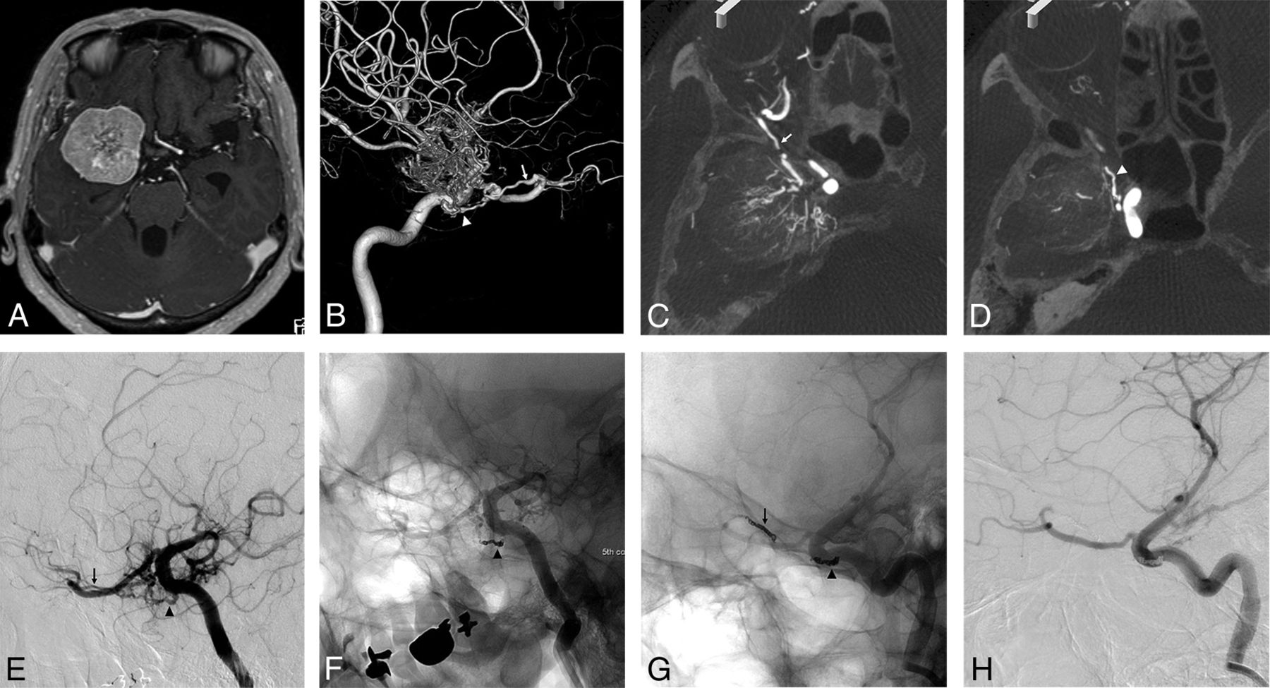
A, Axial view of the gadolinium-enhanced T1-weighted image shows a medial-type sphenoid ridge meningioma. The lateral view of the 3D-DSA (B) and axial views of slab MIP images (C and D) of the right internal carotid angiogram show the feeder of the RMA passing through lateral part of the SOF (arrow) from the lacrimal artery and ILT (arrowhead). There is an anastomosis between the RMA-LSOF and the ILT. E, Oblique view of the internal carotid angiogram before embolization shows both feeders. Internal carotid angiogram after coil embolization of the ILT (F) and RMA-LSOF (G and H). The tumor stain is markedly diminished.

FIG 3.
  • Download figure
  • Open in new tab
  • Download powerpoint
FIG 3.

A, The axial view of the gadolinium-enhanced T1-weighted image shows a medial-type sphenoid ridge meningioma. B, Anterior-posterior view of the left external carotid angiogram shows the feeder from the accessory meningeal artery (large gray arrow) and sphenoid branch (large black arrow) of the MMA. The lateral views of the 2D-DSA (C) and 3D-DSA (D) and sagittal views of slab MIP images (E and F) of the left internal carotid angiogram show the feeder of the RMA passing through the OC (arrow) and MHT (arrowhead). The slab MIP images clearly show that the RMA-OC and OphA (dotted arrow) pass through the OC (E and F).

FIG 4.
  • Download figure
  • Open in new tab
  • Download powerpoint
FIG 4.

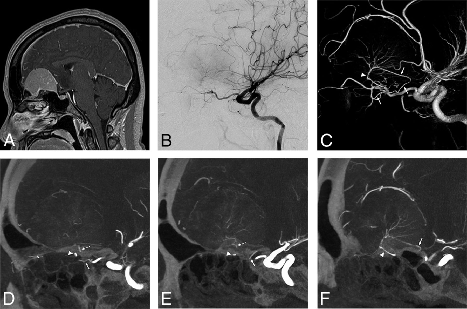
A, Sagittal view of the gadolinium-enhanced T1-weighted image shows an olfactory groove meningioma. The lateral views of the 2D-DSA (B) and 3D-DSA (C) and sagittal views of slab MIP images (D–F) of the right internal carotid angiogram show the feeder from the RMA passing through the OC (arrow) from the third segment of the OphA, posterior EtA, (arrowhead), and TOB (dotted arrow). The slab MIP images show the RMA-OC, and the OphA passes through the OC (E). Feeders from the RMA-OC and EtA configure an arcade shape on the blistering of the planum sphenoidale.

Preoperative Examination

Preoperative DSA was performed with the patient under local anesthesia. After obtaining 2D-DSA of the ECA and ICA, which are ipsilateral to the tumor, 3D-DSA with a 5-second protocol was performed on the branching feeders of the vessels. For 3D-DSA, a 4F diagnostic catheter was inserted into an ICA, and a 100% iodine contrast medium with a 300-mg/mL concentration was infused using an injector (2.5 mL/s flow rate, 1.5- to 2.0-second delay, total 16–18 mL). After obtaining volume-rendered images of 3D-DSA, slab MIP images were derived from rotational angiography data using a workstation to analyze details of the feeder anatomy. An Artis zee biplane (Siemens Healthcare) was used as a flat panel detector, and the syngo X Workplace (Siemens Healthcare) was used as a workstation.

RESULTS

The lesion characteristics and arterial anatomy of meningiomas are summarized in the Table, and details of each case are shown in On-line Table 1. In terms of lesion location, there were 4 sphenoid ridge lateral types, 9 sphenoid ridge medial types, 3 sphenoid ridge diffuse types spreading to the middle and anterior cranial base, and 4 olfactory groove lesions. There were 3 lesions with intraorbital invasion and 1 lesion with invasion to the temporal bone and muscle. The mean maximum diameter was 56 mm. In terms of histopathologic diagnosis, there were 19 WHO grade I meningiomas and 1 WHO grade II meningioma. The 2 most frequent diagnoses were meningothelial (n = 8) and transitional (n = 8) meningiomas.

View this table:
  • View inline
  • View popup

Lesion characteristics and arterial anatomy of meningiomas

We found that 19 (95%), 15 (75%), and 15 (75%) lesions had feeders from the OphA, ICA, and ECA, respectively. As the feeders from the OphA, there were 7 RMA-OCs in 7 lesions, 14 RMA-LSOFs in 13 lesions, 2 RMA-MSOFs in 2 lesions, 4 TOBs in 4 lesions, and 6 EtAs in 6 lesions. Six of 7 RMA-OCs (86%) originated from the first segment of the OphA. Eleven of 14 RMA-LSOFs (79%) originated from the lacrimal artery. Two RMA-MSOFs originated from the lacrimal artery and the second portion of the OphA. All (4/4) TOBs originated from the second or third segment of the OphA. Four of 6 EtAs (67%) originated from the third segment of the OphA. RMAs from the OphA were involved in 18 lesions (90%). All (7/7) RMA-OCs appeared in medial-type sphenoid ridge meningiomas or olfactory groove meningiomas. RMA-LSOFs appeared in all types of sphenoid ridge meningiomas. All sphenoid ridge meningiomas with invasion to other locations (cases 1, 2, 8, 15 in the On-line Table) had multiple feeders from the OphA. One lesion (case 9 in the On-line Table) that had no feeder from the OphA was a psammomatous meningioma (WHO grade I). There were no other relationships between the arterial anatomy and invasion or histopathologic diagnosis.

As the feeders from the ICA, the ILT and MHT were involved in 13 and 9 lesions, respectively. As feeders from the ECA, the sphenoidal branch of the MMA, accessory meningeal artery, artery of the foramen rotundum, and artery of the SOF were involved in 12, 7, 6, and 3 lesions, respectively. Other feeders from the ECA were the petrosal branch of the MMA, deep temporal artery, and sphenopalatine artery. There were 4 lesions with olfactory groove meningiomas. Most of the feeders of the olfactory groove meningioma were from the OphA, and there was no involvement of the ICA branches and MMA. As the feeders from the OphA, the EtAs, RMA-OCs, and TOBs were involved in 4, 3, and 1 lesion, respectively.

We found that 15 lesions (75%) had anastomoses between each feeder. The most frequent anastomosis was between the OphA and the ICA (eg, RMA-LSOF and ILT). We also found anastomoses between feeders from the ipsilateral OphA (eg, RMA-OC and EtA). Five cases had anastomoses between the OphA or ICA and ECA. Four cases had multiple anastomoses, such as the RMA-ILT-MMA-artery of the SOF. In 3 cases of olfactory groove meningiomas, we found anastomosis between the RMA-OC and the EtA. The mean maximum diameter of the meningiomas that had anastomosis of feeders was 45 ± 13 mm and that of meningiomas without anastomosis was 39 ± 16 mm.

DISCUSSION

Detailed arterial anatomy of the sphenoid ridge and the olfactory groove meningiomas is little understood due to the fine and complicated nature of arterial anatomy in the skull base. It is difficult to differentiate these subtle vessels using conventional 2D-DSA, 3D-CTA, or MR imaging. We demonstrated that the latest imaging technique and slab MIP image of 3D rotational angiography can visualize this fine angioarchitecture. In the present study, we found that 95%, 75%, and 75% of lesions have feeders from the OphA, ICA, and ECA, respectively. Moreover, we demonstrated that 75% of lesions have anastomoses between each feeder. Notably, this is the first report to demonstrate the detailed anatomy and frequency of recurrent branches from the OphA and their anastomoses using slab MIP images.

Anatomy of the Dural Arteries

Martins et al1 reported the detailed microsurgical anatomy of dural arteries as follows: The supply to the middle fossa and paracavernous dura derives laterally from the MMA, accessory meningeal artery, and ascending pharyngeal artery and medially from the intracavernous branches (ILT) of the ICA. They also described the contributions to these regions from the recurrent branches of the ophthalmic and lacrimal arteries. There is confusion about the definition of the recurrent ophthalmic arteries. Lasjaunias et al6 reported that both the superficial and deep recurrent ophthalmic arteries pass backward through the lateral and medial parts of the SOF, respectively. Meanwhile, Martins et al1 reported that the superficial recurrent ophthalmic artery, deep recurrent ophthalmic artery, and RMA pass through the optic canal, the medial part of the SOF, and the lateral part of the SOF, respectively. Therefore, descriptions of both authors were competing. In the present article, we describe the RMA as posteriorly recurrent branches from the OphA passing through the OC or SOF, including the superficial and deep recurrent ophthalmic arteries. Martins et al1 and Hayreh3 described the recurrent branch passing through the OC. However, to the best of our knowledge, no articles have shown this recurrent artery passing through the OC in imaging or cadaveric studies.

Yamaki et al7 reported details of feeding arteries in parasellar meningiomas, including 9 sphenoid ridge meningiomas. In this article, they analyzed 2 lesions (22%) in the sphenoid ridge that were supplied from the superficial recurrent ophthalmic artery or recurrent meningeal artery, respectively. The current imaging technology and slab MIP images provide us with the ability to analyze detailed arterial anatomy. We can demonstrate that RMAs from the OphA were involved in most of the lesions (90%) in the present study.

Anastomosis of Each Feeder

Numerous anastomoses between each artery have been reported.2⇓⇓-5 In the setting of hypervascular lesions related to the dura, such as meningiomas or dural arteriovenous fistulas, these feeding arteries can be enlarged and tend to connect.8 In the present study, there were various anastomoses in 75% of cases, and 4 cases had multiple anastomoses.

According to the embryologic concepts of the OphA, the connection between the OphA and the ILT is the remnant of the dorsal ophthalmic artery and the connection between the RMA and the MMA is the remnant of the supraorbital artery (branch of the stapedial artery).9⇓⇓-12 De La Torre and Netsky13 reported the persistent primitive maxillary artery in the human fetus. They reported that this artery arises from the cavernous portion of the ICA and anastomoses with the lacrimal artery and the anterior branch of the MMA. We can comprehend the complicated anastomoses among the OphA, ICA, and MMA using these findings. Therefore, it is certain that there are various anastomoses among branches of the OphA, ICA, and ECA.

Clinical Implications

Preoperative embolization for meningioma is performed as a standard treatment. Although its usefulness is widely accepted, the embolization of target vessels other than the ECA has been reported as a risk for procedural complications in a nationwide surveillance in Japan.14 Rosen et al15 reported their treatment results for preoperative embolization using microparticles to 167 cranial base meningiomas, and 9% of their patients experienced permanent neurologic deficits. They also showed that embolization of the MHT, MMA, and ascending pharyngeal artery involves the risk of a minor or permanent neurologic injury. Meanwhile, Waldron et al16 reported their treatment results for preoperative embolization using microparticles and coils to 119 cranial base meningiomas, with no complications. Some authors have reported the usefulness of embolization of the OphA branches.17,18 In limited cases, embolization may be performed for branches of the OphA, but it is associated with a risk of visual disturbance due to migration of the embolic material to the central retinal artery.17 Appropriate knowledge of anatomy, distal catheterization, and the gentle injection of embolic material without reflux are necessary for safe embolization of the OphA.19

In our opinion, the RMA-OC is inappropriate as the target vessel of preoperative embolization due to the difficulty in catheterization because of its small caliber and short distance from the origin of the OphA with an acute angle. The RMA-LSOF may have a lower risk because of its origins and anastomoses. The RMA-LSOF usually originates from a lacrimal artery and is anastomosed with the sphenoid branch of the MMA. However, if the RMA-LSOF has an anastomosis with the ILT, it involves a risk of the migration of particulate or liquid materials to the ICA. Thus, we recommend embolization of both of the RMA and the ILT using detachable coils in such a case.

Another precaution related to embolization is residual feeding from anastomotic vessels. If there is an anastomosis between each feeder, proximal occlusion of the feeder, which can be easily catheterized, can result in increased blood flow to the tumor from the residual feeder. One should embolize the common trunk of both feeders or the proximal parts of both feeders (Fig 2).

A detailed understanding of arterial microanatomy is also useful in direct surgery. A large sphenoid ridge meningioma requires skull base techniques, such as the transzygomatic approach.20,21 Using a skull base technique and extradural drilling of the sphenoid wing to the SOF, we can devascularize the feeder from the ECA and RMA-LSOF/MSOF.21 After these procedures, we open the dura mater, and we can debulk the intradural tumor, most of which is already devascularized, whereas, we may not be able to find the feeder from the RMA-OC until identification of the optic nerve. Moreover, coagulation to the RMA-OC involves the risk of optic nerve injury. If we identify all of the feeders using preoperative angiography, we can search for feeders on the basis of the anatomic landmarks of the bone and devascularize them in a safe and efficient manner.

Limitations

The current study has some limitations. First, the number of patients was small. Second, we did not confirm the presented feeding arteries with operative findings. In future studies, we should examine the accuracy of the presented results using operative findings or cadaveric research.

CONCLUSIONS

Sphenoid ridge and olfactory groove meningiomas have feeders from the OphA, ICA, and ECA in 95%, 75%, and 75% of lesions, respectively. Furthermore, 75% of lesions have anastomoses between each feeder.

Acknowledgments

The authors thank Dr Ryota Ishibashi (Department of Neurosurgery, Kurashiki Central Hospital, Kurashiki, Japan) for his suggestions on the writing of this article.

Footnotes

  • Disclosures: Masafumi Hiramatsu—UNRELATED: Payment for Lectures Including Service on Speakers Bureaus: Siemens; Travel/Accommodations/Meeting Expenses Unrelated to Activities Listed: Siemens. Kenji Sugiu—UNRELATED: Payment for Lectures Including Service on Speakers Bureaus: Siemens; Travel/Accommodations/Meeting Expenses Unrelated to Activities Listed: Siemens.

References

  1. 1.↵
    1. Martins C,
    2. Yasuda A,
    3. Campero A, et al
    . Microsurgical anatomy of the dural arteries. Neurosurgery 2005;56(2 Suppl):211–51; discussion 211–51 doi:10.1227/01.neu.0000144823.94402.3d pmid:15794820
    CrossRefPubMed
  2. 2.↵
    1. Geibprasert S,
    2. Pongpech S,
    3. Armstrong D, et al
    . Dangerous extracranial-intracranial anastomoses and supply to the cranial nerves: vessels the neurointerventionalist needs to know. AJNR Am J Neuroradiol 2009;30:1459–68 doi:10.3174/ajnr.A1500 pmid:19279274
    Abstract/FREE Full Text
  3. 3.↵
    1. Hayreh SS
    . Orbital vascular anatomy. Eye (Lond) 2006;20:1130–44 doi:10.1038/sj.eye.6702377 pmid:17019411
    CrossRefPubMed
  4. 4.↵
    1. Perrini P,
    2. Cardia A,
    3. Fraser K, et al
    . A microsurgical study of the anatomy and course of the ophthalmic artery and its possibly dangerous anastomoses. J Neurosurg 2007;106:142–50 doi:10.3171/jns.2007.106.1.142 pmid:17236500
    CrossRefPubMed
  5. 5.↵
    1. Toma N
    . Anatomy of the ophthalmic artery: embyrological consideration. Neurol Med Chir (Tokyo) 2016;56:585–91 doi:10.2176/nmc.ra.2015-0324 pmid:27298261
    CrossRefPubMed
  6. 6.↵
    1. Lasjaunias P,
    2. Brismar J,
    3. Moret J, et al
    . Recurrent cavernous branches of the ophthalmic artery. Acta Radiology Diagn (Stockh) 1978;19:553–60 doi:10.1177/028418517801900402 pmid:717011
    CrossRefPubMed
  7. 7.↵
    1. Yamaki T,
    2. Tanabe S,
    3. Sohma T, et al
    . Feeding arteries of parasellar meningiomas: angiographic study of medial sphenoid ridge and tuberculum sellae meningiomas. Neurol Med Chir (Tokyo) 1988;28:553–58 doi:10.2176/nmc.28.553 pmid:2460782
    CrossRefPubMed
  8. 8.↵
    1. Kiyosue H,
    2. Tanoue S,
    3. Hongo N, et al
    . Artery of the superior orbital fissure: an undescribed branch from the pterygopalatine segment of the maxillary artery to the orbital apex connecting with the anteromedial branch of the inferolateral trunk. AJNR Am J Neuroradiol 2015;36:1741–47 doi:10.3174/ajnr.A4331 pmid:26206808
    Abstract/FREE Full Text
  9. 9.↵
    1. Lasjaunias P,
    2. Berenstein A,
    3. terBrugge KG
    1. Lasjaunias P,
    2. Berenstein A,
    3. terBrugge KG
    . Spinal artery. In: Lasjaunias P, Berenstein A, terBrugge KG. Surgical Neuroangiography, Volume 1; Clinical Vascular Anatomy and Variations. 2nd ed. Springer-Verlag; 2001:123
  10. 10.↵
    1. Lasjaunias P,
    2. Moret J,
    3. Mink J
    . The anatomy of the inferolateral trunk (ILT) of the internal carotid artery. Neuroradiology 1977;13:215–20 doi:10.1007/BF00344216 pmid:876454
    CrossRefPubMed
  11. 11.↵
    1. Bonasia S,
    2. Bojanowski M,
    3. Robert T
    . Embryology and anatomical variations of the ophthalmic artery. Neuroradiology 2020;62:139–52 doi:10.1007/s00234-019-02336-4 pmid:31863143
    CrossRefPubMed
  12. 12.↵
    1. Padget DH
    . The development of the cranial arteries in the human embryo. Contrib Embryol 1948;32:205–61
  13. 13.↵
    1. De La Torre E,
    2. Netsky MG
    . Study of persistent primitive maxillary artery in human fetus: Some homologies of cranial arteries in man and dog. Am J Anat 1960;106:185–95 doi:10.1002/aja.1001060302
    CrossRef
  14. 14.↵
    1. Sugiu K,
    2. Hishikawa T,
    3. Murai S, et al
    . Treatment outcome of intracranial tumor embolization in Japan: Japanese Registry of NeuroEndovascular Therapy 3 (JR-NET3). Neurol Med Chir(Tokyo) 2019;59:41–47 doi:10.2176/nmc.st.2018-0220 pmid:30686813
    CrossRefPubMed
  15. 15.↵
    1. Rosen CL,
    2. Ammerman JM,
    3. Sekhar LN, et al
    . Outcome analysis of preoperative embolization in cranial base surgery. Acta Neurochir (Wien) 2002;144:1157–64 doi:10.1007/s00701-002-0965-y pmid:12434172
    CrossRefPubMed
  16. 16.↵
    1. Waldron JS,
    2. Sughrue ME,
    3. Hetts SW, et al
    . Embolization of skull base meningiomas and feeding vessels arising from the internal carotid circulation. Neurosurgery 2011;68:162–69 doi:10.1227/NEU.0b013e3181fe2de9 pmid:21150761
    CrossRefPubMed
  17. 17.↵
    1. Terada T,
    2. Kinoshita Y,
    3. Yokote H, et al
    . Preoperative embolization of meningiomas fed by ophthalmic branch arteries. Surg Neurol 1996;45:161–66 doi:10.1016/S0090-3019(96)80010-3 pmid:8607067
    CrossRefPubMed
  18. 18.↵
    1. Trivelatto F,
    2. Nakiri GS,
    3. Manisor M, et al
    . Preoperative Onyx embolization of meningiomas fed by the ophthalmic artery: a case series. AJNR Am J Neuroradiol 2011;32:1762–66 doi:10.3174/ajnr.A2591 pmid:21835949
    Abstract/FREE Full Text
  19. 19.↵
    1. Matsumaru Y,
    2. Alvarez H,
    3. Rodesch G, et al
    . Embolisation of branches of the ophthalmic artery. Interv Neuroradiol 1997;3:239–45 doi:10.1177/159101999700300306 pmid:20678429
    CrossRefPubMed
  20. 20.↵
    1. Campero A,
    2. Villalonga JF,
    3. Elizalde RL, et al
    . Transzygomatic approach plus mini-peeling of middle fossa for devascularization of sphenoid wing meningiomas: technical note. Surg Neurol Int 2018;9:140 doi:10.4103/sni.sni_135_18 pmid:30105134
    CrossRefPubMed
  21. 21.↵
    1. González-Darder JM
    . Combined extradural and intradural pterional transzygomatic approach to large sphenoid wing meningiomas: operative technique and surgical results. J Neurol Surg B Skull Base 2019;80:244–51 doi:10.1055/s-0038-1668538 pmid:31143566
    CrossRefPubMed
  • Received May 16, 2020.
  • Accepted after revision July 15, 2020.
  • © 2020 by American Journal of Neuroradiology
PreviousNext
Back to top

In this issue

American Journal of Neuroradiology: 41 (11)
American Journal of Neuroradiology
Vol. 41, Issue 11
1 Nov 2020
  • Table of Contents
  • Index by author
  • Complete Issue (PDF)
Advertisement
Print
Download PDF
Email Article

Thank you for your interest in spreading the word on American Journal of Neuroradiology.

NOTE: We only request your email address so that the person you are recommending the page to knows that you wanted them to see it, and that it is not junk mail. We do not capture any email address.

Enter multiple addresses on separate lines or separate them with commas.
Detailed Arterial Anatomy and Its Anastomoses of the Sphenoid Ridge and Olfactory Groove Meningiomas with Special Reference to the Recurrent Branches from the Ophthalmic Artery
(Your Name) has sent you a message from American Journal of Neuroradiology
(Your Name) thought you would like to see the American Journal of Neuroradiology web site.
CAPTCHA
This question is for testing whether or not you are a human visitor and to prevent automated spam submissions.
Cite this article
M. Hiramatsu, K. Sugiu, T. Hishikawa, J. Haruma, Y. Takahashi, S. Murai, K. Nishi, Y. Yamaoka, Y. Shimazu, K. Fujii, M. Kameda, K. Kurozumi, I. Date
Detailed Arterial Anatomy and Its Anastomoses of the Sphenoid Ridge and Olfactory Groove Meningiomas with Special Reference to the Recurrent Branches from the Ophthalmic Artery
American Journal of Neuroradiology Nov 2020, 41 (11) 2082-2087; DOI: 10.3174/ajnr.A6790

Citation Manager Formats

  • BibTeX
  • Bookends
  • EasyBib
  • EndNote (tagged)
  • EndNote 8 (xml)
  • Medlars
  • Mendeley
  • Papers
  • RefWorks Tagged
  • Ref Manager
  • RIS
  • Zotero
0 Responses
Respond to this article
Share
Bookmark this article
Detailed Arterial Anatomy and Its Anastomoses of the Sphenoid Ridge and Olfactory Groove Meningiomas with Special Reference to the Recurrent Branches from the Ophthalmic Artery
M. Hiramatsu, K. Sugiu, T. Hishikawa, J. Haruma, Y. Takahashi, S. Murai, K. Nishi, Y. Yamaoka, Y. Shimazu, K. Fujii, M. Kameda, K. Kurozumi, I. Date
American Journal of Neuroradiology Nov 2020, 41 (11) 2082-2087; DOI: 10.3174/ajnr.A6790
del.icio.us logo Twitter logo Facebook logo Mendeley logo
  • Tweet Widget
  • Facebook Like
  • Google Plus One
Purchase

Jump to section

  • Article
    • Abstract
    • ABBREVIATIONS:
    • MATERIALS AND METHODS
    • RESULTS
    • DISCUSSION
    • CONCLUSIONS
    • Acknowledgments
    • Footnotes
    • References
  • Figures & Data
  • Supplemental
  • Info & Metrics
  • Responses
  • References
  • PDF

Related Articles

  • PubMed
  • Google Scholar

Cited By...

  • No citing articles found.
  • Crossref (10)
  • Google Scholar

This article has been cited by the following articles in journals that are participating in Crossref Cited-by Linking.

  • Midline Skull Base Meningiomas: Transcranial and Endonasal Perspectives
    Ciro Mastantuoni, Luigi Maria Cavallo, Felice Esposito, Elena d’Avella, Oreste de Divitiis, Teresa Somma, Andrea Bocchino, Gianluca Lorenzo Fabozzi, Paolo Cappabianca, Domenico Solari
    Cancers 2022 14 12
  • Cone-Beam Computed Tomography Fusion Technique for Vascular Assessment of Skull Base Meningiomas
    Keisuke Yoshida, Takenori Akiyama, Satoshi Takahashi, Tomoru Miwa, Takashi Horiguchi, Hikaru Sasaki, Masahiro Toda
    World Neurosurgery 2021 151
  • Role of the ophthalmic artery in the endovascular treatment for intracranial vascular diseases
    Kun Hou, Wei Wu, Ying Liu, Lai Qu, Baofeng Xu, Jinlu Yu
    Acta Neurologica Belgica 2021 121 2
  • Variations in the branching patterns of the anterior choroidal artery: an angiographic study with special reference to temporal lobe epilepsy surgery
    Tadashi Hamasaki, Hiroki Uchikawa, Yuki Ohmori, Yasuyuki Kaku, Tomonori Ono, Shuichi Tochihara, Toshinori Hirai, Tatsuya Kawano, Akitake Mukasa
    Acta Neurochirurgica 2022 164 8
  • Immediate procedural safety of adjunctive proximal coil occlusion in middle meningeal artery embolization for chronic subdural hematomas: Experience in 137 cases
    Jessica K Campos, Benjamen M Meyer, David A Zarrin, Muhammad Waqas Khan, Jonathan C Collard de Beaufort, Gizal Amin, Michael B Avery, Kiarash Golshani, Narlin B Beaty, Matthew T Bender, Geoffrey P Colby, Li-Mei Lin, Alexander L Coon
    Interventional Neuroradiology 2024
  • Rare anastomosis between the bilateral internal carotid arteries via the recurrent arteries branching from the first segment of the ophthalmic artery
    Mitsushige Ando, Yoshinori Maki, Masato Hojo, Taketo Hatano
    Neuroradiology 2022 64 7
  • Adult Infratentorial Pial Arteriovenous Fistula Treated with Detachable Coils: A Case Report and a Review of Literature
    Naoya KIDANI, Yusuke INOUE, Shohei NISHIGAKI, Juntaro FUJITA, Yuta SOTOME, Yusuke TOMITA, Kenichiro MURAOKA, Nobuyuki HIROTSUNE, Shigeki NISHINO
    NMC Case Report Journal 2023 10
  • Comprehensive analysis of the distribution of dural artery-derived tumor-feeding arteries in intracranial meningiomas
    Kei Yamashiro, Kazuhide Adachi, Saeko Higashiguchi, Eiji Fujiwara, Motoharu Hayakawa, Mitsushiro Hasegawa, Yuichi Hirose
    Acta Neurochirurgica 2025 167 1
  • Cutting Edge of Micro-vascular Anatomy in Neuroendovascular Therapy
    Kenji Sugiu, Hisanori Edaki, Masafumi Hiramatsu, Tomohito Hishikawa, Jun Haruma, Yu Takahashi, Satoshi Murai, Kazuhiko Nishi, Yoko Yamaoka, Yu Sato, Yuki Ebisudani, Ryu Kimura, Isao Date
    Japanese Journal of Neurosurgery 2022 31 6
  • Preoperative Vascular and Cranial Nerve Imaging in Skull Base Tumors
    Akinari Yamano, Masahide Matsuda, Eiichi Ishikawa
    Cancers 2024 17 1

More in this TOC Section

Adult Brain

  • Diagnostic Neuroradiology of Monoclonal Antibodies
  • ML for Glioma Molecular Subtype Prediction
  • Segmentation of Brain Metastases with BLAST
Show more Adult Brain

Interventional

  • SAVE vs. Solumbra Techniques for Thrombectomy
  • CT Perfusion&Reperfusion in Acute Ischemic Stroke
  • Delayed Reperfusion Post-Thrombectomy&Thrombolysis
Show more Interventional

Similar Articles

Advertisement

Indexed Content

  • Current Issue
  • Accepted Manuscripts
  • Article Preview
  • Past Issues
  • Editorials
  • Editors Choice
  • Fellow Journal Club
  • Letters to the Editor

Cases

  • Case Collection
  • Archive - Case of the Week
  • Archive - Case of the Month
  • Archive - Classic Case

Special Collections

  • Special Collections

Resources

  • News and Updates
  • Turn around Times
  • Submit a Manuscript
  • Author Policies
  • Manuscript Submission Guidelines
  • Evidence-Based Medicine Level Guide
  • Publishing Checklists
  • Graphical Abstract Preparation
  • Imaging Protocol Submission
  • Submit a Case
  • Become a Reviewer/Academy of Reviewers
  • Get Peer Review Credit from Publons

Multimedia

  • AJNR Podcast
  • AJNR SCANtastic
  • Video Articles

About Us

  • About AJNR
  • Editorial Board
  • Not an AJNR Subscriber? Join Now
  • Alerts
  • Feedback
  • Advertise with us
  • Librarian Resources
  • Permissions
  • Terms and Conditions

American Society of Neuroradiology

  • Not an ASNR Member? Join Now

© 2025 by the American Society of Neuroradiology All rights, including for text and data mining, AI training, and similar technologies, are reserved.
Print ISSN: 0195-6108 Online ISSN: 1936-959X

Powered by HighWire