Advertisement

Main menu

  • Home
  • Content
    • Current Issue
    • Accepted Manuscripts
    • Article Preview
    • Past Issue Archive
    • AJNR Case Collection
    • Case of the Week Archive
    • Classic Case Archive
    • Case of the Month Archive
  • Special Collections
    • Spinal CSF Leak Articles (Jan 2020-June 2024)
    • 2024 AJNR Journal Awards
    • Most Impactful AJNR Articles
  • Multimedia
    • AJNR Podcast
    • AJNR Scantastics
    • Video Articles
  • For Authors
    • Submit a Manuscript
    • Author Policies
    • Fast publishing of Accepted Manuscripts
    • Graphical Abstract Preparation
    • Manuscript Submission Guidelines
    • Imaging Protocol Submission
    • Submit a Case for the Case Collection
  • About Us
    • About AJNR
    • Editorial Board
  • More
    • Become a Reviewer/Academy of Reviewers
    • Subscribers
    • Permissions
    • Alerts
    • Feedback
    • Advertisers
    • ASNR Home
  • Other Publications
    • ajnr

User menu

  • Alerts
  • Log in

Search

  • Advanced search
American Journal of Neuroradiology

American Journal of Neuroradiology

ASHNR American Society of Functional Neuroradiology ASHNR American Society of Pediatric Neuroradiology ASSR
  • Alerts
  • Log in

Advanced Search

  • Home
  • Content
    • Current Issue
    • Accepted Manuscripts
    • Article Preview
    • Past Issue Archive
    • AJNR Case Collection
    • Case of the Week Archive
    • Classic Case Archive
    • Case of the Month Archive
  • Special Collections
    • Spinal CSF Leak Articles (Jan 2020-June 2024)
    • 2024 AJNR Journal Awards
    • Most Impactful AJNR Articles
  • Multimedia
    • AJNR Podcast
    • AJNR Scantastics
    • Video Articles
  • For Authors
    • Submit a Manuscript
    • Author Policies
    • Fast publishing of Accepted Manuscripts
    • Graphical Abstract Preparation
    • Manuscript Submission Guidelines
    • Imaging Protocol Submission
    • Submit a Case for the Case Collection
  • About Us
    • About AJNR
    • Editorial Board
  • More
    • Become a Reviewer/Academy of Reviewers
    • Subscribers
    • Permissions
    • Alerts
    • Feedback
    • Advertisers
    • ASNR Home
  • Follow AJNR on Twitter
  • Visit AJNR on Facebook
  • Follow AJNR on Instagram
  • Join AJNR on LinkedIn
  • RSS Feeds

Welcome to the new AJNR, Updated Hall of Fame, and more. Read the full announcements.


AJNR is seeking candidates for the position of Associate Section Editor, AJNR Case Collection. Read the full announcement.


Research ArticleHead & Neck
Open Access

Diagnostic Value of Model-Based Iterative Reconstruction Combined with a Metal Artifact Reduction Algorithm during CT of the Oral Cavity

Y. Kubo, K. Ito, M. Sone, H. Nagasawa, Y. Onishi, N. Umakoshi, T. Hasegawa, T. Akimoto and M. Kusumoto
American Journal of Neuroradiology November 2020, 41 (11) 2132-2138; DOI: https://doi.org/10.3174/ajnr.A6767
Y. Kubo
aFrom the Department of Diagnostic Radiology (Y.K., K.I., M.S., H.N., Y.O., N.U., T.H., M.K.), National Cancer Center Hospital, Tokyo, Japan
bDepartment of Cancer Medicine (Y.K., T.A.), Jikei University Graduate School of Medicine, Tokyo, Japan
  • Find this author on Google Scholar
  • Find this author on PubMed
  • Search for this author on this site
  • ORCID record for Y. Kubo
K. Ito
aFrom the Department of Diagnostic Radiology (Y.K., K.I., M.S., H.N., Y.O., N.U., T.H., M.K.), National Cancer Center Hospital, Tokyo, Japan
  • Find this author on Google Scholar
  • Find this author on PubMed
  • Search for this author on this site
  • ORCID record for K. Ito
M. Sone
aFrom the Department of Diagnostic Radiology (Y.K., K.I., M.S., H.N., Y.O., N.U., T.H., M.K.), National Cancer Center Hospital, Tokyo, Japan
  • Find this author on Google Scholar
  • Find this author on PubMed
  • Search for this author on this site
  • ORCID record for M. Sone
H. Nagasawa
aFrom the Department of Diagnostic Radiology (Y.K., K.I., M.S., H.N., Y.O., N.U., T.H., M.K.), National Cancer Center Hospital, Tokyo, Japan
  • Find this author on Google Scholar
  • Find this author on PubMed
  • Search for this author on this site
  • ORCID record for H. Nagasawa
Y. Onishi
aFrom the Department of Diagnostic Radiology (Y.K., K.I., M.S., H.N., Y.O., N.U., T.H., M.K.), National Cancer Center Hospital, Tokyo, Japan
  • Find this author on Google Scholar
  • Find this author on PubMed
  • Search for this author on this site
  • ORCID record for Y. Onishi
N. Umakoshi
aFrom the Department of Diagnostic Radiology (Y.K., K.I., M.S., H.N., Y.O., N.U., T.H., M.K.), National Cancer Center Hospital, Tokyo, Japan
  • Find this author on Google Scholar
  • Find this author on PubMed
  • Search for this author on this site
  • ORCID record for N. Umakoshi
T. Hasegawa
aFrom the Department of Diagnostic Radiology (Y.K., K.I., M.S., H.N., Y.O., N.U., T.H., M.K.), National Cancer Center Hospital, Tokyo, Japan
  • Find this author on Google Scholar
  • Find this author on PubMed
  • Search for this author on this site
  • ORCID record for T. Hasegawa
T. Akimoto
bDepartment of Cancer Medicine (Y.K., T.A.), Jikei University Graduate School of Medicine, Tokyo, Japan
cDivision of Radiation Oncology and Particle Therapy (T.A.), National Cancer Center Hospital East, Kashiwa, Japan
  • Find this author on Google Scholar
  • Find this author on PubMed
  • Search for this author on this site
  • ORCID record for T. Akimoto
M. Kusumoto
aFrom the Department of Diagnostic Radiology (Y.K., K.I., M.S., H.N., Y.O., N.U., T.H., M.K.), National Cancer Center Hospital, Tokyo, Japan
  • Find this author on Google Scholar
  • Find this author on PubMed
  • Search for this author on this site
  • ORCID record for M. Kusumoto
  • Article
  • Figures & Data
  • Info & Metrics
  • Responses
  • References
  • PDF
Loading

Abstract

BACKGROUND AND PURPOSE: Metal artifacts reduce the quality of CT images and increase the difficulty of interpretation. This study compared the ability of model-based iterative reconstruction and hybrid iterative reconstruction to improve CT image quality in patients with metallic dental artifacts when both techniques were combined with a metal artifact reduction algorithm.

MATERIALS AND METHODS: This retrospective clinical study included 40 patients (men, 31; women, 9; mean age, 62.9 ± 12.3 years) with oral and oropharyngeal cancer who had metallic dental fillings or implants and underwent contrast-enhanced ultra-high-resolution CT of the neck. Axial CT images were reconstructed using hybrid iterative reconstruction and model-based iterative reconstruction, and the metal artifact reduction algorithm was applied to all images. Finally, hybrid iterative reconstruction + metal artifact reduction algorithms and model-based iterative reconstruction + metal artifact reduction algorithm data were obtained. In the quantitative analysis, SDs were measured in ROIs over the apex of the tongue (metal artifacts) and nuchal muscle (no metal artifacts) and were used to calculate the metal artifact indexes. In a qualitative analysis, 3 radiologists blinded to the patients’ conditions assessed the image-quality scores of metal artifact reduction and structural depictions.

RESULTS: Hybrid iterative reconstruction + metal artifact reduction algorithms and model-based iterative reconstruction + metal artifact reduction algorithms yielded significantly different metal artifact indexes of 82.2 and 73.6, respectively (95% CI, 2.6–14.7; P < .01). The latter algorithms resulted in significant reduction in metal artifacts and significantly improved structural depictions(P < .01).

CONCLUSIONS: Model-based iterative reconstruction + metal artifact reduction algorithms significantly reduced the artifacts and improved the image quality of structural depictions on neck CT images.

ABBREVIATIONS:

IR
iterative reconstruction
hybrid-IR+MAR
combination of hybrid iterative reconstruction and metal artifact reduction algorithms
MAR
metal artifact reduction algorithms
MBIR
model-based iterative reconstruction
MBIR+MAR
combination of model-based iterative reconstruction and metal artifact reduction algorithms
U-HRCT
ultra-high-resolution CT

Many patients have metallic dental fillings or implants, which are highly attenuating objects that often cause metal artifacts on CT and thus limit the diagnostic value of these data by reducing the image quality.1 On CT, these artifacts comprise areas of low or high density that appear as streaks or radial foci with variable levels of brightness.2 Metal artifacts can also cause areas of whiteout, where CT numbers around the metallic object exceed the maximum CT number range, or blackout, where no image data are visible. Consequently, several artifact reduction methods have been developed to improve the quality of images produced by modern CT systems.

Iterative reconstruction (IR) was initially developed to preserve the quality of a CT image while reducing the level of noise.3 Although iterative reconstruction methods have been available since 1970, the limited computational power available at that time meant that this option was not feasible in clinical settings due to the overly long duration of image reconstruction. Therefore, a simpler approach (filtered back-projection) was used because it allowed faster processing and greater feasibility in clinical settings. Iterative reconstruction did not reappear until 2009.4 Unlike conventional filtered back-projection, which is based on simpler mathematic assumptions of the tomographic imaging system,5 IR is used to generate a set of synthesized projections by accurately modeling the data-collection process in CT. Hybrid iterative reconstruction (hybrid-IR) approaches apply some noise-reduction techniques to sinograms and image spaces. Hybrid-IR provides much better image quality and potentially enables reductions in radiation doses.6⇓-8 Recently, the evolution of hybrid-IR led to model-based iterative reconstruction (MBIR). This fully iterative algorithm minimizes the difference between the measured original sinogram and the sinogram reproduced by forward projection and uses a more complex system of prediction models that account for scanner hardware parameters, the conebeam trajectory, and the photoelectric trajectory.9,10 Compared with earlier hybrid-IR techniques, MBIR provides superior image resolution at lower radiation doses.11⇓⇓⇓⇓⇓-17 MBIR has also been reported to constitute a useful approach for metal artifact reduction.18⇓⇓-21

Another approach for the reduction of metal artifacts involves the use of dedicated metal artifact reduction algorithms (MAR). With time, researchers have described the remarkable ability of MAR to enhance the visualization of various target lesions by reducing metallic artifacts.18,22⇓⇓⇓⇓⇓⇓-29 Therefore, the intended effect of MAR has been established, and the use of CT with MAR comprises the current clinical standard.

Recent advances have made possible the combination of MBIR and MAR. This study aimed to clarify differences in the degree of metal artifact reduction and depiction of oral cavity structures between hybrid-IR and MBIR when both techniques were combined with MAR and to evaluate the ability of the latter to improve image quality and diagnostic value.

MATERIALS AND METHODS

This retrospective single-institution study was approved by the institutional review board of the National Cancer Center Hospital, Tokyo, Japan. The institutional review board waived the requirement for written informed consent from patients due to the design of the study and the use of anonymized patient records and data.

Patient Characteristics

The inclusion criteria were the diagnosis of oral or oropharyngeal carcinoma with tumors of >2 cm (longest diameter) and evaluation by contrast-enhanced ultra-high-resolution CT (U-HRCT) at our hospital between October 2017 and August 2018. The exclusion criteria were as follows: 1) a history of an oral cavity operation, 2) a lack of dental fillings or implants, and 3) a lack of raw CT data required for reconstruction.

For all patients, the age, sex, tumor location, histopathologic cancer type, and the number and sites of dental fillings or implants were determined. In cases involving the placement of dental fillings on multiple adjacent teeth, such as dental bridges, the number of originally treated teeth was counted. The size of the dental fillings or implants was not considered because these were ≤12 mm per tooth and thus equal to or less than the size of the original tooth.

CT

All images were acquired on a U-HRCT scanner (Aquilion Precision, Canon Medical Systems) in super-high-resolution mode (1792 channels/detector row, 0.25  × 160 rows; matrix size, 1024). The scanning parameters were a rotation time of 0.5 seconds, pitch factor of 0.569, scanning FOV of 24 cm, and voltage of 120 kV. Automatic tube current modulation was used in all examinations, resulting in a mean tube current of 272.4 ± 38.2 mA, mean CT dose index of 14.5 ± 1.3 mGy, and mean dose-length product of 511.9 ± 57.4 mGy. A body weight–adapted volume of iodinated contrast medium (iopromide, Ultravist, 370 mg/mL; Bayer HealthCare) was administered intravenously at a flow rate of 1.6–2.0 mL/s for 50 seconds. The scan was acquired at a delay of 80 seconds after the commencement of contrast injection.

Axial images of the neck were reconstructed from helical scan data using the following algorithms: hybrid-IR (Adaptive Iterative Dose Reduction 3D [AIDR3D]; Canon Medical Systems) and MBIR (Forward projected model-based Iterative Reconstruction SoluTion [FIRST]; Canon Medical Systems); then, MAR (Single Energy Metal Artifact Reduction [SEMAR]; Canon Medical Systems) was applied to all images. Finally, hybrid-IR+MAR and MBIR+MAR were performed. A total of 80 image sets (40 patients) were obtained by reconstruction. AIDR3D reconstructions of areas of soft tissue were created using the standard reconstruction FC13 kernel. FIRST was reconstructed using a BODY kernel equivalent to a standard reconstruction kernel. The following parameters were identical across the reconstruction algorithms: z-axis range, frontal sinus to cricoid cartilage; section thickness, 3.0 mm; section interval, 3.0 mm; and reconstruction FOV, 24 cm.

Quantitative Image Analysis

The quantitative image analyses were performed by a board-certified radiologist (Y.K., with 14 years of experience). Circular ROIs were placed over the tongue apex in an area containing metal artifacts (∼400 mm2) and over the nuchal muscle at the level of the hyoid bone in an area without metal artifacts (∼100 mm2). The copy-paste function was used to ensure that the sizes and locations of the ROIs were identical across both reconstruction algorithms applied to data from a single patient. The mean CT attenuation value (Hounsfield units) and SD of all ROIs were measured twice, and the average value of the 2 corresponding measures was applied.

The SD of the ROI is a widely used measure of noise on radiographic images.30 When used in biologic tissue, the SD represents a combination of the tissue heterogeneity and noise (which may also be caused by metal). In a section containing a metal object, the SD is affected by both the image noise and the metal artifacts. Calculating the index between the SDs of the muscle (no metal artifacts) and tongue (metal artifacts) reduces the influence of tissue heterogeneity, though this assumes a consistent level of tissue heterogeneity. According to previous studies,31,32 the artifact index (AI) used to quantify the severity of metal artifacts is defined as Embedded Image where SDTONGUE and SDMUSCLE denote the SD of the tongue apex and nuchal muscle, respectively.

The number of reconstructed CT slices was counted, and the time required per CT section reconstructed by hybrid-IR+MAR or MBIR+MAR was measured.

Qualitative Analyses

Three board-certified radiologists (N.U., T.H., and Y.O., with 3, 8, and 9 years of experience, respectively) performed a qualitative analysis of the CT images in consensus. All readers were blinded to the patient demographics and CT parameters and given standardized instructions and training on image sets from 5 patients not included in this study. The readers used the same monitor (RadiForce RX440; EIZO), and the study images were presented in random order on a preset soft-tissue window (window width and level, 325 and 60 HU, respectively).

The readers evaluated the image-quality scores of metal artifact reduction and the depictions of representative structures (apex and base of the tongue, parapharyngeal space, superior portion of the internal jugular chain, parotid gland) on a 5-point scale (5, no artifacts/excellent visualization; 4, minimal artifacts/good visualization; 3, moderate metal artifacts/acceptable visualization for diagnosis; 2, severe metal artifacts in a small area/poor visualization; and 1, severe metal artifacts in a large area/no visualization), as shown in Fig 1. Finally, the readers assigned a diagnostic tumor stage according to the criteria of the American Joint Committee on Cancer, Cancer Staging Manual, 8th edition33 and examined the correlation between the qualitative metal artifact reduction score and the number of dental fillings or implants in each patient.

FIG 1.
  • Download figure
  • Open in new tab
  • Download powerpoint
FIG 1.

Representative CT images of the degree of metal artifact reduction. The image quality of metal artifact reduction was scored using the following 5-point scale: 1, severe metal artifacts in a large area; 2, severe metals artifact in a small area; 3, moderate metal artifacts; 4, minimal metal artifacts; and 5, no metal artifacts.

Statistical Analysis

The SPSS Statistics 25 software package (IBM) was used to perform the statistical analyses. The paired Student t test was used to assess quantitative differences among the CT images reconstructed using hybrid-IR+MAR and MBIR+MAR. The Wilcoxon signed rank test was used to evaluate qualitative differences in image-quality scores between the 2 reconstruction algorithms. In the qualitative analysis, we calculated each of the weighted κ statistics for all combinations of the 2 readers to assess the degree of agreement among the 3 readers (ie, interobserver agreement). κ statistics of 0.81–1.00, 0.61–0.80, 0.41–0.60, 0.21–0.40, or 0.00–0.20 were interpreted as excellent, substantial, moderate, fair, or poor agreement, respectively.34 The relationship between the number of dental fillings or implants and the metal artifact reduction image-quality score in the qualitative analysis was analyzed by calculating the Spearman rank correlation coefficient. Specifically, using Excel 2016 (Microsoft), we created an approximation curve by plotting the mean scores of the 3 readers that corresponded to the number of dental fillings or implants on a scatter diagram. The level of statistical significance was set at P < .05.

RESULTS

Patient Characteristics

Figure 2 presents the flow diagram used to determine the inclusion of potentially eligible patients. Overall, 40 patients were included. Table 1 presents the additional demographic and clinical characteristics of the study population. The number of metallic dental fillings or implants per patient ranged from 1 to 23 (median, 11). These fillings or implants were bilateral, right-sided, and left-sided in 37, 2, and 1 patient, respectively.

FIG 2.
  • Download figure
  • Open in new tab
  • Download powerpoint
FIG 2.

Flow diagram of the inclusion of potentially eligible patients.

View this table:
  • View inline
  • View popup
Table 1:

Characteristics of the study population

Quantitative Analyses

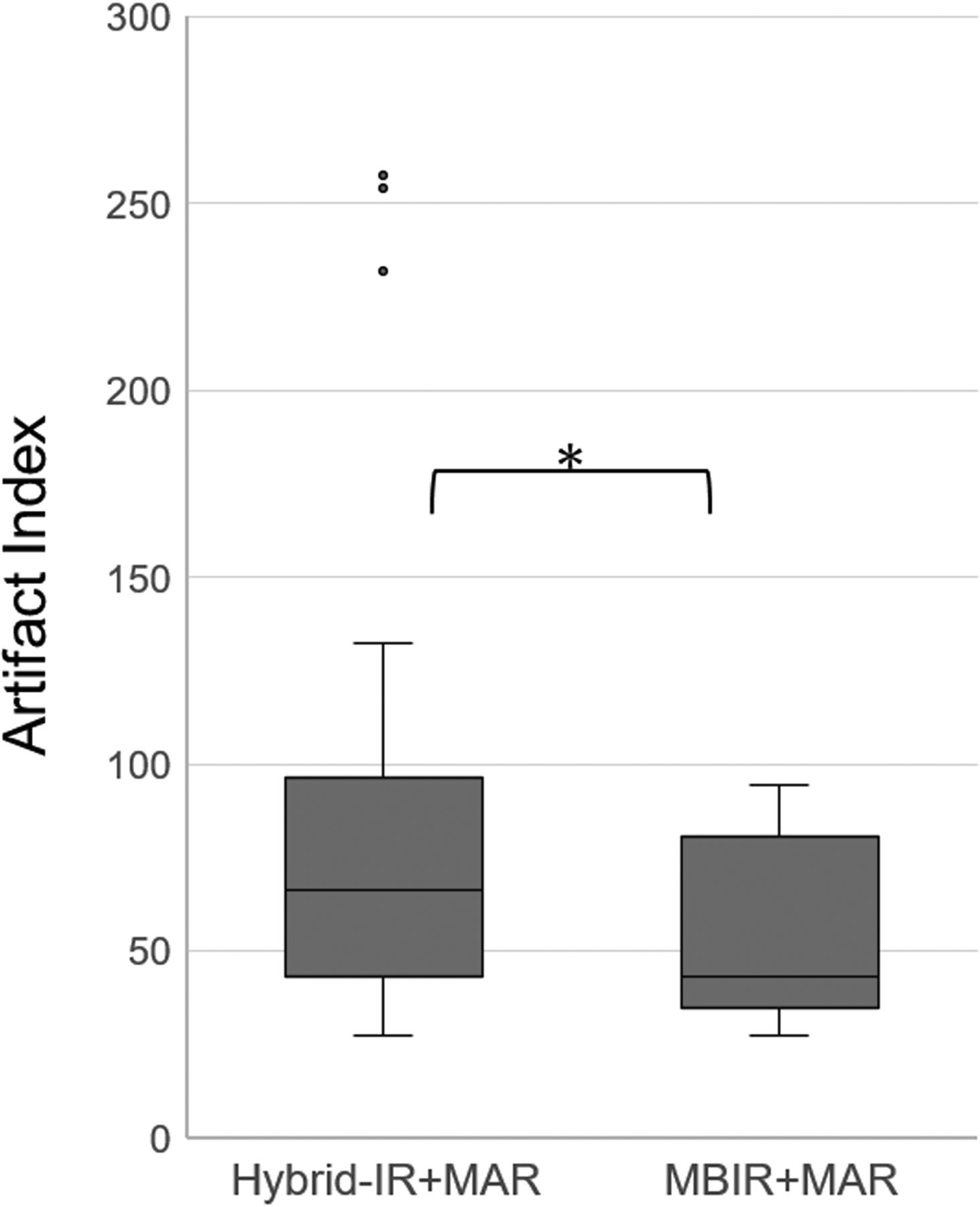
Table 2 presents the findings from quantitative image analyses. At the apex of the tongue and nuchal muscle, the mean CT attenuation value and SD from MBIR+MAR were significantly lower than those from hybrid-IR+MAR (P < .01). At the apex of the tongue, hybrid-IR+MAR and MBIR+MAR yielded mean metal artifact index values of 82.2 (95% CI, 64.2–100.2) and 73.6 (95% CI, 53.0–94.1), respectively, and these values differed significantly between the 2 algorithms (95% CI, 2.6–14.7; P < .01). Notably, MBIR+MAR led to a reduction of 11% in the metal artifact index relative to hybrid-IR+MAR (Fig 3).

FIG 3.
  • Download figure
  • Open in new tab
  • Download powerpoint
FIG 3.

Boxplot comparison of the artifact index values calculated using the 2 reconstruction algorithms: hybrid-IR+MAR and MBIR+MAR. The asterisk indicates a significant difference (P < .01).

View this table:
  • View inline
  • View popup
Table 2:

Quantitative evaluation of CT attenuation (HU) of the apex of the tongue and nuchal muscle

The median number of CT slices reconstructed was 102 (range, 83–118). The reconstruction times by hybrid-IR+MAR and MBIR+MAR were 2.1 and 37.6 seconds per section, respectively. In other words, MBIR+MAR required a reconstruction time >17 times longer than that of hybrid-IR+MAR.

Qualitative Analyses

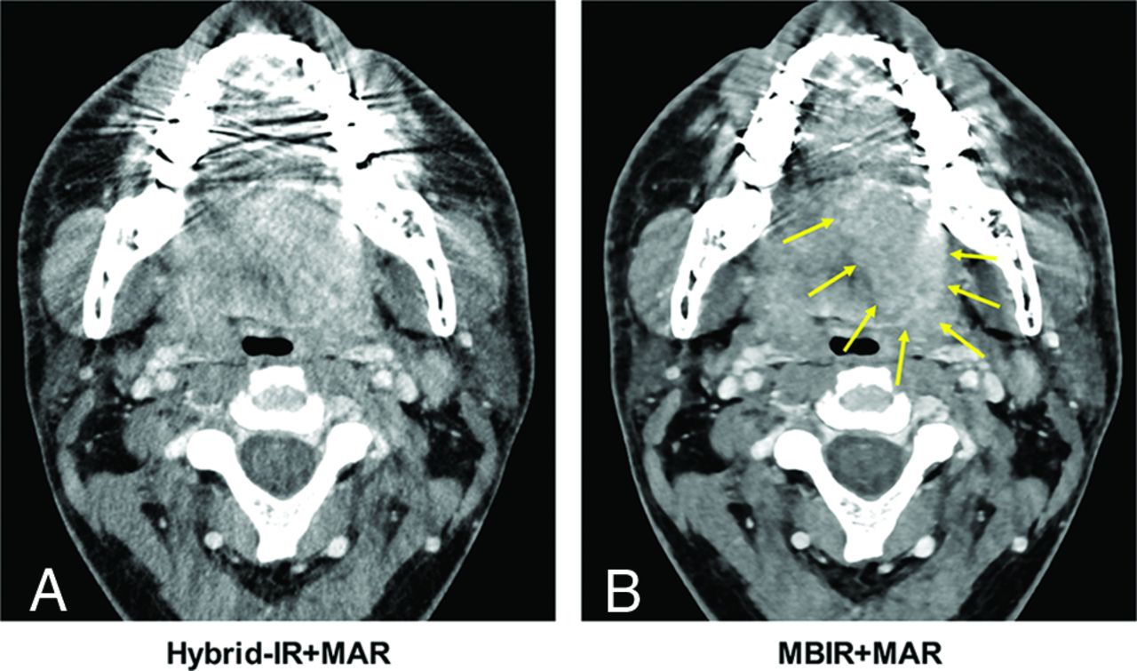
The weighted κ statistic revealed excellent interobserver agreement among the 3 readers (κ range, 0.88–0.98). A comparison of the metal artifact reduction image-quality scores revealed that MBIR+MAR yielded significant reductions in metal artifacts compared with hybrid-IR+MAR (P < .01). Figure 4 demonstrates that MBIR+MAR improved the delineation of the oral structures and oral cavity cancers by significantly reducing metal artifacts relative to those produced by hybrid-IR+MAR.

FIG 4.
  • Download figure
  • Open in new tab
  • Download powerpoint
FIG 4.

Axial CT images of a 32-year-old man with left-tongue cancer. Reconstruction was performed using hybrid-IR+MAR (A) and MBIR+MAR (B). As shown, MBIR+MAR provided superior reduction of dental artifacts and a better depiction of the tumor (arrows).

Table 3 presents the image-quality scores of the depicted neck structures. All representative structures except the apex of the tongue had a mean score of ≥3, which was within the identifiable visual level; the apex of the tongue only had a score of ≥3 using MBIR+MAR. Comparisons revealed significant differences in the average scores obtained using the 2 reconstruction algorithms. MBIR+MAR yielded higher average scores for all representative structural depictions compared with hybrid-IR+MAR.

View this table:
  • View inline
  • View popup
Table 3:

Qualitative evaluation of image-quality scores of representative structures

Finally, the approximate power curve indicated a negative relationship between the number of dental fillings or implants and the metal artifact reduction image-quality score, regardless of the reconstruction algorithm used. Higher scores were achieved with MBIR+MAR than with hybrid-IR+MAR (Fig 5). The cancer tumor stage diagnoses did not differ significantly with respect to the type of reconstruction algorithm.

FIG 5.
  • Download figure
  • Open in new tab
  • Download powerpoint
FIG 5.

Analysis of the correlation between the image-quality score of artifact reduction and the number of dental fillings or implants between the 2 reconstruction algorithms: hybrid-IR+MAR and MBIR+MAR.

DISCUSSION

This study aimed to compare the CT image quality achieved using a combination of MBIR and MAR versus that achieved with a combination of hybrid-IR and MAR in a sample of patients with oral or oropharyngeal cancer. Our quantitative analysis demonstrated that MBIR+MAR significantly reduced both the mean CT attenuation at the apex of the tongue and the metal artifact index compared with hybrid-IR+MAR. Also, our qualitative analysis demonstrated that MBIR+MAR yielded a more significant reduction of metal artifacts in the oral cavity than hybrid-IR+MAR. In other words, objective and subjective in vivo evaluations revealed significantly better artifact reduction and image quality with MBIR+MAR relative to hybrid-IR+MAR.

CT is considered a first-line diagnostic technique for oral cancer because of its broad availability, capacity for whole-body tumor staging, and good overall detection of sensitivity and specificity.35 However, it is essential to achieve an excellent image quality with minimal artifacts despite the presence of metal dental fillings or implants. Prior studies indicated a superior reduction of dental artifacts caused by dental hardware or diverse maxillofacial metal implants when several MAR algorithms from major vendors were used, compared with standard reconstruction.24⇓⇓⇓⇓-29 However, MAR algorithms may introduce new artifacts into the image. These new artifacts can appear as defects or blurring around metal hardware in the bone window.26,27 Recently, several clinical studies reported that the combination of spectral detector CT (or dual-energy CT) with virtual monoenergetic images and MAR provided optimal artifact reduction and improved diagnostic imaging assessments in patients with dental implants and bridges or metallic dental prostheses.36,37

As noted previously, the MBIR algorithm is a revolutionary reconstruction technology that uses various models and repeats the subtraction of original raw data after forward projection to yield a reconstructed image that differs minimally from the raw data. In addition to reducing image noise, only the MBIR algorithm reduces streak and beam-hardening artifacts, respectively.19 In a previous study, MBIR similarly reduced metal artifacts in the pelvis, spine, oral cavity, and extremities, which was to a greater extent than filtered back-projection or hybrid-IR.18⇓-20 Previous reports indicated a better reduction of metal artifacts when hybrid-IR+MAR was used, compared with MBIR without MAR.21,38 These results suggest that both MBIR and MAR can effectively reduce metal artifacts. Therefore, this in vivo study investigated the usefulness of MBIR+MAR for reducing metal artifacts in the oral cavity. We found that compared with hybrid-IR+MAR, MBIR+MAR provided better representations of normal neck structures and reduced metal artifacts. We attribute the improved image quality achieved with MBIR+MAR to the incorporation of MBIR settings such as the focus size, detector size, and voxel size, which would improve the spatial resolution. In previous analyses, Wellenberg et al39 and Neroladaki et al40 reported that MBIR+MAR significantly reduced orthopedic metal artifacts on pelvic CT images produced by other vendors., Although the results of those studies were consistent with our findings, the methods differed from ours in that either phantoms or small numbers of large orthopedic metal implants were used.

A previous phantom study observed that the metal artifacts increased and the CT image accuracy decreased as the number of metal implants in the oral cavity increased.41 From in our vivo study, we also concluded that the metal artifacts worsened as the number of dental fillings increased. MBIR+MAR was relatively less affected by an increased number of dental fillings or implants. Overall, our results demonstrate the specific clinical impact of the combination of MBIR and MAR, even though the metallic artifacts could not be removed completely. Specifically, this in vivo study revealed a reduction of metal artifacts on CT images of the neck region when using MBIR+MAR.

Our study had some limitations: First, U-HRCT, which first became commercially available in 2017, uses smaller detector elements equivalent to a quarter of the area of the elements in a conventional CT detector. In a previous report, U-HRCT with improved spatial resolution was shown to reduce the artifacts associated with calcified lesions in coronary arteries.42 Theoretically, U-HRCT with small detector elements would affect metal artifact reduction by improving the spatial resolution. In our study, however, all patients were scanned using U-HRCT; therefore, we could not evaluate the specific reduction of metal artifacts by U-HRCT. Second, in addition to dental fillings or implants, metal plates and screws used in postoperative applications are a major cause of image degradation and a main obstacle to the follow-up of local recurrence in patients with oral cancer. However, these devices were set as an exclusion criterion in this study. Third, the patient population was relatively small and derived from a single institution, and the study design was retrospective. Further studies with much larger samples are needed to reject null hypotheses with clinically negligible differences.43 Fourth, the inclusion of only patients with oropharyngeal and oral cancers with solid tumors of >2 cm might have led to selection bias. Ideally, we would have included early cancers because these would be most susceptible to metal artifacts. Finally, although MBIR+MAR has some advantages over hybrid-IR+MAR, as demonstrated in our study, it is also disadvantaged by significant computational requirements. Consequently, a longer reconstruction period is required. Future studies should aim to reduce the reconstitution time and increase the practical application of MBIR+MAR.

CONCLUSIONS

The combination of MBIR and MAR enabled the significant reduction of metal artifacts during oral cavity CT. Moreover, this reconstruction algorithm improved the depiction of structures in the neck with a minimal dependence on the number of dental fillings or implants.

Footnotes

  • The study was supported by a grant from Canon Medical Systems.

  • Disclosures: Yuko Kubo—RELATED: Grant: Canon Medical Systems.* Masahiko Kusumoto—RELATED: Grant: Canon Medical Systems*; UNRELATED: Payment for Lectures Including Service on Speakers Bureaus: Ono Pharmaceutical, AstraZeneca KK, MSD KK, Comments: I received honoraria for lecture fees from Ono Pharmaceutical, AstraZeneca KK, and MSD KK. Miyuki Sone—RELATED: Grant: Canon Medical Systems.* *Money paid to the institution.

  • Paper previously presented, in part, at: Annual Meeting of the European Society of Head and Neck Radiology, October 3–5, 2019; Palermo, Italy.

Indicates open access to non-subscribers at www.ajnr.org

References

  1. 1.↵
    1. Goerres GW,
    2. Hany TF,
    3. Kamel E, et al
    . Head and neck imaging with PET and PET/CT: artefacts from dental metallic implants. Eur J Nucl Med Mol Imaging 2002;29:367–70 doi:10.1007/s00259-001-0721-1 pmid:12002712
    CrossRefPubMed
  2. 2.↵
    1. Barrett JF,
    2. Keat N
    . Artifacts in CT: recognition and avoidance. Radiographics 2004;24:1679–91 doi:10.1148/rg.246045065 pmid:15537976
    CrossRefPubMed
  3. 3.↵
    1. Ravishankar S,
    2. Ye JC,
    3. Fessler JA
    . Image reconstruction: from sparsity to data-adaptive methods and machine learning. Proc IEEE Inst Electr Electron Eng 2020;108:86–109 doi:10.1109/JPROC.2019.2936204 pmid:32095024
    CrossRefPubMed
  4. 4.↵
    1. Willemink MJ,
    2. Noel PB
    . The evolution of image reconstruction for CT: from filtered back projection to artificial intelligence. Eur Radiol 2019;29:2185–95 doi:10.1007/s00330-018-5810-7 pmid:30377791
    CrossRefPubMed
  5. 5.↵
    1. Herman GT
    . Fundamentals of computerized tomography: image reconstruction from projections. Springer-Verlag: London, 2009 doi:10.1007/978-1-84628-723-7
  6. 6.↵
    1. Singh S,
    2. Kalra MK,
    3. Hsieh J, et al
    . Abdominal CT: comparison of adaptive statistical iterative and filtered back projection reconstruction techniques. Radiology 2010;257:373–83 doi:10.1148/radiol.10092212 pmid:20829535
    CrossRefPubMed
  7. 7.↵
    1. Gervaise A,
    2. Osemont B,
    3. Lecocq S, et al
    . CT image quality improvement using adaptive iterative dose reduction with wide-volume acquisition on 320-detector CT. Eur Radiol 2012;22:295–301 doi:10.1007/s00330-011-2271-7 pmid:21927791
    CrossRefPubMed
  8. 8.↵
    1. Martinsen ACT,
    2. Sæther HK,
    3. Hol PK, et al
    . Iterative reconstruction reduces abdominal CT dose. Eur J Radiol 2012;81:1483–87 doi:10.1016/j.ejrad.2011.04.021 pmid:21543174
    CrossRefPubMed
  9. 9.↵
    1. Yu Z,
    2. Thibault JB,
    3. Bouman CA, et al
    . Fast model-based X-ray CT reconstruction using spatially nonhomogeneous ICD optimization. IEEE Trans Image Process 2011;20:161–75 doi:10.1109/TIP.2010.2058811 pmid:20643609
    CrossRefPubMed
  10. 10.↵
    1. Fleischmann D,
    2. Boas FE
    . Computed tomograph: old ideas and new technology. Eur Radiol 2011;21:510–17 doi:10.1007/s00330-011-2056-z pmid:21249371
    CrossRefPubMed
  11. 11.↵
    1. Chang W,
    2. Lee JM,
    3. Lee K, et al
    . Assessment of a model-based, iterative reconstruction algorithm (MBIR) regarding image quality and dose reduction in liver computed tomography. Invest Radiol 2013;48:598–606 doi:10.1097/RLI.0b013e3182899104 pmid:23511193
    CrossRefPubMed
  12. 12.↵
    1. Goodenberger MH,
    2. Wagner-Bartak NA,
    3. Gupta S, et al
    . Computed tomography image quality evaluation of a new iterative reconstruction algorithm in the abdomen (adaptive statistical iterative reconstruction-V): a comparison with model-based iterative reconstruction, adaptive statistical iterative reconstruction, and filtered back projection reconstructions. J Comput Assist Tomogr 2018;42:184–90 doi:10.1097/RCT.0000000000000666 pmid:28806318
    CrossRefPubMed
  13. 13.↵
    1. Noda Y,
    2. Goshima S,
    3. Koyasu H, et al
    . Renovascular CT: comparison between adaptive statistical iterative reconstruction and model-based iterative reconstruction. Clin Radiol 2017;72:901.e913–19 doi:10.1016/j.crad.2017.06.002 pmid:28673448
    CrossRefPubMed
  14. 14.↵
    1. Smith EA,
    2. Dillman JR,
    3. Goodsitt MM, et al
    . Model-based iterative reconstruction: effect on patient radiation dose and image quality in pediatric body CT. Radiology 2014;270:526–34 doi:10.1148/radiol.13130362 pmid:24091359
    CrossRefPubMed
  15. 15.↵
    1. Taguchi N,
    2. Oda S,
    3. Imuta M, et al
    . Model-based iterative reconstruction in low-radiation-dose computed tomography colonography: preoperative assessment in patients with colorectal cancer. Acad Radiol 2018;25:415–22 doi:10.1016/j.acra.2017.10.008 pmid:29191684
    CrossRefPubMed
  16. 16.↵
    1. Yasaka K,
    2. Katsura M,
    3. Akahane M, et al
    . Model-based iterative reconstruction and adaptive statistical iterative reconstruction: dose-reduced CT for detecting pancreatic calcification. Acta Radiol Open 2016;5:205846011662834 doi:10.1177/2058460116628340 pmid:27110389
    CrossRefPubMed
  17. 17.↵
    1. Yuki H,
    2. Oda S,
    3. Utsunomiya D, et al
    . Clinical impact of model-based type iterative reconstruction with fast reconstruction time on image quality of low-dose screening chest CT. Acta Radiol 2016;57:295–302 doi:10.1177/0284185115575537 pmid:25817455
    CrossRefPubMed
  18. 18.↵
    1. De Crop A,
    2. Casselman J,
    3. Van Hoof T, et al
    . Analysis of metal artifact reduction tools for dental hardware in CT scans of the oral cavity: kVp, iterative reconstruction, dual-energy CT, metal artifact reduction software—does it make a difference? Neuroradiology 2015;57:841–49 doi:10.1007/s00234-015-1537-1 pmid:25929982
    CrossRefPubMed
  19. 19.↵
    1. Boudabbous S,
    2. Arditi D,
    3. Paulin E, et al
    . Model-based iterative reconstruction (MBIR) for the reduction of metal artifacts on CT. AJR Am J Roentgenol 2015;205:380–85 doi:10.2214/AJR.14.13334 pmid:26204291
    CrossRefPubMed
  20. 20.↵
    1. Kuya K,
    2. Shinohara Y,
    3. Kato A, et al
    . Reduction of metal artifacts due to dental hardware in computed tomography angiography: assessment of the utility of model-based iterative reconstruction. Neuroradiology 2017;59:231–35 doi:10.1007/s00234-017-1811-5 pmid:28255903
    CrossRefPubMed
  21. 21.↵
    1. Yasaka K,
    2. Kamiya K,
    3. Irie R, et al
    . Metal artefact reduction for patients with metallic dental fillings in helical neck computed tomography: comparison of adaptive iterative dose reduction 3D (AIDR 3D), forward-projected model-based iterative reconstruction solution (FIRST) and AIDR 3D with single-energy metal artefact reduction (SEMAR). Dentomaxillofac Radiol 2016;45:20160114 doi:10.1259/dmfr.20160114 pmid:27268082
    CrossRefPubMed
  22. 22.↵
    1. Li H,
    2. Noel C,
    3. Chen H, et al
    . Clinical evaluation of a commercial orthopedic metal artifact reduction tool for CT simulations in radiation therapy. Med Phys 2012;39:7507–17 doi:10.1118/1.4762814 pmid:23231300
    CrossRefPubMed
  23. 23.↵
    1. Brook OR,
    2. Gourtsoyianni S,
    3. Brook A, et al
    . Spectral CT with metal artifacts reduction software for improvement of tumor visibility in the vicinity of gold fiducial markers. Radiology 2012;263:696–705 doi:10.1148/radiol.12111170 pmid:22416251
    CrossRefPubMed
  24. 24.↵
    1. Lell MM,
    2. Meyer E,
    3. Kuefner MA, et al
    . Normalized metal artifact reduction in head and neck computed tomography. Invest Radiol 2012;47:415–21 doi:10.1097/RLI.0b013e3182532f17 pmid:22659592
    CrossRefPubMed
  25. 25.↵
    1. Kidoh M,
    2. Nakaura T,
    3. Nakamura S, et al
    . Reduction of dental metallic artefacts in CT: value of a newly developed algorithm for metal artefact reduction (O-MAR). Clin Radiol 2014;69:e11–16 doi:10.1016/j.crad.2013.08.008 pmid:24156796
    CrossRefPubMed
  26. 26.↵
    1. Diehn FE,
    2. Michalak GJ,
    3. DeLone DR, et al
    . CT dental artifact: comparison of an iterative metal artifact reduction technique with weighted filtered back-projection. Acta Radiol Open 2017;6:205846011774327 doi:10.1177/2058460117743279 pmid:29225924
    CrossRefPubMed
  27. 27.↵
    1. Hakim A,
    2. Slotboom J,
    3. Lieger O, et al
    . Clinical evaluation of the iterative metal artefact reduction algorithm for post-operative CT examination after maxillofacial surgery. Dentomaxillofac Radiol 2017;46:20160355 doi:10.1259/dmfr.20160355 pmid:28112538
    CrossRefPubMed
  28. 28.↵
    1. Weiß J,
    2. Schabel C,
    3. Bongers M, et al
    . Impact of iterative metal artifact reduction on diagnostic image quality in patients with dental hardware. Acta Radiol 2017;58:279–85 doi:10.1177/0284185116646144 pmid:27166346
    CrossRefPubMed
  29. 29.↵
    1. Niehues SM,
    2. Vahldiek JL,
    3. Tröltzsch D, et al
    . Impact of single-energy metal artifact reduction on CT image quality in patients with dental hardware. Comput Biol Med 2018;103:161–66 doi:10.1016/j.compbiomed.2018.10.023 pmid:30384174
    CrossRefPubMed
  30. 30.↵
    1. Lubner MG,
    2. Pickhardt PJ,
    3. Tang J, et al
    . Reduced image noise at low-dose multidetector CT of the abdomen with prior image constrained compressed sensing algorithm. Radiology 2011;260:248–56 doi:10.1148/radiol.11101380 pmid:21436086
    CrossRefPubMed
  31. 31.↵
    1. Lin XZ,
    2. Miao F,
    3. Li JY, et al
    . High-definition CT Gemstone spectral imaging of the brain: initial results of selecting optimal monochromatic image for beam-hardening artifacts and image noise reduction. J Comput Assist Tomogr 2011;35:294–97 doi:10.1097/RCT.0b013e3182058d5c
    CrossRefPubMed
  32. 32.↵
    1. Wang Y,
    2. Qian B,
    3. Li B, et al
    . Metal artifacts reduction using monochromatic images from spectral CT: evaluation of pedicle screws in patients with scoliosis. Eur J Radiol 2013;82:e360–66 doi:10.1016/j.ejrad.2013.02.024 pmid:23518146
    CrossRefPubMed
  33. 33.↵
    1. Lydiatt WM,
    2. Patel SG,
    3. O’Sullivan B, et al
    . Head and neck cancers: major changes in the American Joint Committee on Cancer Eighth Edition Cancer Staging Manual. CA Cancer J Clin 2017;67:122–37 doi:10.3322/caac.21389 pmid:28128848
    CrossRefPubMed
  34. 34.↵
    1. Svanholm H,
    2. Starklint H,
    3. Gundersen HJ, et al
    . Reproducibility of histomorphologic diagnoses with special reference to the kappa statistic. APMIS 1989;97:689–98 doi:10.1111/j.1699-0463.1989.tb00464.x pmid:2669853
    CrossRefPubMed
  35. 35.↵
    1. Blatt S,
    2. Ziebart T,
    3. Krüger M, et al
    . Diagnosing oral squamous cell carcinoma: How much imaging do we really need? A review of the current literature. J Craniomaxillofac Surg 2016;44:538–49 doi:10.1016/j.jcms.2016.02.003 pmid:26976698
    CrossRefPubMed
  36. 36.↵
    1. Cha J,
    2. Kim HJ,
    3. Kim ST, et al
    . Dual-energy CT with virtual monochromatic images and metal artifact reduction software for reducing metallic dental artifacts. Acta Radiol 2017;58:1312–19 doi:10.1177/0284185117692174 pmid:28273739
    CrossRefPubMed
  37. 37.↵
    1. Laukamp KR,
    2. Zopfs D,
    3. Lennartz S, et al
    . Metal artifacts in patients with large dental implants and bridges: combination of metal artifact reduction algorithms and virtual monoenergetic images provides an approach to handle even strongest artifacts. Eur Radiol 2019;29:4228–38 doi:10.1007/s00330-018-5928-7 pmid:30649598
    CrossRefPubMed
  38. 38.↵
    1. Toso S,
    2. Laurent M,
    3. Lozeron ED, et al
    . Iterative algorithms for metal artifact reduction in children with orthopedic prostheses: preliminary results. Pediatr Radiol 2018;48:1884–90 doi:10.1007/s00247-018-4217-6 pmid:30056564
    CrossRefPubMed
  39. 39.↵
    1. Wellenberg RH,
    2. Boomsma MF,
    3. van Osch JA, et al
    . Computed tomography imaging of a hip prosthesis using iterative model-based reconstruction and orthopaedic metal artefact reduction: a quantitative analysis. J Comput Assist Tomogr 2016;40:971–78 doi:10.1097/RCT.0000000000000449 pmid:27331924
    CrossRefPubMed
  40. 40.↵
    1. Neroladaki A,
    2. Martin SP,
    3. Bagetakos I, et al
    . Metallic artifact reduction by evaluation of the additional value of iterative reconstruction algorithms in hip prosthesis computed tomography imaging. Medicine 2019;98:e14341 doi:10.1097/MD.0000000000014341 pmid:30732160
    CrossRefPubMed
  41. 41.↵
    1. Tsuchida Y,
    2. Takahashi H,
    3. Watanabe H, et al
    . Effects of number of metal restorations and mandibular position during computed tomography imaging on accuracy of maxillofacial models. J Prosthodont Res 2019;63:239–44 doi:10.1016/j.jpor.2018.12.006 pmid:30658907
    CrossRefPubMed
  42. 42.↵
    1. Motoyama S,
    2. Ito H,
    3. Sarai M, et al
    . Ultra-high-resolution computed tomography angiography for assessment of coronary artery stenosis. Circ J 2018;82:1844–51 doi:10.1253/circj.CJ-17-1281 pmid:29743388
    CrossRefPubMed
  43. 43.↵
    1. Faber J,
    2. Fonseca LM
    . How sample size influences research outcomes. Dental Press J Orthod 2014;19:27–29 doi:10.1590/2176-9451.19.4.027-029.ebo pmid:25279518
    CrossRefPubMed
  • Received April 13, 2020.
  • Accepted after revision July 7, 2020.
  • © 2020 by American Journal of Neuroradiology
PreviousNext
Back to top

In this issue

American Journal of Neuroradiology: 41 (11)
American Journal of Neuroradiology
Vol. 41, Issue 11
1 Nov 2020
  • Table of Contents
  • Index by author
  • Complete Issue (PDF)
Advertisement
Print
Download PDF
Email Article

Thank you for your interest in spreading the word on American Journal of Neuroradiology.

NOTE: We only request your email address so that the person you are recommending the page to knows that you wanted them to see it, and that it is not junk mail. We do not capture any email address.

Enter multiple addresses on separate lines or separate them with commas.
Diagnostic Value of Model-Based Iterative Reconstruction Combined with a Metal Artifact Reduction Algorithm during CT of the Oral Cavity
(Your Name) has sent you a message from American Journal of Neuroradiology
(Your Name) thought you would like to see the American Journal of Neuroradiology web site.
CAPTCHA
This question is for testing whether or not you are a human visitor and to prevent automated spam submissions.
Cite this article
Y. Kubo, K. Ito, M. Sone, H. Nagasawa, Y. Onishi, N. Umakoshi, T. Hasegawa, T. Akimoto, M. Kusumoto
Diagnostic Value of Model-Based Iterative Reconstruction Combined with a Metal Artifact Reduction Algorithm during CT of the Oral Cavity
American Journal of Neuroradiology Nov 2020, 41 (11) 2132-2138; DOI: 10.3174/ajnr.A6767

Citation Manager Formats

  • BibTeX
  • Bookends
  • EasyBib
  • EndNote (tagged)
  • EndNote 8 (xml)
  • Medlars
  • Mendeley
  • Papers
  • RefWorks Tagged
  • Ref Manager
  • RIS
  • Zotero
0 Responses
Respond to this article
Share
Bookmark this article
Diagnostic Value of Model-Based Iterative Reconstruction Combined with a Metal Artifact Reduction Algorithm during CT of the Oral Cavity
Y. Kubo, K. Ito, M. Sone, H. Nagasawa, Y. Onishi, N. Umakoshi, T. Hasegawa, T. Akimoto, M. Kusumoto
American Journal of Neuroradiology Nov 2020, 41 (11) 2132-2138; DOI: 10.3174/ajnr.A6767
del.icio.us logo Twitter logo Facebook logo Mendeley logo
  • Tweet Widget
  • Facebook Like
  • Google Plus One
Purchase

Jump to section

  • Article
    • Abstract
    • ABBREVIATIONS:
    • MATERIALS AND METHODS
    • RESULTS
    • DISCUSSION
    • CONCLUSIONS
    • Footnotes
    • References
  • Figures & Data
  • Info & Metrics
  • Responses
  • References
  • PDF

Related Articles

  • No related articles found.
  • PubMed
  • Google Scholar

Cited By...

  • No citing articles found.
  • Crossref (14)
  • Google Scholar

This article has been cited by the following articles in journals that are participating in Crossref Cited-by Linking.

  • Advances in metal artifact reduction in CT images: A review of traditional and novel metal artifact reduction techniques
    Mark Selles, Jochen A.C. van Osch, Mario Maas, Martijn F. Boomsma, Ruud H.H. Wellenberg
    European Journal of Radiology 2024 170
  • Ultra-High-Resolution CT of the Head and Neck with Deep Learning Reconstruction—Assessment of Image Quality and Radiation Exposure and Intraindividual Comparison with Normal-Resolution CT
    Sebastian Altmann, Mario A. Abello Mercado, Felix A. Ucar, Andrea Kronfeld, Bilal Al-Nawas, Anirban Mukhopadhyay, Christian Booz, Marc A. Brockmann, Ahmed E. Othman
    Diagnostics 2023 13 9
  • Iterative Metal Artifact Reduction in Head and Neck CT Facilitates Tumor Visualization of Oral and Oropharyngeal Cancer Obscured by Artifacts From Dental Hardware
    Nadine Bayerl, Matthias Stefan May, Wolfgang Wuest, Jan-Peter Roth, Manuel Kramer, Christian Hofmann, Bernhard Schmidt, Michael Uder, Stephan Ellmann
    Academic Radiology 2023 30 12
  • A projection-domain iterative algorithm for metal artifact reduction by minimizing the total-variation norm and the negative-pixel energy
    Gengsheng L. Zeng
    Visual Computing for Industry, Biomedicine, and Art 2022 5 1
  • Reconstruction Algorithm-Based CT Imaging for the Diagnosis of Hepatic Ascites
    Huitao Zhang, Wenhao Lv, Haofeng Diao, Li Shang, Ahmed Faeq Hussein
    Computational and Mathematical Methods in Medicine 2022 2022
  • Deep Learning Reconstruction Plus Single-Energy Metal Artifact Reduction for Supra Hyoid Neck CT in Patients With Dental Metals
    Masumi Mizuki, Koichiro Yasaka, Rintaro Miyo, Yuta Ohtake, Akiyoshi Hamada, Reina Hosoi, Osamu Abe
    Canadian Association of Radiologists Journal 2024 75 1
  • Effectiveness of Radiological Technologists in Reporting Computed Tomography Findings in After-hour Emergencies
    Norikazu Koori, Yusuke Yoshida, Akari Noda, Akiko Maeda, Fuminari Nishikawa, Mayumi Yasui, Kazuma Kurata, Yudai Suzuki, Hiroki Kamekawa, Hiroko Nishikawa
    Japanese Journal of Radiological Technology 2022 78 6
  • A comprehensive dual energy method for CBCT metal artifact reduction
    Weiwei Ge, Zihao Liu, Hehe Cui, Xiaogang Yuan, Yidong Yang
    Physics in Medicine & Biology 2025 70 1
  • CT Image Features Based on the Reconstruction Algorithm for Continuous Blood Purification Combined with Nursing Intervention in the Treatment of Severe Acute Pancreatitis
    Yanyan Liu, Mingli Gu, Liping Liu, Lunmeng Cui, Aimin Xing, M Pallikonda Rajasekaran
    Contrast Media & Molecular Imaging 2022 2022 1
  • Efficacy of a radiographic film holder adapter in generating radiographs of dental implants with improved geometric accuracy and sharpness
    Eduarda Alberti Bonadiman, Eduarda Lins Fachetti, Francisco Haiter-Neto, Teresa Cristina Rangel Pereira, Sergio Lins de-Azevedo-Vaz
    The Journal of Prosthetic Dentistry 2024 132 2

More in this TOC Section

  • Chondrosarcoma vs Synovial Chondromatosis: Imaging
  • NI-RADS for HEAD&NECK Cancer Recurrence
  • WHO Classification Update: Nasal&Skull Base Tumors
Show more HEAD & NECK

Similar Articles

Advertisement

Indexed Content

  • Current Issue
  • Accepted Manuscripts
  • Article Preview
  • Past Issues
  • Editorials
  • Editors Choice
  • Fellow Journal Club
  • Letters to the Editor

Cases

  • Case Collection
  • Archive - Case of the Week
  • Archive - Case of the Month
  • Archive - Classic Case

Special Collections

  • Special Collections

Resources

  • News and Updates
  • Turn around Times
  • Submit a Manuscript
  • Author Policies
  • Manuscript Submission Guidelines
  • Evidence-Based Medicine Level Guide
  • Publishing Checklists
  • Graphical Abstract Preparation
  • Imaging Protocol Submission
  • Submit a Case
  • Become a Reviewer/Academy of Reviewers
  • Get Peer Review Credit from Publons

Multimedia

  • AJNR Podcast
  • AJNR SCANtastic
  • Video Articles

About Us

  • About AJNR
  • Editorial Board
  • Not an AJNR Subscriber? Join Now
  • Alerts
  • Feedback
  • Advertise with us
  • Librarian Resources
  • Permissions
  • Terms and Conditions

American Society of Neuroradiology

  • Not an ASNR Member? Join Now

© 2025 by the American Society of Neuroradiology All rights, including for text and data mining, AI training, and similar technologies, are reserved.
Print ISSN: 0195-6108 Online ISSN: 1936-959X

Powered by HighWire