Advertisement

Main menu

  • Home
  • Content
    • Current Issue
    • Accepted Manuscripts
    • Article Preview
    • Past Issue Archive
    • AJNR Case Collection
    • Case of the Week Archive
    • Classic Case Archive
    • Case of the Month Archive
  • Special Collections
    • Spinal CSF Leak Articles (Jan 2020-June 2024)
    • 2024 AJNR Journal Awards
    • Most Impactful AJNR Articles
  • Multimedia
    • AJNR Podcast
    • AJNR Scantastics
    • Video Articles
  • For Authors
    • Submit a Manuscript
    • Author Policies
    • Fast publishing of Accepted Manuscripts
    • Graphical Abstract Preparation
    • Manuscript Submission Guidelines
    • Imaging Protocol Submission
    • Submit a Case for the Case Collection
  • About Us
    • About AJNR
    • Editorial Board
  • More
    • Become a Reviewer/Academy of Reviewers
    • Subscribers
    • Permissions
    • Alerts
    • Feedback
    • Advertisers
    • ASNR Home
  • Other Publications
    • ajnr

User menu

  • Alerts
  • Log in

Search

  • Advanced search
American Journal of Neuroradiology

American Journal of Neuroradiology

ASHNR American Society of Functional Neuroradiology ASHNR American Society of Pediatric Neuroradiology ASSR
  • Alerts
  • Log in

Advanced Search

  • Home
  • Content
    • Current Issue
    • Accepted Manuscripts
    • Article Preview
    • Past Issue Archive
    • AJNR Case Collection
    • Case of the Week Archive
    • Classic Case Archive
    • Case of the Month Archive
  • Special Collections
    • Spinal CSF Leak Articles (Jan 2020-June 2024)
    • 2024 AJNR Journal Awards
    • Most Impactful AJNR Articles
  • Multimedia
    • AJNR Podcast
    • AJNR Scantastics
    • Video Articles
  • For Authors
    • Submit a Manuscript
    • Author Policies
    • Fast publishing of Accepted Manuscripts
    • Graphical Abstract Preparation
    • Manuscript Submission Guidelines
    • Imaging Protocol Submission
    • Submit a Case for the Case Collection
  • About Us
    • About AJNR
    • Editorial Board
  • More
    • Become a Reviewer/Academy of Reviewers
    • Subscribers
    • Permissions
    • Alerts
    • Feedback
    • Advertisers
    • ASNR Home
  • Follow AJNR on Twitter
  • Visit AJNR on Facebook
  • Follow AJNR on Instagram
  • Join AJNR on LinkedIn
  • RSS Feeds

Welcome to the new AJNR, Updated Hall of Fame, and more. Read the full announcements.


AJNR is seeking candidates for the position of Associate Section Editor, AJNR Case Collection. Read the full announcement.


  • Getting new auth cookie, if you see this message a lot, tell someone!
  • Getting new auth cookie, if you see this message a lot, tell someone!
Research ArticleInterventional

Differential Contribution of ASPECTS Regions to Clinical Outcome after Thrombectomy for Acute Ischemic Stroke

S.M. Seyedsaadat, A.A. Neuhaus, P.J. Nicholson, E.C. Polley, C.A. Hilditch, D.C. Mihal, T. Krings, J. Benson, I. Mark, D.F. Kallmes, W. Brinjikji and J.D. Schaafsma
American Journal of Neuroradiology June 2021, 42 (6) 1104-1108; DOI: https://doi.org/10.3174/ajnr.A7096
S.M. Seyedsaadat
aFrom the Department of Radiology (S.M.S., E.C.P., J.B., I.M., D.F.K., W.B.), Mayo Clinic, Rochester, Minnesota
bDepartment of Radiology (S.M.S.), Mayo Clinic, Jacksonville, Florida
  • Find this author on Google Scholar
  • Find this author on PubMed
  • Search for this author on this site
  • ORCID record for S.M. Seyedsaadat
A.A. Neuhaus
cRadcliffe Department of Medicine (A.A.N.), University of Oxford, Oxford, UK
  • Find this author on Google Scholar
  • Find this author on PubMed
  • Search for this author on this site
  • ORCID record for A.A. Neuhaus
P.J. Nicholson
dDepartment of Medicine (P.J.N., C.A.H., T.K., J.D.S.), Division of Neurology. University Health Network/University of Toronto, Toronto, Ontario, Canada
  • Find this author on Google Scholar
  • Find this author on PubMed
  • Search for this author on this site
  • ORCID record for P.J. Nicholson
E.C. Polley
aFrom the Department of Radiology (S.M.S., E.C.P., J.B., I.M., D.F.K., W.B.), Mayo Clinic, Rochester, Minnesota
  • Find this author on Google Scholar
  • Find this author on PubMed
  • Search for this author on this site
  • ORCID record for E.C. Polley
C.A. Hilditch
dDepartment of Medicine (P.J.N., C.A.H., T.K., J.D.S.), Division of Neurology. University Health Network/University of Toronto, Toronto, Ontario, Canada
eDepartment of Neuroradiology (C.A.H.), Manchester Centre for Clinical Neurosciences, Salford Royal National Health Service Foundation Trust, Salford, UK
  • Find this author on Google Scholar
  • Find this author on PubMed
  • Search for this author on this site
  • ORCID record for C.A. Hilditch
D.C. Mihal
fDepartment of Radiology (D.C.M.), Division of Neuroradiology, Cleveland Clinic, Cleveland, Ohio
  • Find this author on Google Scholar
  • Find this author on PubMed
  • Search for this author on this site
  • ORCID record for D.C. Mihal
T. Krings
dDepartment of Medicine (P.J.N., C.A.H., T.K., J.D.S.), Division of Neurology. University Health Network/University of Toronto, Toronto, Ontario, Canada
  • Find this author on Google Scholar
  • Find this author on PubMed
  • Search for this author on this site
  • ORCID record for T. Krings
J. Benson
aFrom the Department of Radiology (S.M.S., E.C.P., J.B., I.M., D.F.K., W.B.), Mayo Clinic, Rochester, Minnesota
  • Find this author on Google Scholar
  • Find this author on PubMed
  • Search for this author on this site
  • ORCID record for J. Benson
I. Mark
aFrom the Department of Radiology (S.M.S., E.C.P., J.B., I.M., D.F.K., W.B.), Mayo Clinic, Rochester, Minnesota
  • Find this author on Google Scholar
  • Find this author on PubMed
  • Search for this author on this site
  • ORCID record for I. Mark
D.F. Kallmes
aFrom the Department of Radiology (S.M.S., E.C.P., J.B., I.M., D.F.K., W.B.), Mayo Clinic, Rochester, Minnesota
  • Find this author on Google Scholar
  • Find this author on PubMed
  • Search for this author on this site
  • ORCID record for D.F. Kallmes
W. Brinjikji
aFrom the Department of Radiology (S.M.S., E.C.P., J.B., I.M., D.F.K., W.B.), Mayo Clinic, Rochester, Minnesota
gDepartment of Neurosurgery (W.B.), Mayo Clinic, Rochester, Minnesota
  • Find this author on Google Scholar
  • Find this author on PubMed
  • Search for this author on this site
  • ORCID record for W. Brinjikji
J.D. Schaafsma
dDepartment of Medicine (P.J.N., C.A.H., T.K., J.D.S.), Division of Neurology. University Health Network/University of Toronto, Toronto, Ontario, Canada
  • Find this author on Google Scholar
  • Find this author on PubMed
  • Search for this author on this site
  • ORCID record for J.D. Schaafsma
  • Article
  • Figures & Data
  • Info & Metrics
  • Responses
  • References
  • PDF
Loading

Abstract

BACKGROUND AND PURPOSE: Ischemic stroke is the leading cause of long-term disability in adults, but our ability to prognosticate from baseline imaging data is limited. The ASPECTS measures ischemic change in the middle cerebral artery territory on noncontrast CT based on 10 anatomic regions. Here, we investigated whether infarction in particular regions was associated with worse long-term outcome.

MATERIALS AND METHODS: We identified consecutive patients receiving mechanical thrombectomy for ICA/MCA occlusion at 2 comprehensive stroke centers. Pretreatment ASPECTS was assessed by 2 blinded reviewers. Clinical data including demographics, baseline NIHSS score, and 90-day mRS were collected. The relationship between individual ASPECTS regions and the mRS score (0–2 versus 3–6) was assessed using multivariable logistic regression.

RESULTS: Three hundred fifty-three patients were included (mean age, 70 years; 46% men), of whom 214 had poor outcome (mRS = 3–6). Caudate (OR = 3.26; 95% CI, 1.33–8.82), M4 region (OR = 2.94; 95% CI, 1.09–9.46), and insula (OR = 1.75; 95% CI, 1.08–2.85) infarcts were associated with significantly greater odds of poor outcome, whereas M1 region infarction reduced the odds of poor outcome (OR = 0.38; 95% CI, 0.14–0.99). This finding remained unchanged when restricted to only patients with good recanalization. No significant associations were found by laterality. Similarly, no region was predictive of neurologic improvement during the first 24 hours or of symptomatic intracerebral hemorrhage.

CONCLUSIONS: Our results indicate that ASPECTS regions are not equal in their contribution to functional outcome. This finding suggests that patient selection based on total ASPECTS alone might be insufficient, and infarct topography should be considered when deciding eligibility for thrombectomy.

ABBREVIATION:

sICH
symptomatic intracranial hemorrhage

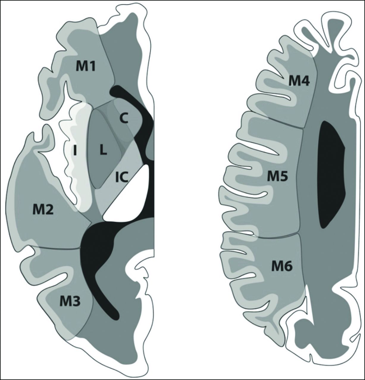
Recent advances in the field of endovascular thrombectomy have led to a sea change in the management of large-vessel-occlusion acute ischemic stroke, with several initial trials showing benefit with new-generation endovascular approaches.1⇓⇓⇓⇓-6 The time window for thrombectomy has subsequently expanded to up to 24 hours from onset.7,8 In all these trials, imaging was crucial to identify patients likely to benefit. Most trials in 2015 used lesion size on CT as part of their selection criteria, quantified as the ASPECTS. ASPECTS was first described in 2000 and separates the middle cerebral artery territory into 10 regions (6 superficial, 4 deep; Figure). These are then assigned a value of 0 if there are early ischemic changes—parenchymal hypoattenuation, loss of gray-white differentiation, and focal swelling—and a value of 2 if the region is normal in appearance.9 Correspondingly, lower scores imply more extensive ischemia and intuitively suggest that the outcome is more likely to be poor; indeed, the ASPECTS is known to have value in long-term prognostication after stroke,10 and has previously been shown to correlate with functional independence in intra-arterial thrombolysis.11 Thus, low ASPECTS values continue to be used as an exclusion criterion for thrombectomy because these patients are assumed to have a low likelihood of meaningful improvement.

FIGURE.
  • Download figure
  • Open in new tab
  • Download powerpoint
FIGURE.

Illustration of ASPECTS, showing 10 regions in 1 hemisphere. C indicates caudate; IC, internal capsule; L, lentiform; I, insula. Reproduced from Neuhaus et al25 with permission from BMJ Publishing Group Ltd.

However, there are a number of disadvantages in using ASPECTS. First, although it significantly correlates with long-term function on a group level, individual outcomes are discriminated less accurately, particularly when the ASPECTS is moderate to high (eg, 6–10, implying limited ischemic change).12 Second, it is known that involvement of specific regions leads to particular functional deficits, eg, the angular gyrus in language13 and multiple cortical and subcortical areas in motor function.14 The ASPECTS treats all 10 areas equally; therefore, a composite ASPECTS of 7 may reflect very different lesion patterns, and there is no a priori reason to think these would be equivalent in terms of functional consequences. Third, the volumes of ASPECTS regions are not equal, and the loss of a single point can reflect a wide range of ischemic volumes, depending on which areas are affected. Indeed, it has been previously reported that some regions confer a greater risk of poor long-term outcome.15 Variation in outcome based on the affected area has also been described with ASPECTS regions from baseline CT,15⇓⇓-18 though with inconsistent findings.

The implication of this finding is that a significant number of patients with a poor composite ASPECTS might, in fact, have a greater likelihood of good outcome than the total score would suggest, which may influence treatment decisions. In this study, we sought to estimate regional contributions to long-term function using pretreatment ASPECTS data in a thrombectomy cohort.

MATERIALS AND METHODS

Study Subjects

We conducted a retrospective analysis of 353 consecutively treated patients with a diagnosis of acute ischemic stroke related to an emergent large-vessel occlusion from January 2015 to December 2018. One hundred seventy-eight patients were treated at the first participating center, and 175 patients, at the second (Mayo Clinic and Toronto Western Hospital). ASPECTS ≥6 was a prerequisite for proceeding with thrombectomy at our institutions.

The inclusion criteria were as follows:

  1. Clinical diagnosis of acute stroke in the anterior circulation

  2. Eighteen years of age or older

  3. Availability of NCCT images acquired within 24 hours of onset of symptoms

  4. Patients with intracranial ICA, M1, or M2 occlusion identified on CTA who were treated by endovascular thrombectomy.

Among the exclusion criteria were nonischemic pathology found on baseline imaging (eg, intracerebral hemorrhage), poor quality of baseline scans (eg, excessive artifacts preventing regional ASPECTS estimation), and denial of consent following checking for research authorization.

Baseline clinical characteristics collected from subjects included age, sex, NIHSS score, and time of last known well; posttreatment characteristics included the mRS score at 90 days and recanalization status after thrombectomy. The study was approved by the institutional review boards at both institutions; because this was a retrospective study, informed consent was waived. However, per Minnesota state law, after checking the status of all patients residing in the state of Minnesota, 21 patients who denied access to their medical records for research use were not included in this study.

CT Acquisition and Analysis

Noncontrast CT scans with 5-mm section thickness were obtained at admission, with a maximal delay of <24 hours from stroke onset. ASPECTS was independently determined by 2 neuroradiologists at each site, for a total of 4 raters who were blinded to clinical outcome. In case of any disagreement, a third neuroradiologist in each center reviewed and adjudicated the score. 9,19 In contrast to the original methodology of using 1 supraganglionic and 1 ganglionic section for ASPECTS, assessors had access to the entire NCCT scan. Evidence of early ischemic change was defined as the presence of hypoattenuation and/or loss of gray matter–white matter differentiation, with or without cortical swelling. Assessors were aware of the presumed laterality of the stroke but blinded to other clinical details.

Statistical Analysis

A summary of clinical and imaging characteristics is presented in the Online Supplemental Data as mean (SD) (age, NIHSS) or median with interquartile range (ASPECTS). The relationship between individual ASPECTS regions and the ordinal functional outcome score (mRS) was assessed using multivariable logistic regression. mRS was dichotomized as good (0–2) or poor (3–6). Ten-fold cross-validation was then used to estimate the predictive value of each region, and a multivariable logistic regression with the 10 regions in a single model was fitted. This was also used for subgroup analyses based on laterality (left and right hemispheres separately) and recanalization status based on angiographic findings (dichotomized as TICI 2b/3 for successful recanalization and also examining TICI 3 separately). We also explored the predictive value of regional ASPECTS for symptomatic intracranial hemorrhage following thrombectomy and for NIHSS change within the first day following stroke onset (calculated simply as NIHSS24 hours – NIHSSbaseline) as an indicator of acute neurologic deterioration or improvement. Data were collected in Excel (Microsoft) and analyzed using R statistical and computing software (http://www.r-project.org/).

RESULTS

Baseline Characteristics

The baseline characteristics of the patient population are presented in the Online Supplemental Data. The mean age of patients was 70 years, and 46% were men. Baseline imaging showed a median total ASPECTS of 9; early ischemic changes were most commonly seen in the insula (172 patients, 48.7%) and lentiform nucleus (96 patients, 27.2%) and least frequently in M6 (16 patients, 4.5%).

Primary Outcome

Logistic regression for regional ASPECTS irrespective of laterality (Table 1) showed that the regions significantly associated with poor outcome were the caudate nucleus (ORcaudate = 3.26; 95% CI, 1.33–8.82), M4 (ORM4 = 2.94; 95% CI, 1.09–9.46), and insula (ORinsula = 1.75; 95% CI, 1.08–2.85). Conversely, M1 was associated with reduced odds of poor outcome (ORM1 = 0.38; 95% CI, 0.14–0.99). The remaining 6 areas (M2, M3, M5, M6, lentiform nucleus, and internal capsule) did not reach statistical significance. When we separated them by the side of the stroke, no regions reached statistical significance, though similar trends were observed; on the right, there was an additional trend for M6 in particular (ORM6 = 3.71; 95% CI, 0.72–29.4) and on the left for M5 (ORM5 = 2.80; 95% CI, 0.81–13.2).

View this table:
  • View inline
  • View popup
Table 1:

Predictive value of ASPECTS regions for mRS at 90 days

Subgroup Analyses

The average total ASPECTS for patients without insular or caudate infarcts was higher than for those with insula or caudate involvement (P value < .0001) (Table 2).

View this table:
  • View inline
  • View popup
Table 2:

Comparison of total ASPECTS between patients with caudate or insular infarct with the rest of cohort

Of the studied population, 302 (85.6%) had successful recanalization, defined as TICI 2b/3. When this cohort was analyzed as a subgroup, the results were effectively unchanged from the total population (Table 3), with the caudate nucleus (ORcaudate = 4.02; 95% CI, 1.51–12.2), M4 (ORM4 = 3.54; 95% CI, 1.26–11.9), and insula (ORinsula = 1.69; 95% CI, 1.01–2.85) being associated with poor outcome; again, M1 infarction was associated with lower odds of poor outcome (ORM1 = 0.28; 95% CI, 0.09–0.83). When this cohort was restricted to only TICI 3 (n = 154), suggesting complete reperfusion, only the caudate nucleus (ORcaudate = 14.86; 95% CI, 3.22–114.94) reached statistical significance. The group of patients with unsuccessful recanalization (TICI 0/1/2a, n = 51) was too small to reliably estimate ORs.

View this table:
  • View inline
  • View popup
Table 3:

Predictive value of ASPECTS regions for mRS at 90 days, stratified by recanalization status

ASPECTS Regions and Short-Term Outcomes

We then repeated the analyses using symptomatic intracranial hemorrhage (sICH) as an outcome. In our sample, 27 patients (7.6%) developed sICH within the first 24 hours following thrombectomy. Due to the small sample size, there was no significant association with any ASPECTS region; because the lowest P value was .299, it was not possible to characterize any trends in the data (Table 4). We also used changes in the NIHSS score during the first 24 hours as the outcome metric to explore the impact of individual regions on short-term neurologic improvement. No individual ASPECTS region showed significant correlation with NIHSS change (Online Supplemental Data), with a slight trend toward the lentiform nucleus (ORlentiform = 6.89; 95% CI, 0.57–83.10).

View this table:
  • View inline
  • View popup
Table 4:

Predictive value of ASPECTS regions for sICH

DISCUSSION

In this study, we have characterized the long-term functional impact of ischemia in specific regions of the ASPECTS system in patients who underwent thrombectomy after baseline imaging. Our main finding is that infarction in the caudate, M4, and insula areas is significantly associated with poor outcome (mRS = 3–6) following thrombectomy, whereas patients with M1 infarctions have reduced odds of poor outcome. The same observation was made when analyses were restricted to only those patients in whom good recanalization was achieved. Analyses by laterality or looking at neurologic improvement and sICH as outcomes did not reach statistical significance. The implication of these results may be that the use of a composite ASPECTS should be re-evaluated because the total score may be less informative than individual territories, as discussed below.

Previous studies have examined the contribution of individual ASPECTS regions to outcome. However, the results reported in the literature show significant disagreement. For example, Khan et al18 reported that M3 and M6 infarction on either side, M2 on the right, and M5 on the left were associated with poor outcome. Beare et al17 also reported M2 and M5 infarction as a poor prognostic factor, without specifying side. In the largest study looking at admission NCCT, Payabvash et al20 found M5, caudate, lentiform, and insula infarction to be associated with poor outcome in patients treated with intravenous thrombolysis. Phan et al16 only found significant results from interaction terms between age and lentiform, and age and M6. There have also been studies using the ASPECTS template with modalities other than NCCT. Haranhalli et al21 found that insula, M2, and M5 infarcts on CT perfusion were associated with poor outcome. Payabvash et al15 also reported that insula infarction predicted poor outcome, using DWI at admission.

One important difference with the results reported here is the clinical care provided to the patient population. Most patients in the aforementioned studies were either managed conservatively or treated with intravenous thrombolysis. Payabvash et al15,20 and Haranhalli et al21 did include patients with subsequent intra-arterial therapy, but given that data collection occurred before 2015, these likely represented intra-arterial thrombolysis and thrombectomy with first-generation devices, which were notably less effective. In contrast, our study included only patients who underwent stent retriever or aspiration thrombectomy and is, therefore, more reflective of the current criterion standard approach to management of large-vessel-occlusion stroke.

We can only speculate on why the regions identified by our study were associated with worse outcomes. M4 represents the frontal cortex on the supraganglionic section, whereas M1 is more the caudal frontal cortex. In terms of function, infarcts involving M1 might lead to deficits in behavioral planning due to involvement of the operculum and may also lead to nonfluent aphasia if the dominant hemisphere (ie, left in most individuals) is affected. Hemiplegia, in particular, might be expected to have a substantial effect on mRS because the scale is largely based on mobility. M4 might similarly be associated with aphasia and apraxia on the dominant side, though limb weakness is less likely. Similarly, the caudate nucleus is part of the basal ganglia and crucial for motor control, as well as being associated with cognitive changes. The insula is primarily associated with autonomic function, but also with motor control, cognition, and emotion.22,23

Limitations

There are limitations to this study. First, although our total cohort was relatively large, subgroup analyses for sICH, stroke laterality, and outcome in poorly recanalized patients were limited by small sample sizes. In addition, we had relatively few cases with stroke in some of the cortical areas (most notably M6) and the internal capsule. This spectrum bias is probably the consequence of selection bias for endovascular thrombectomy. Second, the median ASPECTS in this study was 9, suggesting only limited ischemic changes. Although this was also the median ASPECTS value in the meta-analysis of the 2015 trials1 and is therefore representative of patients receiving endovascular thrombectomy, it, nonetheless, complicates applying these findings to patients with more extensive ischemic changes and lower total ASPECTS. Third, our analyses currently assume that regional ischemic changes were independent of each other, and we have not adjusted for collinearity; however, preliminary analyses did not show significant collinearity in our dataset. Although in our cohort, patients with infarcts in the insula or caudate nucleus had poorer total ASPECTS, this still warrants further follow-up studies. Fourth, there is potential interobserver variability, as was reported by Nicholson et al24 in their study of 375 patients with acute ischemic stroke, which showed substantial interobserver variability among readers in scoring different ASPECTS regions. They found that readers had the highest agreement in scoring the insula (κ = 0.56; range, 0.48–0.64) and the lowest agreement in scoring M3 (κ = 0.34; range, 0.12–0.56).

CONCLUSIONS

We have characterized outcome based on regional infarction as determined by the ASPECTS template on baseline NCCT. This work is hypothesis-generating and may be of potential use in the development of scoring systems that take infarct topography into account and allow improved patient-selection algorithms for thrombectomy. Therefore, larger datasets are required to validate our findings and provide more definitive quantification of the impact of ischemia in different ASPECTS areas on outcome.

Footnotes

  • W. Brinjikji and J.D. Schaafsma contributed equally as senior and supervising authors.

  • This work was supported by an Award from the American Heart Association (19POST34381067) to S.M. Seyedsaadat. A.A. Neuhaus was supported by an award from the Oxford University Clinical Academic Graduate School.

  • Disclosures: Seyed Mohammad Seyedsaadat—RELATED: Grant: American Heart Association, Comments: 2 years postdoctoral fellowship American Heart Association research grant. Timo Krings—UNRELATED: Consultancy: Stryker, Medtronic, Penumbra; Royalties: Thieme; Stock/Stock Options: Marblehead Medical LLC. Ain A. Neuhaus—RELATED: Grant: Oxford University Clinical Academic Graduate School, Comments: internal grant from the Oxford University Clinical Academic Graduate School to support travel and living expenses for medical elective at the Mayo Clinic. David F. Kallmes—UNRELATED: Stock/Stock Options: Superior Medical Experts LLC, Marblehead Medical LLC.

References

  1. 1.↵
    1. Goyal M,
    2. Menon BK,
    3. van Zwam WH, et al
    . HERMES Collaborators. Endovascular thrombectomy after large-vessel ischaemic stroke: a meta-analysis of individual patient data from five randomised trials. Lancet 2016;387:1723–31 doi:10.1016/S0140-6736(16)00163-X pmid:26898852
    CrossRefPubMed
  2. 2.↵
    1. Campbell BC,
    2. Mitchell PJ,
    3. Kleinig TJ, et al
    . EXTEND-IA Investigators. Endovascular therapy for ischemic stroke with perfusion-imaging selection. N Engl J Med 2015;372:1009–18 doi:10.1056/NEJMoa1414792 pmid:25671797
    CrossRefPubMed
  3. 3.↵
    1. Jovin TG,
    2. Chamorro A,
    3. Cobo E, et al
    . REVASCAT Trial Investigators. Thrombectomy within 8 hours after symptom onset in ischemic stroke. N Engl J Med 2015;372:2296–2306 doi:10.1056/NEJMoa1503780 pmid:25882510
    CrossRefPubMed
  4. 4.↵
    1. Saver JL,
    2. Goyal M,
    3. Bonafe A, et al
    . SWIFT PRIME Investigators. Stent-retriever thrombectomy after intravenous t-PA vs. t-PA alone in stroke. N Engl J Med 2015;372:2285–95 doi:10.1056/NEJMoa1415061 pmid:25882376
    CrossRefPubMed
  5. 5.↵
    1. Goyal M,
    2. Demchuk AM,
    3. Menon BK, et al
    . ESCAPE Trial Investigators. Randomized assessment of rapid endovascular treatment of ischemic stroke. N Engl J Med 2015;372:1019–30 doi:10.1056/NEJMoa1414905
    CrossRefPubMed
  6. 6.↵
    1. Berkhemer OA,
    2. Fransen PS,
    3. Beumer D, et al
    . MR CLEAN Investigators. A randomized trial of intraarterial treatment for acute ischemic stroke. N Engl J Med 2015;372:11–20 doi:10.1056/NEJMoa1411587 pmid:25517348
    CrossRefPubMed
  7. 7.↵
    1. Albers GW,
    2. Marks MP,
    3. Kemp S, et al
    . DEFUSE 3 Investigators. Thrombectomy for stroke at 6 to 16 hours with selection by perfusion imaging. N Engl J Med 2018;378:708–18 doi:10.1056/NEJMoa1713973 pmid:29364767
    CrossRefPubMed
  8. 8.↵
    1. Nogueira RG,
    2. Jadhav AP,
    3. Haussen DC, et al
    . DAWN Trial Investigators. Thrombectomy 6 to 24 hours after stroke with a mismatch between deficit and infarct. N Engl J Med 2018;378:11–21 doi:10.1056/NEJMoa1706442 pmid:29129157
    CrossRefPubMed
  9. 9.↵
    1. Barber PA,
    2. Demchuk AM,
    3. Zhang J, et al
    . Validity and reliability of a quantitative computed tomography score in predicting outcome of hyperacute stroke before thrombolytic therapy: ASPECTS Study Group—Alberta Stroke Programme Early CT Score. Lancet 2000;355:1670–74 doi:10.1016/S0140-6736(00)02237-6 pmid:10905241
    CrossRefPubMed
  10. 10.↵
    1. Hill MD,
    2. Buchan AM
    ; Canadian Alteplase for Stroke Effectiveness Study (CASES) Investigators. Canadian Alteplase for Stroke Effectiveness Study I: thrombolysis for acute ischemic stroke—results of the Canadian Alteplase for Stroke Effectiveness Study. CMAJ 2005;172:1307–12 doi:10.1503/cmaj.1041561 pmid:15883405
    Abstract/FREE Full Text
  11. 11.↵
    1. Hill MD,
    2. Rowley HA,
    3. Adler F, et al
    . PROACT-II Investigators. Selection of acute ischemic stroke patients for intra-arterial thrombolysis with pro-urokinase by using ASPECTS. Stroke 2003;34:1925–31 doi:10.1161/01.STR.0000082483.37127.D0 pmid:12843342
    Abstract/FREE Full Text
  12. 12.↵
    1. Weir NU,
    2. Pexman JH,
    3. Hill MD, et al
    . CASES Investigators. How well does ASPECTS predict the outcome of acute stroke treated with IV tPA? Neurology 2006;67:516–18 doi:10.1212/01.wnl.0000228221.44334.73 pmid:16894120
    Abstract/FREE Full Text
  13. 13.↵
    1. Payabvash S,
    2. Kamalian S,
    3. Fung S, et al
    . Predicting language improvement in acute stroke patients presenting with aphasia: a multivariate logistic model using location-weighted atlas-based analysis of admission CT perfusion scans. AJNR Am J Neuroradiol 2010;31:1661–68 doi:10.3174/ajnr.A2125 pmid:20488905
    Abstract/FREE Full Text
  14. 14.↵
    1. Payabvash S,
    2. Souza LC,
    3. Kamalian S, et al
    . Location-weighted CTP analysis predicts early motor improvement in stroke: a preliminary study. Neurology 2012;78:1853–59 doi:10.1212/WNL.0b013e318258f799 pmid:22573641
    Abstract/FREE Full Text
  15. 15.↵
    1. Payabvash S,
    2. Benson JC,
    3. Tyan AE, et al
    . Multivariate prognostic model of acute stroke combining admission infarct location and symptom severity: a proof-of-concept study. J Stroke Cerebrovasc Dis 2018;27:936–44 doi:10.1016/j.jstrokecerebrovasdis.2017.10.034 pmid:29198948
    CrossRefPubMed
  16. 16.↵
    1. Phan TG,
    2. Demchuk A,
    3. Srikanth V, et al
    . Proof of concept study: relating infarct location to stroke disability in the NINDS rt-PA trial. Cerebrovasc Dis 2013;35:560–65 doi:10.1159/000351147 pmid:23838753
    CrossRefPubMed
  17. 17.↵
    1. Beare R,
    2. Chen J,
    3. Phan TG
    ; VISTA-Acute Collaboration. Googling stroke ASPECTS to determine disability: exploratory analysis from VISTA-Acute Collaboration. PLoS One 2015;10:e0125687 doi:10.1371/journal.pone.0125687 pmid:25961856
    CrossRefPubMed
  18. 18.↵
    1. Khan M,
    2. Baird GL,
    3. Goddeau RP Jr., et al
    . Alberta Stroke Program Early CT Score infarct location predicts outcome following M2 occlusion. Front Neurol 2017;8:98 doi:10.3389/fneur.2017.00098 pmid:28352248
    CrossRefPubMed
  19. 19.↵
    1. Pexman JH,
    2. Barber PA,
    3. Hill MD, et al
    . Use of the Alberta Stroke Program Early CT Score (ASPECTS) for assessing CT scans in patients with acute stroke. AJNR Am J Neuroradiol 2001;22:1534–42 pmid:11559501
    Abstract/FREE Full Text
  20. 20.↵
    1. Payabvash S,
    2. Noorbaloochi S,
    3. Qureshi AI
    . Topographic assessment of acute ischemic changes for prognostication of anterior circulation stroke. J Neuroimaging 2017;27:227–31 doi:10.1111/jon.12383 pmid:27593522
    CrossRefPubMed
  21. 21.↵
    1. Haranhalli N,
    2. Mbabuike N,
    3. Grewal SS, et al
    . Topographic correlation of infarct area on CT perfusion with functional outcome in acute ischemic stroke. J Neurosurg 2019;132:33–41 doi:10.3171/2018.8.JNS181095 pmid:30641833
    CrossRefPubMed
  22. 22.↵
    1. Berman SA,
    2. Hayman LA,
    3. Hinck VC
    . Correlation of CT cerebral vascular territories with function, 3: middle cerebral artery. AJR Am J Roentgenol 1984;142:1035–40 doi:10.2214/ajr.142.5.1035 pmid:6609554
    CrossRefPubMed
  23. 23.↵
    1. Fink JN,
    2. Selim MH,
    3. Kumar S, et al
    . Insular cortex infarction in acute middle cerebral artery territory stroke: predictor of stroke severity and vascular lesion. Arch Neurol 2005;62:1081–85 doi:10.1001/archneur.62.7.1081 pmid:16009763
    CrossRefPubMed
  24. 24.↵
    1. Nicholson P,
    2. Hilditch CA,
    3. Neuhaus A, et al
    . Per-region interobserver agreement of Alberta Stroke Program Early CT Scores (ASPECTS). J Neurointerv Surg 2020;12:1069–71 doi:10.1136/neurintsurg-2019-015473 pmid:32024784
    Abstract/FREE Full Text
  25. 25.↵
    1. Neuhaus A,
    2. Seyedsaadat SM,
    3. Mihal D, et al
    . Region-specific agreement in ASPECTS estimation between neuroradiologists and e-ASPECTS software. J Neurointerv Surg 2020;12:720–23 doi:10.1136/neurintsurg-2019-015442 pmid:31818971
    Abstract/FREE Full Text
  • Received November 14, 2019.
  • Accepted after revision December 17, 2020.
  • © 2021 by American Journal of Neuroradiology
View Abstract
PreviousNext
Back to top

In this issue

American Journal of Neuroradiology: 42 (6)
American Journal of Neuroradiology
Vol. 42, Issue 6
1 Jun 2021
  • Table of Contents
  • Index by author
  • Complete Issue (PDF)
Advertisement
Print
Download PDF
Email Article

Thank you for your interest in spreading the word on American Journal of Neuroradiology.

NOTE: We only request your email address so that the person you are recommending the page to knows that you wanted them to see it, and that it is not junk mail. We do not capture any email address.

Enter multiple addresses on separate lines or separate them with commas.
Differential Contribution of ASPECTS Regions to Clinical Outcome after Thrombectomy for Acute Ischemic Stroke
(Your Name) has sent you a message from American Journal of Neuroradiology
(Your Name) thought you would like to see the American Journal of Neuroradiology web site.
CAPTCHA
This question is for testing whether or not you are a human visitor and to prevent automated spam submissions.
Cite this article
S.M. Seyedsaadat, A.A. Neuhaus, P.J. Nicholson, E.C. Polley, C.A. Hilditch, D.C. Mihal, T. Krings, J. Benson, I. Mark, D.F. Kallmes, W. Brinjikji, J.D. Schaafsma
Differential Contribution of ASPECTS Regions to Clinical Outcome after Thrombectomy for Acute Ischemic Stroke
American Journal of Neuroradiology Jun 2021, 42 (6) 1104-1108; DOI: 10.3174/ajnr.A7096

Citation Manager Formats

  • BibTeX
  • Bookends
  • EasyBib
  • EndNote (tagged)
  • EndNote 8 (xml)
  • Medlars
  • Mendeley
  • Papers
  • RefWorks Tagged
  • Ref Manager
  • RIS
  • Zotero
0 Responses
Respond to this article
Share
Bookmark this article
Differential Contribution of ASPECTS Regions to Clinical Outcome after Thrombectomy for Acute Ischemic Stroke
S.M. Seyedsaadat, A.A. Neuhaus, P.J. Nicholson, E.C. Polley, C.A. Hilditch, D.C. Mihal, T. Krings, J. Benson, I. Mark, D.F. Kallmes, W. Brinjikji, J.D. Schaafsma
American Journal of Neuroradiology Jun 2021, 42 (6) 1104-1108; DOI: 10.3174/ajnr.A7096
del.icio.us logo Twitter logo Facebook logo Mendeley logo
  • Tweet Widget
  • Facebook Like
  • Google Plus One
Purchase

Jump to section

  • Article
    • Abstract
    • ABBREVIATION:
    • MATERIALS AND METHODS
    • RESULTS
    • DISCUSSION
    • CONCLUSIONS
    • Footnotes
    • References
  • Figures & Data
  • Info & Metrics
  • Responses
  • References
  • PDF

Related Articles

  • No related articles found.
  • PubMed
  • Google Scholar

Cited By...

  • Association between ASPECTS region of infarction and clinical outcome in non-acute large vessel occlusion ischaemic stroke after endovascular recanalisation
  • Penumbra salvage in extensive stroke: exploring limits for reperfusion therapy
  • Factors associated with unfavorable outcome after anterior circulation stroke despite successful reperfusion and early neurological improvement
  • Crossref (13)
  • Google Scholar

This article has been cited by the following articles in journals that are participating in Crossref Cited-by Linking.

  • Baseline clinical and neuroradiological predictors of outcome in patients with large ischemic core undergoing mechanical thrombectomy: A retrospective multicenter study
    Andrea M Alexandre, Mauro Monforte, Valerio Brunetti, Luca Scarcia, Luigi Cirillo, Andrea Zini, Irene Scala, Vincenzo Nardelli, Francesco Arbia, Giuseppe Arbia, Giovanni Frisullo, Erwah Kalsoum, Arianna Camilli, Davide De Leoni, Francesca Colò, Serena Abruzzese, Mariangela Piano, Claudia Rollo, Antonio Macera, Maria Ruggiero, Elvis Lafe, Joseph D Gabrieli, Giacomo Cester, Nicola Limbucci, Francesco Arba, Simone Ferretti, Valerio Da Ros, Luigi Bellini, Giancarlo Salsano, Nicola Mavilio, Riccardo Russo, Mauro Bergui, Antonio A Caragliano, Sergio L Vinci, Daniele G Romano, Giulia Frauenfelder, Vittorio Semeraro, Maria P Ganimede, Emilio Lozupone, Andrea Romi, Anna Cavallini, Luca Milonia, Massimo Muto, Paolo Candelaresi, Paolo Calabresi, Alessandro Pedicelli, Aldobrando Broccolini
    International Journal of Stroke 2024 19 7
  • Endovascular therapy in acute ischaemic stroke with large infarction with matched or mismatched clinical-radiological severities: a post-hoc analysis of the ANGEL-ASPECT trial
    Lina Zheng, Ximing Nie, Mengxing Wang, Xin Liu, Wanying Duan, Zhe Zhang, Jingyi Liu, Yufei Wei, Miao Wen, Zhonghua Yang, Thomas W. Leung, Gaoting Ma, Xiaochuan Huo, Yuesong Pan, Thanh N. Nguyen, Xinyi Leng, Zhongrong Miao, Liping Liu, Yongjun Wang, Yilong Wang, Liping Liu, David S. Liebeskind, Zhongrong Miao, Zeguang Ren, Vitor Mendes Pereira, Xunming Ji, Qiang Dong, Anding Xu, Xinfeng Liu, Qingwu Yang, Jing Jing (Chair), Zhe Zhang, Yingkui Zhang, Wei Wu, Dapeng Sun, Zhongqi Qi, Shuo Li, Zhenqiang Liu, Zequan Yu, Jingyu Zhang, Fangguang Chen, Kangyue Li, Kai Zhang, Mingkai Hu, Jianmin Liu (Chair), Chen Yao, Kangning Chen, Kun Fang (Chair), Bo Song, Yi Dong, Zhongrong Miao, Xiaochuan Huo, Gaoting Ma, Guangxiong Yuan, Hongxing Han, Wenhuo Chen, Ming Wei, Jiangang Zhang, Zhiming Zhou, Xiaoxi Yao, Guoqing Wang, Weigen Song, Xueli Cai, Guangxian Nan, Di Li, Yizhou Wang, Wentong Ling, Chuwei Cai, Changming Wen, En Wang, Liyong Zhang, Changchun Jiang, Yajie Liu, Geng Liao, Xiaohui Chen, Tianxiao Li, Shudong Liu, Jinglun Li, Yaxuan Sun, Na Xu, Zong’en Gao, Dongsheng Ju, Cunfeng Song, Jinggang Xuan, Feng Zhou, Qing Shi, Jun Luo, Yan Liu, Zaiyu Guo, Tong Li, Hongbo Zheng, Linzhi Dai, Junfeng Zhao, Liqiang Gui, Xiaokun Geng, Yufeng Tang, Congguo Yin, Hua Yang, Xiaochuan Huo, Gaoting Ma, Ruiyang An, Yuying Sun, Yanan Wu, Chunlai Yu, Shuangcheng Zheng, Yuesong Pan, Aoming Jin, Xianglong Xiang, Mengxing Wang, Hongyi Yan, Yuanling He, Chunyang Li, Weixia Kong, Yuhuan Chen, Chenhao Guo, Fengjie Ji, Pengshan Ji, Lei Liu, Xinghua Lu, Guangkuo Luo, Nanjing Wang, Yu Zhang, Bo Liu, Jian Yang, Jingjing Deng, Juan Wang, Wanru Wang, Hang Yu, Le Cui, Wenwen Liu, Ziyong Wang, Xia Zhao, Zhou Zhou, Zhongrong Miao, Guangxiong Yuan, Hongxing Han, Wenhuo Chen, Ming Wei, Jiangang Zhang, Zhiming Zhou, Xiaoxi Yao, Guoqing Wang, Weigen Song, Xueli Cai, Guangxian Nan, Di Li, Alvin Yi-Chou Wang, Wentong Ling, Chuwei Cai, Changming Wen, En Wang, Liyong Zhang, Changchun Jiang, Yajie Liu, Geng Liao, Xiaohui Chen, Tianxiao Li, Shudong Liu, Jinglun Li, Yaxuan Sun, Na Xu, Zong’en Gao, Dongsheng Ju, Cunfeng Song, Jinggang Xuan, Feng Zhou, Qing Shi, Jun Luo, Yan Liu, Zaiyu Guo, Tong Li, Hongbo Zheng, Linzhi Dai, Junfeng Zhao, Liqiang Gui, Xiaokun Geng, Yufeng Tang, Congguo Yin, Hua Yang
    eClinicalMedicine 2024 72
  • Intra- and inter-rater consistency of dual assessment by radiologist and neurologist for evaluating DWI-ASPECTS in ischemic stroke
    F. Bing, I. Berger, A. Fabry, A.-L. Moroni, C. Casile, N. Morel, S. M’Biene, J. Guellerin, C. Pignal-Jacquard, W. Vadot, G. Rodier, T. Delory, J. Jund
    Revue Neurologique 2022 178 3
  • Predictive value of computed tomography perfusion for acute ischemic stroke patients with ASPECTS &lt; 6 in an early time window
    Yiming Gu, Yu Hang, Zhenyu Jia, Linbo Zhao, Haibin Shi, Yuezhou Cao, Sheng Liu
    Clinical Neurology and Neurosurgery 2023 225
  • Predictive value of ischemia location on multimodal CT in thrombectomy-treated patients
    Clara Aguirre, Santiago Trillo, Carmen Ramos, Gustavo Zapata-Wainberg, Ancor Sanz-García, Álvaro Ximénez-Carrillo, Antonio Barbosa, José L Caniego, José Vivancos
    The Neuroradiology Journal 2023 36 3
  • Unequal Efficacy of Different Infarct Location in Predicting Futile Recanalization of Patients With Acute Ischemic Stroke
    Zhao-shuo Li, Hai-long Zhong, Teng-fei Zhou, Ying-kun He, Qiang Li, Zi-liang Wang, Liang-fu Zhu, Chang-ming Wen, Jian-feng Han, Tian-xiao Li
    Frontiers in Neurology 2022 13
  • Penumbra salvage in extensive stroke: exploring limits for reperfusion therapy
    Gabriel Broocks, Lukas Meyer, Matthias Bechstein, Sarah Elsayed, Gerhard Schön, Helge Kniep, Andre Kemmling, Uta Hanning, Jens Fiehler, Rosalie V McDonough
    Journal of NeuroInterventional Surgery 2023 15 e3
  • Selection of patients with acute vertebrobasilar artery occlusion for endovascular treatment by magnetic resonance imaging
    Jun Chen, Jiwei Zhang, Xianjin Zhu, Xuebin Zhang, Bin Jiang, Qi Liu, Zheng Z. Wei
    Frontiers in Neurology 2023 14
  • Age-specific ASPECTS atlas of Chinese subjects across different age groups for assessing acute ischemic stroke
    Qi Sun, Guan Wang, Jinzhu Yang, Yimo Zhou, Yuliang Yuan, Yan Huang, Ziyu Fu
    Scientific Data 2024 11 1
  • Association between ASPECTS region of infarction and clinical outcome in non-acute large vessel occlusion ischaemic stroke after endovascular recanalisation
    Jialiang Lu, Ziwei Lu, Ye Li, Fangcun Li, Yuxuan Feng, Meijuan Dang, Yang Yang, Fan Tang, Tao Li, Lili Zhao, Yating Jian, Xiaoya Wang, Lei Zhang, Hong Fan, Guilian Zhang
    Stroke and Vascular Neurology 2024

More in this TOC Section

  • SAVE vs. Solumbra Techniques for Thrombectomy
  • CT Perfusion&Reperfusion in Acute Ischemic Stroke
  • Delayed Reperfusion Post-Thrombectomy&Thrombolysis
Show more Interventional

Similar Articles

Advertisement

Indexed Content

  • Current Issue
  • Accepted Manuscripts
  • Article Preview
  • Past Issues
  • Editorials
  • Editors Choice
  • Fellow Journal Club
  • Letters to the Editor

Cases

  • Case Collection
  • Archive - Case of the Week
  • Archive - Case of the Month
  • Archive - Classic Case

Special Collections

  • Special Collections

Resources

  • News and Updates
  • Turn around Times
  • Submit a Manuscript
  • Author Policies
  • Manuscript Submission Guidelines
  • Evidence-Based Medicine Level Guide
  • Publishing Checklists
  • Graphical Abstract Preparation
  • Imaging Protocol Submission
  • Submit a Case
  • Become a Reviewer/Academy of Reviewers
  • Get Peer Review Credit from Publons

Multimedia

  • AJNR Podcast
  • AJNR SCANtastic
  • Video Articles

About Us

  • About AJNR
  • Editorial Board
  • Not an AJNR Subscriber? Join Now
  • Alerts
  • Feedback
  • Advertise with us
  • Librarian Resources
  • Permissions
  • Terms and Conditions

American Society of Neuroradiology

  • Not an ASNR Member? Join Now

© 2025 by the American Society of Neuroradiology All rights, including for text and data mining, AI training, and similar technologies, are reserved.
Print ISSN: 0195-6108 Online ISSN: 1936-959X

Powered by HighWire