Advertisement

Main menu

  • Home
  • Content
    • Current Issue
    • Accepted Manuscripts
    • Article Preview
    • Past Issue Archive
    • AJNR Case Collection
    • Case of the Week Archive
    • Classic Case Archive
    • Case of the Month Archive
  • Special Collections
    • Spinal CSF Leak Articles (Jan 2020-June 2024)
    • 2024 AJNR Journal Awards
    • Most Impactful AJNR Articles
  • Multimedia
    • AJNR Podcast
    • AJNR Scantastics
    • Video Articles
  • For Authors
    • Submit a Manuscript
    • Author Policies
    • Fast publishing of Accepted Manuscripts
    • Graphical Abstract Preparation
    • Manuscript Submission Guidelines
    • Imaging Protocol Submission
    • Submit a Case for the Case Collection
  • About Us
    • About AJNR
    • Editorial Board
  • More
    • Become a Reviewer/Academy of Reviewers
    • Subscribers
    • Permissions
    • Alerts
    • Feedback
    • Advertisers
    • ASNR Home
  • Other Publications
    • ajnr

User menu

  • Alerts
  • Log in

Search

  • Advanced search
American Journal of Neuroradiology

American Journal of Neuroradiology

ASHNR American Society of Functional Neuroradiology ASHNR American Society of Pediatric Neuroradiology ASSR
  • Alerts
  • Log in

Advanced Search

  • Home
  • Content
    • Current Issue
    • Accepted Manuscripts
    • Article Preview
    • Past Issue Archive
    • AJNR Case Collection
    • Case of the Week Archive
    • Classic Case Archive
    • Case of the Month Archive
  • Special Collections
    • Spinal CSF Leak Articles (Jan 2020-June 2024)
    • 2024 AJNR Journal Awards
    • Most Impactful AJNR Articles
  • Multimedia
    • AJNR Podcast
    • AJNR Scantastics
    • Video Articles
  • For Authors
    • Submit a Manuscript
    • Author Policies
    • Fast publishing of Accepted Manuscripts
    • Graphical Abstract Preparation
    • Manuscript Submission Guidelines
    • Imaging Protocol Submission
    • Submit a Case for the Case Collection
  • About Us
    • About AJNR
    • Editorial Board
  • More
    • Become a Reviewer/Academy of Reviewers
    • Subscribers
    • Permissions
    • Alerts
    • Feedback
    • Advertisers
    • ASNR Home
  • Follow AJNR on Twitter
  • Visit AJNR on Facebook
  • Follow AJNR on Instagram
  • Join AJNR on LinkedIn
  • RSS Feeds

Welcome to the new AJNR, Updated Hall of Fame, and more. Read the full announcements.


AJNR is seeking candidates for the position of Associate Section Editor, AJNR Case Collection. Read the full announcement.


Research ArticleInterventional

Learning Curve for Flow Diversion of Posterior Circulation Aneurysms: A Long-Term International Multicenter Cohort Study

N. Adeeb, M. Dibas, C.J. Griessenauer, H.H. Cuellar, M.M. Salem, S. Xiang, A. Enriquez-Marulanda, T. Hong, H. Zhang, P. Taussky, R. Grandhi, M. Waqas, A.S. Aldine, V.M. Tutino, A. Aslan, A.H. Siddiqui, E.I. Levy, C.S. Ogilvy, A.J. Thomas, C. Ulfert, M.A. Möhlenbruch, L. Renieri, J.D. Bengzon Diestro, G. Lanzino, W. Brinjikji, J. Spears, J.E. Vranic, R.W. Regenhardt, J.D. Rabinov, P. Harker, E. Müller-Thies-Broussalis, M. Killer-Oberpfalzer, C. Islak, N. Kocer, M. Sonnberger, T. Engelhorn, A. Kapadia, V.X.D Yang, A. Salehani, M.R. Harrigan, T. Krings, C.C. Matouk, S. Mirshahi, K.S. Chen, M.A. Aziz-Sultan, M. Ghorbani, C.M. Schirmer, O. Goren, S.S. Dalal, T. Finkenzeller, M. Holtmannspötter, J.-H. Buhk, P.M. Foreman, M.C. Cress, R.A. Hirschl, W. Reith, A. Simgen, H. Janssen, T.R. Marotta, C.J. Stapleton, A.B. Patel and A.A. Dmytriw
American Journal of Neuroradiology November 2022, 43 (11) 1615-1620; DOI: https://doi.org/10.3174/ajnr.A7679
N. Adeeb
aFrom the Departments of Neurosurgery and Interventional Neuroradiology (N.A., M.D., H.H.C., A.S.A., A.A.), Louisiana State University Hospital, Shreveport, Louisiana
  • Find this author on Google Scholar
  • Find this author on PubMed
  • Search for this author on this site
  • ORCID record for N. Adeeb
M. Dibas
aFrom the Departments of Neurosurgery and Interventional Neuroradiology (N.A., M.D., H.H.C., A.S.A., A.A.), Louisiana State University Hospital, Shreveport, Louisiana
  • Find this author on Google Scholar
  • Find this author on PubMed
  • Search for this author on this site
  • ORCID record for M. Dibas
C.J. Griessenauer
bDepartments of Neurosurgery and Radiology (C.J.G., C.M.S., O.G., S.S.D.), Geisinger, Danville, Pennsylvania
cDepartment of Neurology/Institut of Neurointervention (C.J.G., E.M.-T.-B., M.K.-O.), University Hospital Salzburg, Paracelsus Medical University, Salzburg, Austria
  • Find this author on Google Scholar
  • Find this author on PubMed
  • Search for this author on this site
  • ORCID record for C.J. Griessenauer
H.H. Cuellar
aFrom the Departments of Neurosurgery and Interventional Neuroradiology (N.A., M.D., H.H.C., A.S.A., A.A.), Louisiana State University Hospital, Shreveport, Louisiana
  • Find this author on Google Scholar
  • Find this author on PubMed
  • Search for this author on this site
  • ORCID record for H.H. Cuellar
M.M. Salem
dNeurosurgical Service (M.M.S., A.E.-M., P.T., C.S.O.), Beth Israel Deaconess Medical Center, Harvard Medical School, Boston, Massachusetts
  • Find this author on Google Scholar
  • Find this author on PubMed
  • Search for this author on this site
  • ORCID record for M.M. Salem
S. Xiang
fDepartment of Neurosurgery (S.X., H.Z., T.H.), Xuanwu Hospital, Capital Medical University, Beijing, China
  • Find this author on Google Scholar
  • Find this author on PubMed
  • Search for this author on this site
A. Enriquez-Marulanda
dNeurosurgical Service (M.M.S., A.E.-M., P.T., C.S.O.), Beth Israel Deaconess Medical Center, Harvard Medical School, Boston, Massachusetts
  • Find this author on Google Scholar
  • Find this author on PubMed
  • Search for this author on this site
  • ORCID record for A. Enriquez-Marulanda
T. Hong
fDepartment of Neurosurgery (S.X., H.Z., T.H.), Xuanwu Hospital, Capital Medical University, Beijing, China
  • Find this author on Google Scholar
  • Find this author on PubMed
  • Search for this author on this site
  • ORCID record for T. Hong
H. Zhang
fDepartment of Neurosurgery (S.X., H.Z., T.H.), Xuanwu Hospital, Capital Medical University, Beijing, China
  • Find this author on Google Scholar
  • Find this author on PubMed
  • Search for this author on this site
  • ORCID record for H. Zhang
P. Taussky
dNeurosurgical Service (M.M.S., A.E.-M., P.T., C.S.O.), Beth Israel Deaconess Medical Center, Harvard Medical School, Boston, Massachusetts
gDepartment of Neurosurgery (P.T., R.G.), University of Utah, Salt Lake City, Utah
  • Find this author on Google Scholar
  • Find this author on PubMed
  • Search for this author on this site
  • ORCID record for P. Taussky
R. Grandhi
gDepartment of Neurosurgery (P.T., R.G.), University of Utah, Salt Lake City, Utah
  • Find this author on Google Scholar
  • Find this author on PubMed
  • Search for this author on this site
  • ORCID record for R. Grandhi
M. Waqas
hDepartment of Neurosurgery (M.W., V.M.T., A.H.S., E.I.L.), State University of New York at Buffalo, Buffalo, New York
  • Find this author on Google Scholar
  • Find this author on PubMed
  • Search for this author on this site
  • ORCID record for M. Waqas
A.S. Aldine
aFrom the Departments of Neurosurgery and Interventional Neuroradiology (N.A., M.D., H.H.C., A.S.A., A.A.), Louisiana State University Hospital, Shreveport, Louisiana
  • Find this author on Google Scholar
  • Find this author on PubMed
  • Search for this author on this site
  • ORCID record for A.S. Aldine
V.M. Tutino
hDepartment of Neurosurgery (M.W., V.M.T., A.H.S., E.I.L.), State University of New York at Buffalo, Buffalo, New York
  • Find this author on Google Scholar
  • Find this author on PubMed
  • Search for this author on this site
  • ORCID record for V.M. Tutino
A. Aslan
aFrom the Departments of Neurosurgery and Interventional Neuroradiology (N.A., M.D., H.H.C., A.S.A., A.A.), Louisiana State University Hospital, Shreveport, Louisiana
  • Find this author on Google Scholar
  • Find this author on PubMed
  • Search for this author on this site
  • ORCID record for A. Aslan
A.H. Siddiqui
hDepartment of Neurosurgery (M.W., V.M.T., A.H.S., E.I.L.), State University of New York at Buffalo, Buffalo, New York
  • Find this author on Google Scholar
  • Find this author on PubMed
  • Search for this author on this site
  • ORCID record for A.H. Siddiqui
E.I. Levy
hDepartment of Neurosurgery (M.W., V.M.T., A.H.S., E.I.L.), State University of New York at Buffalo, Buffalo, New York
  • Find this author on Google Scholar
  • Find this author on PubMed
  • Search for this author on this site
  • ORCID record for E.I. Levy
C.S. Ogilvy
dNeurosurgical Service (M.M.S., A.E.-M., P.T., C.S.O.), Beth Israel Deaconess Medical Center, Harvard Medical School, Boston, Massachusetts
  • Find this author on Google Scholar
  • Find this author on PubMed
  • Search for this author on this site
  • ORCID record for C.S. Ogilvy
A.J. Thomas
eDepartment of Neurological Surgery (A.J.T.), Cooper University Health Care, Cooper Medical School of Rowan University, Camden, New Jersey
  • Find this author on Google Scholar
  • Find this author on PubMed
  • Search for this author on this site
  • ORCID record for A.J. Thomas
C. Ulfert
iDepartment of Neuroradiology (C.U., M.A.M.), Universitätsklinikum Heidelberg, Heidelberg, Germany
  • Find this author on Google Scholar
  • Find this author on PubMed
  • Search for this author on this site
  • ORCID record for C. Ulfert
M.A. Möhlenbruch
iDepartment of Neuroradiology (C.U., M.A.M.), Universitätsklinikum Heidelberg, Heidelberg, Germany
  • Find this author on Google Scholar
  • Find this author on PubMed
  • Search for this author on this site
  • ORCID record for M.A. Möhlenbruch
L. Renieri
jDepartment of Interventional Neuroradiology (L.R.), University of Florence, Florence, Italy
  • Find this author on Google Scholar
  • Find this author on PubMed
  • Search for this author on this site
  • ORCID record for L. Renieri
J.D. Bengzon Diestro
kDivision of Diagnostic and Therapeutic Neuroradiology (J.D.B.D., J.S., T.R.M.), St. Michael’s Hospital, Toronto, Ontario, Canada
  • Find this author on Google Scholar
  • Find this author on PubMed
  • Search for this author on this site
  • ORCID record for J.D. Bengzon Diestro
G. Lanzino
lDepartment of Neurological Surgery (G.L., W.B.), Mayo Clinic, Rochester, Minnesota
  • Find this author on Google Scholar
  • Find this author on PubMed
  • Search for this author on this site
  • ORCID record for G. Lanzino
W. Brinjikji
lDepartment of Neurological Surgery (G.L., W.B.), Mayo Clinic, Rochester, Minnesota
  • Find this author on Google Scholar
  • Find this author on PubMed
  • Search for this author on this site
  • ORCID record for W. Brinjikji
J. Spears
kDivision of Diagnostic and Therapeutic Neuroradiology (J.D.B.D., J.S., T.R.M.), St. Michael’s Hospital, Toronto, Ontario, Canada
  • Find this author on Google Scholar
  • Find this author on PubMed
  • Search for this author on this site
  • ORCID record for J. Spears
J.E. Vranic
uNeuroendovascular Program (J.E.V., R.W.R., J.D.R., P.H., S.M., K.S.C., M.A.A.-S., C.J.S., A.B.P., A.A.D.), Massachusetts General Hospital & Brigham and Women’s Hospital, Harvard Medical School, Boston, Massachusetts
  • Find this author on Google Scholar
  • Find this author on PubMed
  • Search for this author on this site
  • ORCID record for J.E. Vranic
R.W. Regenhardt
uNeuroendovascular Program (J.E.V., R.W.R., J.D.R., P.H., S.M., K.S.C., M.A.A.-S., C.J.S., A.B.P., A.A.D.), Massachusetts General Hospital & Brigham and Women’s Hospital, Harvard Medical School, Boston, Massachusetts
  • Find this author on Google Scholar
  • Find this author on PubMed
  • Search for this author on this site
  • ORCID record for R.W. Regenhardt
J.D. Rabinov
uNeuroendovascular Program (J.E.V., R.W.R., J.D.R., P.H., S.M., K.S.C., M.A.A.-S., C.J.S., A.B.P., A.A.D.), Massachusetts General Hospital & Brigham and Women’s Hospital, Harvard Medical School, Boston, Massachusetts
  • Find this author on Google Scholar
  • Find this author on PubMed
  • Search for this author on this site
  • ORCID record for J.D. Rabinov
P. Harker
uNeuroendovascular Program (J.E.V., R.W.R., J.D.R., P.H., S.M., K.S.C., M.A.A.-S., C.J.S., A.B.P., A.A.D.), Massachusetts General Hospital & Brigham and Women’s Hospital, Harvard Medical School, Boston, Massachusetts
  • Find this author on Google Scholar
  • Find this author on PubMed
  • Search for this author on this site
  • ORCID record for P. Harker
E. Müller-Thies-Broussalis
cDepartment of Neurology/Institut of Neurointervention (C.J.G., E.M.-T.-B., M.K.-O.), University Hospital Salzburg, Paracelsus Medical University, Salzburg, Austria
  • Find this author on Google Scholar
  • Find this author on PubMed
  • Search for this author on this site
  • ORCID record for E. Müller-Thies-Broussalis
M. Killer-Oberpfalzer
cDepartment of Neurology/Institut of Neurointervention (C.J.G., E.M.-T.-B., M.K.-O.), University Hospital Salzburg, Paracelsus Medical University, Salzburg, Austria
  • Find this author on Google Scholar
  • Find this author on PubMed
  • Search for this author on this site
  • ORCID record for M. Killer-Oberpfalzer
C. Islak
mDepartment of Neuroradiology (C.I., N.K.), Cerrahpasa Medical School, Istanbul University, Istanbul, Turkey
  • Find this author on Google Scholar
  • Find this author on PubMed
  • Search for this author on this site
  • ORCID record for C. Islak
N. Kocer
mDepartment of Neuroradiology (C.I., N.K.), Cerrahpasa Medical School, Istanbul University, Istanbul, Turkey
  • Find this author on Google Scholar
  • Find this author on PubMed
  • Search for this author on this site
  • ORCID record for N. Kocer
M. Sonnberger
nDepartment of Neuroradiology (M.S.), Kepler Universitätsklinikum Linz, Linz, Austria
  • Find this author on Google Scholar
  • Find this author on PubMed
  • Search for this author on this site
  • ORCID record for M. Sonnberger
T. Engelhorn
oDepartment of Neuroradiology (T.E.), University Hospital Erlangen, Erlangen, Germany
  • Find this author on Google Scholar
  • Find this author on PubMed
  • Search for this author on this site
  • ORCID record for T. Engelhorn
A. Kapadia
pDepartments of Medical Imaging and Neurosurgery (A.K.), Sunnybrook Health Sciences Centre, University of Toronto, Toronto, Ontario, Canada
  • Find this author on Google Scholar
  • Find this author on PubMed
  • Search for this author on this site
  • ORCID record for A. Kapadia
V.X.D Yang
qNeurointerventional Program (V.X.D.Y., A.A.D.), Departments of Medical Imaging & Clinical Neurological Sciences, London Health Sciences Centre, Western University, Ontario, Canada
  • Find this author on Google Scholar
  • Find this author on PubMed
  • Search for this author on this site
  • ORCID record for V.X.D Yang
A. Salehani
rDepartment of Neurosurgery (A. Salehani, M.R.H.), University of Alabama at Birmingham, Birmingham, Alabama
  • Find this author on Google Scholar
  • Find this author on PubMed
  • Search for this author on this site
  • ORCID record for A. Salehani
M.R. Harrigan
rDepartment of Neurosurgery (A. Salehani, M.R.H.), University of Alabama at Birmingham, Birmingham, Alabama
  • Find this author on Google Scholar
  • Find this author on PubMed
  • Search for this author on this site
  • ORCID record for M.R. Harrigan
T. Krings
sDivision of Interventional Neuroradiology (T.K.), Toronto Western Hospital, University Health Network, Toronto, Ontario, Canada
  • Find this author on Google Scholar
  • Find this author on PubMed
  • Search for this author on this site
  • ORCID record for T. Krings
C.C. Matouk
tDepartment of Neurosurgery (C.C.M.), Yale School of Medicine, New Haven, Connecticut
  • Find this author on Google Scholar
  • Find this author on PubMed
  • Search for this author on this site
  • ORCID record for C.C. Matouk
S. Mirshahi
uNeuroendovascular Program (J.E.V., R.W.R., J.D.R., P.H., S.M., K.S.C., M.A.A.-S., C.J.S., A.B.P., A.A.D.), Massachusetts General Hospital & Brigham and Women’s Hospital, Harvard Medical School, Boston, Massachusetts
  • Find this author on Google Scholar
  • Find this author on PubMed
  • Search for this author on this site
  • ORCID record for S. Mirshahi
K.S. Chen
uNeuroendovascular Program (J.E.V., R.W.R., J.D.R., P.H., S.M., K.S.C., M.A.A.-S., C.J.S., A.B.P., A.A.D.), Massachusetts General Hospital & Brigham and Women’s Hospital, Harvard Medical School, Boston, Massachusetts
  • Find this author on Google Scholar
  • Find this author on PubMed
  • Search for this author on this site
  • ORCID record for K.S. Chen
M.A. Aziz-Sultan
uNeuroendovascular Program (J.E.V., R.W.R., J.D.R., P.H., S.M., K.S.C., M.A.A.-S., C.J.S., A.B.P., A.A.D.), Massachusetts General Hospital & Brigham and Women’s Hospital, Harvard Medical School, Boston, Massachusetts
  • Find this author on Google Scholar
  • Find this author on PubMed
  • Search for this author on this site
M. Ghorbani
vDivision of Vascular and Endovascular Neurosurgery (M.G.), Firoozgar Hospital, Iran University of Medical Sciences, Tehran, Iran
  • Find this author on Google Scholar
  • Find this author on PubMed
  • Search for this author on this site
  • ORCID record for M. Ghorbani
C.M. Schirmer
bDepartments of Neurosurgery and Radiology (C.J.G., C.M.S., O.G., S.S.D.), Geisinger, Danville, Pennsylvania
  • Find this author on Google Scholar
  • Find this author on PubMed
  • Search for this author on this site
  • ORCID record for C.M. Schirmer
O. Goren
bDepartments of Neurosurgery and Radiology (C.J.G., C.M.S., O.G., S.S.D.), Geisinger, Danville, Pennsylvania
  • Find this author on Google Scholar
  • Find this author on PubMed
  • Search for this author on this site
  • ORCID record for O. Goren
S.S. Dalal
bDepartments of Neurosurgery and Radiology (C.J.G., C.M.S., O.G., S.S.D.), Geisinger, Danville, Pennsylvania
  • Find this author on Google Scholar
  • Find this author on PubMed
  • Search for this author on this site
  • ORCID record for S.S. Dalal
T. Finkenzeller
wInstitute of Radiology and Neuroradiology (T.F., M.H.), Klinikum Nuernberg Sued, Paracelsus Medical University Nuernberg, Nuernberg, Germany
  • Find this author on Google Scholar
  • Find this author on PubMed
  • Search for this author on this site
  • ORCID record for T. Finkenzeller
M. Holtmannspötter
wInstitute of Radiology and Neuroradiology (T.F., M.H.), Klinikum Nuernberg Sued, Paracelsus Medical University Nuernberg, Nuernberg, Germany
xDepartment of Neuroradiology (M.H.), Klinikum Weiden, Weiden, Germany
  • Find this author on Google Scholar
  • Find this author on PubMed
  • Search for this author on this site
  • ORCID record for M. Holtmannspötter
J.-H. Buhk
yDepartment of Neuroradiology (J.-H.B.), University Hospital Hamburg Eppendorf, Hamburg, Germany
  • Find this author on Google Scholar
  • Find this author on PubMed
  • Search for this author on this site
  • ORCID record for J.-H. Buhk
P.M. Foreman
aNeuroscience and Rehabilitation Institute (P.M.F., M.C.C., R.A.H.), Orlando Health, Orlando, Florida
  • Find this author on Google Scholar
  • Find this author on PubMed
  • Search for this author on this site
  • ORCID record for P.M. Foreman
M.C. Cress
aNeuroscience and Rehabilitation Institute (P.M.F., M.C.C., R.A.H.), Orlando Health, Orlando, Florida
  • Find this author on Google Scholar
  • Find this author on PubMed
  • Search for this author on this site
  • ORCID record for M.C. Cress
R.A. Hirschl
aNeuroscience and Rehabilitation Institute (P.M.F., M.C.C., R.A.H.), Orlando Health, Orlando, Florida
  • Find this author on Google Scholar
  • Find this author on PubMed
  • Search for this author on this site
  • ORCID record for R.A. Hirschl
W. Reith
aaClinic for Diagnostic and Interventional Neuroradiology (W.R., A. Simgen), Universitätsklinikum des Saarlandes, Homburg/Saar, Germany
  • Find this author on Google Scholar
  • Find this author on PubMed
  • Search for this author on this site
  • ORCID record for W. Reith
A. Simgen
aaClinic for Diagnostic and Interventional Neuroradiology (W.R., A. Simgen), Universitätsklinikum des Saarlandes, Homburg/Saar, Germany
  • Find this author on Google Scholar
  • Find this author on PubMed
  • Search for this author on this site
  • ORCID record for A. Simgen
H. Janssen
bbInstitute for Neuroradiology (H.J.), Klinikum Ingolstadt, Ingolstadt, Germany
  • Find this author on Google Scholar
  • Find this author on PubMed
  • Search for this author on this site
  • ORCID record for H. Janssen
T.R. Marotta
kDivision of Diagnostic and Therapeutic Neuroradiology (J.D.B.D., J.S., T.R.M.), St. Michael’s Hospital, Toronto, Ontario, Canada
  • Find this author on Google Scholar
  • Find this author on PubMed
  • Search for this author on this site
  • ORCID record for T.R. Marotta
C.J. Stapleton
uNeuroendovascular Program (J.E.V., R.W.R., J.D.R., P.H., S.M., K.S.C., M.A.A.-S., C.J.S., A.B.P., A.A.D.), Massachusetts General Hospital & Brigham and Women’s Hospital, Harvard Medical School, Boston, Massachusetts
  • Find this author on Google Scholar
  • Find this author on PubMed
  • Search for this author on this site
  • ORCID record for C.J. Stapleton
A.B. Patel
uNeuroendovascular Program (J.E.V., R.W.R., J.D.R., P.H., S.M., K.S.C., M.A.A.-S., C.J.S., A.B.P., A.A.D.), Massachusetts General Hospital & Brigham and Women’s Hospital, Harvard Medical School, Boston, Massachusetts
  • Find this author on Google Scholar
  • Find this author on PubMed
  • Search for this author on this site
  • ORCID record for A.B. Patel
A.A. Dmytriw
qNeurointerventional Program (V.X.D.Y., A.A.D.), Departments of Medical Imaging & Clinical Neurological Sciences, London Health Sciences Centre, Western University, Ontario, Canada
uNeuroendovascular Program (J.E.V., R.W.R., J.D.R., P.H., S.M., K.S.C., M.A.A.-S., C.J.S., A.B.P., A.A.D.), Massachusetts General Hospital & Brigham and Women’s Hospital, Harvard Medical School, Boston, Massachusetts
  • Find this author on Google Scholar
  • Find this author on PubMed
  • Search for this author on this site
  • ORCID record for A.A. Dmytriw
  • Article
  • Figures & Data
  • Info & Metrics
  • Responses
  • References
  • PDF
Loading

Abstract

BACKGROUND AND PURPOSE: Flow diversion has gradually become a standard treatment for intracranial aneurysms of the anterior circulation. Recently, the off-label use of the flow diverters to treat posterior circulation aneurysms has also increased despite initial concerns of rupture and the suboptimal results. This study aimed to explore the change in complication rates and treatment outcomes across time for posterior circulation aneurysms treated using flow diversion and to further evaluate the mechanisms and variables that could potentially explain the change and outcomes.

MATERIALS AND METHODS: A retrospective review using a standardized data set at multiple international academic institutions was performed to identify patients with ruptured and unruptured posterior circulation aneurysms treated with flow diversion during a decade spanning January 2011 to January 2020. This period was then categorized into 4 intervals.

RESULTS: A total of 378 procedures were performed during the study period. Across time, there was an increasing tendency to treat more vertebral artery and fewer large vertebrobasilar aneurysms (P = .05). Moreover, interventionalists have been increasingly using fewer overlapping flow diverters per aneurysm (P = .07). There was a trend toward a decrease in the rate of thromboembolic complications from 15.8% in 2011–13 to 8.9% in 2018–19 (P = .34).

CONCLUSIONS: This multicenter experience revealed a trend toward treating fewer basilar aneurysms, smaller aneurysms, and increased usage of a single flow diverter, leading to a decrease in the rate of thromboembolic and hemorrhagic complications.

Flow diversion has become an established treatment for intracranial aneurysms. The initial FDA approval for the Pipeline Embolization Device (PED; Covidien) was to treat large and giant wide-neck intracranial aneurysms in the ICA, from the petrous to the superior hypophyseal segments.1 This indication was expanded to include wide-neck ICA aneurysms up to the carotid terminus of all sizes in February 2019.2 In December 2019, the Flow Redirection Endoluminal Device (FRED; MicroVention) was approved by the FDA with indications similar to those of the PED.

Despite the remarkable advancements in technology, flow diverters were being used reluctantly to treat posterior circulation aneurysms. However, due to the challenging nature of posterior circulation aneurysms, including their high risk of rupture and the suboptimal results associated with the use of standard techniques, the off-label use of flow diverters in the treatment of these aneurysms has gradually increased, with several studies attempting to evaluate the risks and benefits.3⇓⇓⇓⇓⇓⇓⇓⇓⇓⇓-14 In a propensity-matched comparison between the PED and FRED for the treatment of posterior circulation aneurysms, Griessenauer et al15 reported no significant differences in aneurysm occlusion or neurologic complications between the devices.

A recent multicenter study investigated the change in complication rates across time for anterior circulation aneurysms treated by the PED.16 A significant decline in complications was noted, which was attributed to the continuous improvement in clinical practice and experience with the PED, including the increased use of platelet function testing before the procedures.17 In this study, we aimed to examine changes in complication rates and outcomes with time for posterior circulation aneurysms treated by the PED and FRED, given that both devices had no significant difference in treatment outcomes.15 We further sought to evaluate the mechanisms and covariates that could explain these changes.

MATERIALS AND METHODS

Patient Population

A retrospective review of prospectively maintained databases at multiple academic institutions in the United States, Canada, Europe, and Asia was performed to identify patients with posterior circulation aneurysms treated with flow diversion using the PED or FRED during a decade spanning January 2011 to January 2020. Inclusion criteria consisted of adult patients (18 years of age or older) with the pathology and treatment mentioned above. Both ruptured and unruptured aneurysms with all morphologies (ie, saccular and fusiform) were included. All consecutive patients who fit the inclusion criteria at the participating center were included. Then, these patients were categorized on the basis of the treatment year to four 2-year intervals, except for the first 3 years due to low case numbers: 2011–2013, 2014–2015, 2016–2017, and 2018–2019. We collected the following information retrospectively: patient demographics, aneurysm characteristics, antiplatelet regimen, procedural details, complications, and angiographic and functional outcomes. Institutional review board approval was obtained at all centers. Patient consent was not required for this study, given that it was a retrospective analysis of de-identified data.

Complications and Outcomes

Thromboembolic complications occurring from the date of the procedure to the last follow-up were included. Intraprocedural thromboembolic complications were identified on DSA as either thrombus formation, slow filling of a previously normal-filling vessel, or complete vessel occlusion. Intraprocedural thromboembolism was treated at the discretion of the interventionalist performing the procedure. Postprocedural thromboembolic complications were identified using a combination of clinical and radiographic findings. Postprocedural imaging was performed at the discretion of the individual institutions. Routine screening for clinically silent ischemic stroke was not performed in all centers. Postprocedural imaging performed to detect an ischemic stroke could include any combination of a noncontrast CT, CTA, or MR imaging. Only ischemic strokes in the territory of the treated vessel were included. An ischemic complication was considered symptomatic if the patient reported symptoms attributable to thromboembolism or demonstrated signs attributable to thromboembolism, including both transient and permanent signs and symptoms. Hemorrhagic complications were identified intraoperatively as contrast extravasation on DSA or on postprocedural imaging. Hemorrhagic complications occurring from the time of the procedure until the last follow-up were included. Hemorrhages were counted as symptomatic if the patient reported symptoms or demonstrated signs attributable to hemorrhage. In contrast to ischemic complications, all vascular territories were included. Minor complications were defined as intraprocedural technical complications and vascular-access complications, which did not result in permanent deficits.

The angiographic outcome was assessed using DSA, MRA, or CTA. Aneurysm occlusion was categorized as complete occlusion (100%), near-complete occlusion (90%–100%), and partial occlusion (< 90%). Functional outcome was assessed using the mRS at the last follow-up. An mRS of 0–2 was considered a favorable outcome.

Statistical Analysis

Statistical analysis was conducted using R statistical and computing software (Version 4.0.2; http://www.r-project.org/). Numeric variables were compared using the Mann-Whitney U test or the Kruskal-Wallis test, depending on the number of groups, while categoric variables were compared using the χ2 test. Univariable and multivariable logistic regression analyses were performed to identify predictors of good outcome (mRS 0–2) and thromboembolic and hemorrhagic complications. The examined covariates included age, sex, smoking, pretreatment mRS, multiple aneurysms, aneurysm location, aneurysm shape, aneurysm size, previous SAH, prior treatment, number of devices, antiplatelet regimen changed, and adjunctive coiling. Covariates that had a P value ≤ 0.1 in the univariable analysis were included in the multivariable analysis. A P value <.05 was considered statistically significant.

RESULTS

Patient and Aneurysm Characteristics

A total of 378 procedures using either the PED or FRED were performed to treat posterior circulation aneurysms during the study period. The median age of the patients was 57 years, with an observed female/male ratio of 1:1. There was no significant difference in patient characteristics among the different periods as seen in the Online Supplemental Data.

Across time, there was an increasing tendency to treat vertebral artery, posterior cerebral artery, and PICA aneurysms, with a lower rate of treating large vertebrobasilar fusiform aneurysms and superior cerebellar artery aneurysms (P = .05). Also, there was a tendency to treat smaller aneurysms across time, but this tendency was not found to be statistically significant (P = .27) (Online Supplemental Data).

Treatment Outcome

There was an increasing rate of using a single device for aneurysm treatment across time, from 72.4% in 2011–13 to 87.8% in 2018–19 (P = .07). At a mean follow-up of 13 months, complete or near-complete occlusion (>90%) was achieved in 84.5% of aneurysms. There was no significant change in the occlusion rate during 9 years of flow-diversion experience. However, the rate of retreatment decreased with time from 12.2% in 2011–13 to 3.5% in 2018–19 (P = .09). Similarly, the percentage of patients with reported worsening of their clinical outcome had declined across the years, from 16% in 2011–13 to 11.4% in 2018–19 (P = .03).

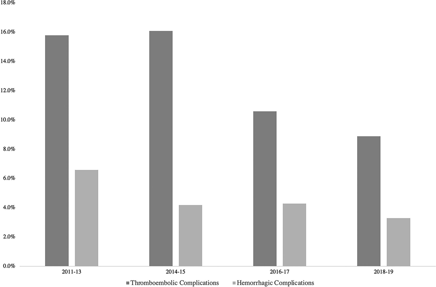
Thromboembolic complications occurred in 13% of procedures, of which 8.7% were symptomatic. There was a trend toward a decrease in the rate of thromboembolic complications from 15.8% in 2011–13 to 8.9% in 2018–19 (P = .3). There was also a nonsignificant decline in the rate of hemorrhagic complications (P = .78) and mortality (P = .88) (Table 1 and Figure).

View this table:
  • View inline
  • View popup
Table 1:

Change in treatment outcome with timea

FIGURE.
  • Download figure
  • Open in new tab
  • Download powerpoint
FIGURE.

The rates of thromboembolic and hemorrhagic complications during the study years.

Factors Associated with Complication Rates

Univariable analyses for determinants of thromboembolic complications are shown in Table 2. In a multivariable analysis, having a pretreatment mRS of 3–5 (adjusted OR, 3.00; 95% CI, 1.30–6.68; P = .008) and multiple aneurysms (adjusted OR, 2.55; 95% CI, 1.16–5.42; P = .017) was significantly associated with a higher risk of thromboembolic complications. On the other hand, vertebral artery aneurysms (as opposed to the basilar artery) (OR, 0.41; 95% CI, 0.18–0.90; P = .025) and saccular shape (as opposed to fusiform aneurysms) (OR, 0.43; 95% CI, 0.18–0.95; P = .045) were significantly associated with a lower risk of thromboembolic complications.

View this table:
  • View inline
  • View popup
Table 2:

Predictors of thromboembolic complicationsa

Univariable and multivariable logistic regression analyses for predictors of hemorrhagic complications are shown in the Online Supplemental Data. In a multivariable analysis, only a pretreatment mRS of 3–5 (OR, 10.15; 95% CI, 3.51–30.71; P < .001) was found to be associated with a higher risk of hemorrhagic complications (Online Supplemental Data).

DISCUSSION

This study reports a multicenter experience with flow diversion of posterior circulation aneurysms and the learning curve built on that experience. During the study period, there was a significant increase in the tendency to treat vertebral artery, posterior cerebral artery, and PICA aneurysms. Conversely, there was a lower tendency to treat large vertebrobasilar aneurysms. Moreover, interventionalists have increasingly used fewer overlapping flow diverters per aneurysm, which could also be related to treating smaller aneurysms. There was also a decline in the rate of symptomatic thromboembolic complications and retreatment rates across time. This was significantly correlated with treating saccular aneurysms and smaller aneurysms and the increased use of a single flow diverter.

To evaluate the performance of flow diversion for the treatment of posterior circulation aneurysms, Griessenauer et al18 reported the largest cohort of posterior circulation aneurysms treated with the PED. A total of 129 consecutive patients (median age, 58 years; male/female ratio of 1:1.7) underwent 129 procedures to treat 131 aneurysms. Complete or near-complete occlusion (>90%) was achieved in 79% of cases. Major (≥2 points of mRS change) and minor complications (<2 of mRS change), including thromboembolic and hemorrhagic strokes, occurred in 8.5% and 16.3% of patients, respectively.18 The same group also performed a propensity-matched comparison between the PED and FRED for the treatment of posterior circulation aneurysms and reported no significant differences in aneurysm occlusion or neurologic complications.15

As for the concern about the fate of posterior circulation branches following flow diversion, Adeeb et al19 found that major branching arteries in the posterior circulation including the PICA, anterior inferior cerebellar artery, and superior cerebellar artery had a low incidence of branch occlusion after coverage with flow diverters. However, occlusion of these terminal branches may carry a risk of ischemic complications, particularly when the anterior inferior cerebellar artery is affected. On the other hand, the vertebral and the posterior cerebral arteries had relatively higher incidences of occlusions, 35% and 24%, respectively, which were attributed to the rich collateral supply. Neither branch occlusion nor ischemic complications were associated with aneurysm morphology. There was also no significant effect of the number of flow-diverting devices on branch occlusion.19

Changes in Practice to Reduce Complications

Flow diverters are designed to divert the blood flow away from the aneurysm, therefore allowing intra-aneurysmal thrombus formation followed by neointimal growth across the neck of the aneurysm. This mechanism theoretically presents an ideal treatment for large, partially thrombosed fusiform vertebrobasilar aneurysms.20 In the multicenter study by Griessenauer et al,17 the rate of major complications (≥2 points in mRS score change) in fusiform aneurysms was 11.5%. However, a study by Natarajan et al10 showed decreased morbidity (14%–8.3%) and mortality rates (57%–0%) following treatment of those subtypes of aneurysms in their practice across time. One of the proposed reasons behind this decline in complication rates is related to moving away from the treatment of holobasilar aneurysms that are partially thrombosed because these aneurysms have a higher risk of occluding critical perforators that may only be supplied through tenuous channels crossing the thrombus. Other proposed reasons included careful attention to antiplatelet therapy, limiting the number of PEDs, and use of adjunctive coiling.10

In a meta-analysis of posterior nonsaccular aneurysms treated with flow diversion, Kiyofuji et al11 added that treatment of aneurysms of <10 mm was associated with fewer complications compared with those larger than 10 mm (18% versus 29%).They also found that aneurysms located within the vertebral artery (83%) had a better outcome compared with the vertebrobasilar junction and proximal basilar artery (33%), mid-/distal basilar artery, and holobasilar artery (18%).11 This finding is potentially related to the abundance of perforators along the basilar artery compared with the vertebral artery. Additionally, the holobasilar fusiform dolichoectatic aneurysm is the product of the unique and poorly understood pathophysiology distinct from other aneurysms. In our study, we have noticed significant changes across the years that align with these recommendations. Additionally, basilar artery aneurysms and fusiform shape were independent predictors of thromboembolic complications compared with their vertebral artery aneurysms and saccular counterparts. Thus, interventionalists transitioned to treating more vertebral artery aneurysms and fewer large vertebrobasilar aneurysms across the years, particularly asymptomatic ones. Also, the median size of aneurysms treated declined from 9 mm in 2011–13 to 7.7 mm in 2018–19. Despite the significant change in the size and location of aneurysms treated, there was no significant change in the rate of fusiform aneurysms treated, per se, across time.

One of the other factors suggested by Natarajan et al10 to reduce complications was limiting the number of overlapping flow diverters, because more devices were associated with an increased risk of perforator occlusion due to greater metal coverage. In our study, there was a significant shift to using only 1 device across time, from 72.4% of procedures in 2011–13 to 87.8% in 2018–19.

Moreover, as implied in the previous study on anterior circulation aneurysms, careful monitoring of platelet testing before procedures and switching to appropriate antiplatelet regimens in cases of clopidogrel nonresponders may have played a role in the drop of thromboembolic complications.16,17 The rate of platelet function testing in this study (72.9%) was lower than the ones reported by previous PED studies (96.1% and 88.5%).16,21

Poor clinical status (mRS 3–5) at presentation was an independent predictor of treatment complications. These patients were more likely to present with a ruptured aneurysm (70.2%) compared with patients with an mRS of 0–2 (19.3%, P < .001). Patients with poor clinical status and ruptured aneurysms were more prone to complications related to brain injury and delayed cerebral ischemia.22 Those patients were also more likely to present with fusiform aneurysms (76.6%) than patients with an mRS of 0–2 (64.5%, P = .07). Moreover, 17% of these patients had large or giant aneurysms (>20 mm) compared with 11.7% of patients with an mRS of 0–2 (P = .2). All these factors might have contributed to the increased rate of complications in this subgroup of patients.

Limitations

The primary limitations of the current study include its retrospective design and variability in the management of patients across centers. Retrospective studies are subject to incomplete data sets, selection bias, and unidentified confounders. The inclusion of multiple institutions improves the generalizability of the findings but introduces variability in patient management. This also introduces variation in aneurysm measurement and the device compaction rate. However, the use of a standardized datasheet among all centers and the large number of cases included should improve the generalization of the results. Although the study addresses improvement in patient and aneurysm selection, it does not account for improvement in the catheters and implants across the years. Screening of silent ischemic complications postoperatively was not routinely performed, which might underestimate the true thromboembolic rate. Moreover, the variability in the follow-up imaging protocol, especially with the use of noninvasive modalities (ie, CTA and MRA), introduces another bias, given that those modalities are less reliable in assessing endoleaks in fusiform aneurysms.

CONCLUSIONS

This is the largest study that evaluates the real-world practice trends in the treatment of posterior circulation aneurysms using flow diversion. Across the years, fewer basilar and vertebrobasilar junction aneurysms were treated, but more aneurysms of the vertebral artery. The average diameter of treated aneurysms has also decreased. These practice changes align with prior studies that showed a dire outcome from treatment of large and partially thrombosed fusiform basilar aneurysms. This led to a gradual decline in the rate of thromboembolic and hemorrhagic complications.

Footnotes

  • Disclosure forms provided by the authors are available with the full text and PDF of this article at www.ajnr.org.

References

  1. 1.↵
    1. Nelson PK,
    2. Lylyk P,
    3. Szikora I, et al
    . The Pipeline Embolization Device for the intracranial treatment of aneurysms trial. AJNR Am J Neuroradiol 2011;32:34–40 doi:10.3174/ajnr.A2421 pmid:21148256
    Abstract/FREE Full Text
  2. 2.↵
    Prospective Study on Embolization of Intracranial Aneurysms With PipelineTM Embolization Device. Full Text View. ClinicalTrials.gov. https://clinicaltrials.gov/ct2/show/NCT02186561. Accessed October 1, 2022
  3. 3.↵
    1. Griessenauer CJ,
    2. Ogilvy CS,
    3. Adeeb N, et al
    . Pipeline embolization of posterior circulation aneurysms: a multicenter study of 131 aneurysms. J Neurosurg 2019;130:923–35 doi:10.3171/2017.9.JNS171376 pmid:29726768
    CrossRefPubMed
  4. 4.↵
    1. Chalouhi N,
    2. Tjoumakaris S,
    3. Dumont AS, et al
    . Treatment of posterior circulation aneurysms with the Pipeline Embolization Device. Neurosurgery 2013;72:883–89 doi:10.1227/NEU.0b013e31828ba984 pmid:23407289
    CrossRefPubMed
  5. 5.↵
    1. Albuquerque FC,
    2. Park MS,
    3. Abla AA, et al
    . A reappraisal of the Pipeline Embolization Device for the treatment of posterior circulation aneurysms. J Neurointerv Surg 2015;7:641–45 doi:10.1136/neurintsurg-2014-011340 pmid:25092926
    Abstract/FREE Full Text
  6. 6.↵
    1. Ducruet AF,
    2. Crowley RW,
    3. Albuquerque FC, et al
    . Reconstructive endovascular treatment of a ruptured vertebral artery dissecting aneurysm using the Pipeline Embolization Device. J Neurointerv Surg 2013;5:e20 doi:10.1136/neurintsurg-2012-010358 pmid:22717921
    Abstract/FREE Full Text
  7. 7.↵
    1. Mazur MD,
    2. Kilburg C,
    3. Wang V, et al
    . Pipeline Embolization Device for the treatment of vertebral artery aneurysms: the fate of covered branch vessels. J Neurointerv Surg 2016;8:1041–47 doi:10.1136/neurintsurg-2015-012040 pmid:26491041
    Abstract/FREE Full Text
  8. 8.↵
    1. Phillips TJ,
    2. Wenderoth JD,
    3. Phatouros CC, et al
    . Safety of the Pipeline Embolization Device in treatment of posterior circulation aneurysms. AJNR Am J Neuroradiol 2012;33:1225–31 doi:10.3174/ajnr.A3166 pmid:22678845
    Abstract/FREE Full Text
  9. 9.↵
    1. Siddiqui AH,
    2. Abla AA,
    3. Kan P, et al
    . Panacea or problem: flow diverters in the treatment of symptomatic large or giant fusiform vertebrobasilar aneurysms. J Neurosurg 2012;116:1258–66 doi:10.3171/2012.2.JNS111942 pmid:22404673
    CrossRefPubMed
  10. 10.↵
    1. Natarajan SK,
    2. Lin N,
    3. Sonig A, et al
    . The safety of Pipeline flow diversion in fusiform vertebrobasilar aneurysms: a consecutive case series with longer-term follow-up from a single US center. J Neurosurg 2016;125:111–19 doi:10.3171/2015.6.JNS1565 pmid:26654175
    CrossRefPubMed
  11. 11.↵
    1. Kiyofuji S,
    2. Graffeo CS,
    3. Perry A, et al
    . Meta-analysis of treatment outcomes of posterior circulation non-saccular aneurysms by flow diverters. J Neurointerv Surg 2018;10:493–99 doi:10.1136/neurintsurg-2017-013312 pmid:28965108
    Abstract/FREE Full Text
  12. 12.↵
    1. Bhogal P,
    2. Pérez MA,
    3. Ganslandt O, et al
    . Treatment of posterior circulation non-saccular aneurysms with flow diverters: a single-center experience and review of 56 patients. J Neurointerv Surg 2017;9:471–81 doi:10.1136/neurintsurg-2016-012781 pmid:27836994
    Abstract/FREE Full Text
  13. 13.↵
    1. Bender MT,
    2. Colby GP,
    3. Jiang B, et al
    . Flow diversion of posterior circulation cerebral aneurysms: a single-institution series of 59 cases. Neurosurgery 2019;84:206–16 doi:10.1093/neuros/nyy076 pmid:29608702
    CrossRefPubMed
  14. 14.↵
    1. Adeeb N,
    2. Ogilvy CS,
    3. Griessenauer CJ, et al
    . Expanding the indications for flow diversion: treatment of posterior circulation aneurysms. Neurosurgery 2020;86:S76–84 doi:10.1093/neuros/nyz344 pmid:31838535
    CrossRefPubMed
  15. 15.↵
    1. Griessenauer CJ,
    2. Enriquez-Marulanda A,
    3. Xiang S, et al
    . Comparison of PED and FRED flow diverters for posterior circulation aneurysms: a propensity score matched cohort study. J Neurointerv Surg 2021;13:153–58 doi:10.1136/neurintsurg-2020-016055 pmid:32611622
    Abstract/FREE Full Text
  16. 16.↵
    1. Dmytriw AA,
    2. Phan K,
    3. Salem MM, et al
    . The Pipeline Embolization Device: changes in practice and reduction of complications in the treatment of anterior circulation aneurysms in a multicenter cohort. Clin Neurosurg 2020;86:266–71 doi:10.1093/neuros/nyz059 pmid:30860254
    CrossRefPubMed
  17. 17.↵
    1. Adeeb N,
    2. Griessenauer CJ,
    3. Foreman PM, et al
    . Use of platelet function testing before Pipeline Embolization Device placement: a multicenter cohort study. Stroke 2017;48:1322–30 doi:10.1161/STROKEAHA.116.015308 pmid:28411263
    Abstract/FREE Full Text
  18. 18.↵
    1. Griessenauer CJ,
    2. Ogilvy CS,
    3. Adeeb N, et al
    . Pipeline embolization of posterior circulation aneurysms: a multicenter study of 131 aneurysms. J Neurosurg 2018:130:923–35 doi:10.3171/2017.9.JNS171376 pmid:29726768
    CrossRefPubMed
  19. 19.↵
    1. Adeeb N,
    2. Griessenauer CJ,
    3. Dmytriw AA, et al
    . Risk of branch occlusion and ischemic complications with the Pipeline Embolization Device in the treatment of posterior circulation aneurysms. AJNR Am J Neuroradiol 2018;39:1303–09 doi:10.3174/ajnr.A5696 pmid:29880475
    Abstract/FREE Full Text
  20. 20.↵
    1. Szikora I,
    2. Turányi E,
    3. Marosfoi M
    . Evolution of flow-diverter endothelialization and thrombus organization in giant fusiform aneurysms after flow diversion: a histopathologic study. AJNR Am J Neuroradiol 2015;36:1716–20 doi:10.3174/ajnr.A4336 pmid:26251428
    Abstract/FREE Full Text
  21. 21.↵
    1. Gupta R,
    2. Moore JM,
    3. Griessenauer CJ, et al
    . Assessment of dual-antiplatelet regimen for Pipeline Embolization Device placement: a survey of major academic neurovascular centers in the United States. World Neurosurg 2016;96:285–92 doi:10.1016/j.wneu.2016.09.013 pmid:27641263
    CrossRefPubMed
  22. 22.↵
    1. de Oliveira Manoel AL,
    2. Goffi A,
    3. Marotta TR, et al
    . The critical care management of poor-grade subarachnoid haemorrhage. Crit Care 2016;20:21 doi:10.1186/s13054-016-1193-9 pmid:26801901
    CrossRefPubMed
  • Received February 6, 2022.
  • Accepted after revision June 28, 2022.
  • © 2022 by American Journal of Neuroradiology
PreviousNext
Back to top

In this issue

American Journal of Neuroradiology: 43 (11)
American Journal of Neuroradiology
Vol. 43, Issue 11
1 Nov 2022
  • Table of Contents
  • Index by author
  • Complete Issue (PDF)
Advertisement
Print
Download PDF
Email Article

Thank you for your interest in spreading the word on American Journal of Neuroradiology.

NOTE: We only request your email address so that the person you are recommending the page to knows that you wanted them to see it, and that it is not junk mail. We do not capture any email address.

Enter multiple addresses on separate lines or separate them with commas.
Learning Curve for Flow Diversion of Posterior Circulation Aneurysms: A Long-Term International Multicenter Cohort Study
(Your Name) has sent you a message from American Journal of Neuroradiology
(Your Name) thought you would like to see the American Journal of Neuroradiology web site.
CAPTCHA
This question is for testing whether or not you are a human visitor and to prevent automated spam submissions.
Cite this article
N. Adeeb, M. Dibas, C.J. Griessenauer, H.H. Cuellar, M.M. Salem, S. Xiang, A. Enriquez-Marulanda, T. Hong, H. Zhang, P. Taussky, R. Grandhi, M. Waqas, A.S. Aldine, V.M. Tutino, A. Aslan, A.H. Siddiqui, E.I. Levy, C.S. Ogilvy, A.J. Thomas, C. Ulfert, M.A. Möhlenbruch, L. Renieri, J.D. Bengzon Diestro, G. Lanzino, W. Brinjikji, J. Spears, J.E. Vranic, R.W. Regenhardt, J.D. Rabinov, P. Harker, E. Müller-Thies-Broussalis, M. Killer-Oberpfalzer, C. Islak, N. Kocer, M. Sonnberger, T. Engelhorn, A. Kapadia, V.X.D Yang, A. Salehani, M.R. Harrigan, T. Krings, C.C. Matouk, S. Mirshahi, K.S. Chen, M.A. Aziz-Sultan, M. Ghorbani, C.M. Schirmer, O. Goren, S.S. Dalal, T. Finkenzeller, M. Holtmannspötter, J.-H. Buhk, P.M. Foreman, M.C. Cress, R.A. Hirschl, W. Reith, A. Simgen, H. Janssen, T.R. Marotta, C.J. Stapleton, A.B. Patel, A.A. Dmytriw
Learning Curve for Flow Diversion of Posterior Circulation Aneurysms: A Long-Term International Multicenter Cohort Study
American Journal of Neuroradiology Nov 2022, 43 (11) 1615-1620; DOI: 10.3174/ajnr.A7679

Citation Manager Formats

  • BibTeX
  • Bookends
  • EasyBib
  • EndNote (tagged)
  • EndNote 8 (xml)
  • Medlars
  • Mendeley
  • Papers
  • RefWorks Tagged
  • Ref Manager
  • RIS
  • Zotero
0 Responses
Respond to this article
Share
Bookmark this article
Learning Curve in Flow Diversion for Aneurysms
N. Adeeb, M. Dibas, C.J. Griessenauer, H.H. Cuellar, M.M. Salem, S. Xiang, A. Enriquez-Marulanda, T. Hong, H. Zhang, P. Taussky, R. Grandhi, M. Waqas, A.S. Aldine, V.M. Tutino, A. Aslan, A.H. Siddiqui, E.I. Levy, C.S. Ogilvy, A.J. Thomas, C. Ulfert, M.A. Möhlenbruch, L. Renieri, J.D. Bengzon Diestro, G. Lanzino, W. Brinjikji, J. Spears, J.E. Vranic, R.W. Regenhardt, J.D. Rabinov, P. Harker, E. Müller-Thies-Broussalis, M. Killer-Oberpfalzer, C. Islak, N. Kocer, M. Sonnberger, T. Engelhorn, A. Kapadia, V.X.D Yang, A. Salehani, M.R. Harrigan, T. Krings, C.C. Matouk, S. Mirshahi, K.S. Chen, M.A. Aziz-Sultan, M. Ghorbani, C.M. Schirmer, O. Goren, S.S. Dalal, T. Finkenzeller, M. Holtmannspötter, J.-H. Buhk, P.M. Foreman, M.C. Cress, R.A. Hirschl, W. Reith, A. Simgen, H. Janssen, T.R. Marotta, C.J. Stapleton, A.B. Patel, A.A. Dmytriw
American Journal of Neuroradiology Nov 2022, 43 (11) 1615-1620; DOI: 10.3174/ajnr.A7679
del.icio.us logo Twitter logo Facebook logo Mendeley logo
  • Tweet Widget
  • Facebook Like
  • Google Plus One
Purchase

Jump to section

  • Article
    • Abstract
    • MATERIALS AND METHODS
    • RESULTS
    • DISCUSSION
    • CONCLUSIONS
    • Footnotes
    • References
  • Figures & Data
  • Info & Metrics
  • Responses
  • References
  • PDF

Related Articles

  • No related articles found.
  • PubMed
  • Google Scholar

Cited By...

  • No citing articles found.
  • Crossref (5)
  • Google Scholar

This article has been cited by the following articles in journals that are participating in Crossref Cited-by Linking.

  • Assessment of the flow-diverter efficacy for intracranial aneurysm treatment considering pre- and post-interventional hemodynamics
    Janneck Stahl, Laurel Morgan Miller Marsh, Maximilian Thormann, Andreas Ding, Sylvia Saalfeld, Daniel Behme, Philipp Berg
    Computers in Biology and Medicine 2023 156
  • Flow-Diverting Devices in the Treatment of Vertebral Artery Aneurysms: Insights into Efficacy and Safety from a Systematic Review and Meta-analysis
    Changya Liu, Xinxin Wu, Kaikai Guo, Yuting Sun, Cai Yike, Xuebin Hu, Bangjiang Fang
    Translational Stroke Research 2025 16 3
  • Flow-diverter stents combined with flow-T stenting-assisted coiling for the treatment of a large basilar apex aneurysm: a case report with a 9-month follow-up
    Hao Wang, Jingyu Zhang, Huajiang Yang, Shi Zeng, Tengyun Guo, Lunshan Xu, Donghong Yang
    Frontiers in Neurology 2024 14
  • Intracranial hemorrhagic events associated with flow diversion treatment: a retrospective analysis from a single academic institution
    Aryan Wadhwa, Felipe Ramirez-Velandia, Alejandro Enriquez-Marulanda, Jean Filo, Thomas B. Fodor, Daniel Sconzo, Michael Young, Sandeep Muram, Justin H. Granstein, Max Shutran, Philipp Taussky, Christopher S. Ogilvy
    Neurosurgical Review 2025 48 1
  • Treatment of basilar artery aneurysms with two braided stents: Two centers experience of low-profile visualized intraluminal support stents versus Pipeline flow diverters
    Bin Luo, Chao Wang, Jian Liu, Yisen Zhang, Kun Wang, Wenqiang Li, Ying Zhang
    The Neuroradiology Journal 2024 37 4

More in this TOC Section

  • SAVE vs. Solumbra Techniques for Thrombectomy
  • CT Perfusion&Reperfusion in Acute Ischemic Stroke
  • Delayed Reperfusion Post-Thrombectomy&Thrombolysis
Show more Interventional

Similar Articles

Advertisement

Indexed Content

  • Current Issue
  • Accepted Manuscripts
  • Article Preview
  • Past Issues
  • Editorials
  • Editors Choice
  • Fellow Journal Club
  • Letters to the Editor

Cases

  • Case Collection
  • Archive - Case of the Week
  • Archive - Case of the Month
  • Archive - Classic Case

Special Collections

  • Special Collections

Resources

  • News and Updates
  • Turn around Times
  • Submit a Manuscript
  • Author Policies
  • Manuscript Submission Guidelines
  • Evidence-Based Medicine Level Guide
  • Publishing Checklists
  • Graphical Abstract Preparation
  • Imaging Protocol Submission
  • Submit a Case
  • Become a Reviewer/Academy of Reviewers
  • Get Peer Review Credit from Publons

Multimedia

  • AJNR Podcast
  • AJNR SCANtastic
  • Video Articles

About Us

  • About AJNR
  • Editorial Board
  • Not an AJNR Subscriber? Join Now
  • Alerts
  • Feedback
  • Advertise with us
  • Librarian Resources
  • Permissions
  • Terms and Conditions

American Society of Neuroradiology

  • Not an ASNR Member? Join Now

© 2025 by the American Society of Neuroradiology All rights, including for text and data mining, AI training, and similar technologies, are reserved.
Print ISSN: 0195-6108 Online ISSN: 1936-959X

Powered by HighWire