Advertisement

Main menu

  • Home
  • Content
    • Current Issue
    • Accepted Manuscripts
    • Article Preview
    • Past Issue Archive
    • AJNR Case Collection
    • Case of the Week Archive
    • Classic Case Archive
    • Case of the Month Archive
  • Special Collections
    • Spinal CSF Leak Articles (Jan 2020-June 2024)
    • 2024 AJNR Journal Awards
    • Most Impactful AJNR Articles
  • Multimedia
    • AJNR Podcast
    • AJNR Scantastics
    • Video Articles
  • For Authors
    • Submit a Manuscript
    • Author Policies
    • Fast publishing of Accepted Manuscripts
    • Graphical Abstract Preparation
    • Manuscript Submission Guidelines
    • Imaging Protocol Submission
    • Submit a Case for the Case Collection
  • About Us
    • About AJNR
    • Editorial Board
  • More
    • Become a Reviewer/Academy of Reviewers
    • Subscribers
    • Permissions
    • Alerts
    • Feedback
    • Advertisers
    • ASNR Home
  • Other Publications
    • ajnr

User menu

  • Alerts
  • Log in

Search

  • Advanced search
American Journal of Neuroradiology

American Journal of Neuroradiology

ASHNR American Society of Functional Neuroradiology ASHNR American Society of Pediatric Neuroradiology ASSR
  • Alerts
  • Log in

Advanced Search

  • Home
  • Content
    • Current Issue
    • Accepted Manuscripts
    • Article Preview
    • Past Issue Archive
    • AJNR Case Collection
    • Case of the Week Archive
    • Classic Case Archive
    • Case of the Month Archive
  • Special Collections
    • Spinal CSF Leak Articles (Jan 2020-June 2024)
    • 2024 AJNR Journal Awards
    • Most Impactful AJNR Articles
  • Multimedia
    • AJNR Podcast
    • AJNR Scantastics
    • Video Articles
  • For Authors
    • Submit a Manuscript
    • Author Policies
    • Fast publishing of Accepted Manuscripts
    • Graphical Abstract Preparation
    • Manuscript Submission Guidelines
    • Imaging Protocol Submission
    • Submit a Case for the Case Collection
  • About Us
    • About AJNR
    • Editorial Board
  • More
    • Become a Reviewer/Academy of Reviewers
    • Subscribers
    • Permissions
    • Alerts
    • Feedback
    • Advertisers
    • ASNR Home
  • Follow AJNR on Twitter
  • Visit AJNR on Facebook
  • Follow AJNR on Instagram
  • Join AJNR on LinkedIn
  • RSS Feeds

Welcome to the new AJNR, Updated Hall of Fame, and more. Read the full announcements.


AJNR is seeking candidates for the position of Associate Section Editor, AJNR Case Collection. Read the full announcement.


Research ArticleAdult Brain
Open Access

Clinical Evaluation of Scout Accelerated Motion Estimation and Reduction Technique for 3D MR Imaging in the Inpatient and Emergency Department Settings

M. Lang, A. Tabari, D. Polak, J. Ford, B. Clifford, W.-C. Lo, K. Manzoor, D.N. Splitthoff, L.L. Wald, O. Rapalino, P. Schaefer, J. Conklin, S. Cauley and S.Y. Huang
American Journal of Neuroradiology February 2023, 44 (2) 125-133; DOI: https://doi.org/10.3174/ajnr.A7777
M. Lang
aFrom the Department of Radiology (M.L., A.T., D.P., J.F., K.M., L.L.W., O.R., P.S., J.C., S.C., S.Y.H.), Athinoula A. Martinos Center for Biomedical Imaging, Massachusetts General Hospital, Charlestown, Massachusetts
bHarvard Medical School (M.L., A.T., J.F., K.M., L.L.W., O.R., P.S., J.C., S.C., S.Y.H.), Boston, Massachusetts
  • Find this author on Google Scholar
  • Find this author on PubMed
  • Search for this author on this site
  • ORCID record for M. Lang
A. Tabari
aFrom the Department of Radiology (M.L., A.T., D.P., J.F., K.M., L.L.W., O.R., P.S., J.C., S.C., S.Y.H.), Athinoula A. Martinos Center for Biomedical Imaging, Massachusetts General Hospital, Charlestown, Massachusetts
bHarvard Medical School (M.L., A.T., J.F., K.M., L.L.W., O.R., P.S., J.C., S.C., S.Y.H.), Boston, Massachusetts
  • Find this author on Google Scholar
  • Find this author on PubMed
  • Search for this author on this site
  • ORCID record for A. Tabari
D. Polak
aFrom the Department of Radiology (M.L., A.T., D.P., J.F., K.M., L.L.W., O.R., P.S., J.C., S.C., S.Y.H.), Athinoula A. Martinos Center for Biomedical Imaging, Massachusetts General Hospital, Charlestown, Massachusetts
cSiemens Healthcare GmbH (D.P., D.N.S.), Erlangen, Germany
  • Find this author on Google Scholar
  • Find this author on PubMed
  • Search for this author on this site
J. Ford
aFrom the Department of Radiology (M.L., A.T., D.P., J.F., K.M., L.L.W., O.R., P.S., J.C., S.C., S.Y.H.), Athinoula A. Martinos Center for Biomedical Imaging, Massachusetts General Hospital, Charlestown, Massachusetts
bHarvard Medical School (M.L., A.T., J.F., K.M., L.L.W., O.R., P.S., J.C., S.C., S.Y.H.), Boston, Massachusetts
  • Find this author on Google Scholar
  • Find this author on PubMed
  • Search for this author on this site
  • ORCID record for J. Ford
B. Clifford
dSiemens Medical Solutions (B.C., W.-C.L.), Boston, Massachusetts
  • Find this author on Google Scholar
  • Find this author on PubMed
  • Search for this author on this site
  • ORCID record for B. Clifford
W.-C. Lo
dSiemens Medical Solutions (B.C., W.-C.L.), Boston, Massachusetts
  • Find this author on Google Scholar
  • Find this author on PubMed
  • Search for this author on this site
K. Manzoor
aFrom the Department of Radiology (M.L., A.T., D.P., J.F., K.M., L.L.W., O.R., P.S., J.C., S.C., S.Y.H.), Athinoula A. Martinos Center for Biomedical Imaging, Massachusetts General Hospital, Charlestown, Massachusetts
bHarvard Medical School (M.L., A.T., J.F., K.M., L.L.W., O.R., P.S., J.C., S.C., S.Y.H.), Boston, Massachusetts
  • Find this author on Google Scholar
  • Find this author on PubMed
  • Search for this author on this site
  • ORCID record for K. Manzoor
D.N. Splitthoff
cSiemens Healthcare GmbH (D.P., D.N.S.), Erlangen, Germany
  • Find this author on Google Scholar
  • Find this author on PubMed
  • Search for this author on this site
  • ORCID record for D.N. Splitthoff
L.L. Wald
aFrom the Department of Radiology (M.L., A.T., D.P., J.F., K.M., L.L.W., O.R., P.S., J.C., S.C., S.Y.H.), Athinoula A. Martinos Center for Biomedical Imaging, Massachusetts General Hospital, Charlestown, Massachusetts
bHarvard Medical School (M.L., A.T., J.F., K.M., L.L.W., O.R., P.S., J.C., S.C., S.Y.H.), Boston, Massachusetts
eHarvard-MIT Health Sciences and Technology (L.L.W.), Massachusetts Institute of Technology, Cambridge, Massachusetts
  • Find this author on Google Scholar
  • Find this author on PubMed
  • Search for this author on this site
  • ORCID record for L.L. Wald
O. Rapalino
aFrom the Department of Radiology (M.L., A.T., D.P., J.F., K.M., L.L.W., O.R., P.S., J.C., S.C., S.Y.H.), Athinoula A. Martinos Center for Biomedical Imaging, Massachusetts General Hospital, Charlestown, Massachusetts
bHarvard Medical School (M.L., A.T., J.F., K.M., L.L.W., O.R., P.S., J.C., S.C., S.Y.H.), Boston, Massachusetts
  • Find this author on Google Scholar
  • Find this author on PubMed
  • Search for this author on this site
  • ORCID record for O. Rapalino
P. Schaefer
aFrom the Department of Radiology (M.L., A.T., D.P., J.F., K.M., L.L.W., O.R., P.S., J.C., S.C., S.Y.H.), Athinoula A. Martinos Center for Biomedical Imaging, Massachusetts General Hospital, Charlestown, Massachusetts
bHarvard Medical School (M.L., A.T., J.F., K.M., L.L.W., O.R., P.S., J.C., S.C., S.Y.H.), Boston, Massachusetts
  • Find this author on Google Scholar
  • Find this author on PubMed
  • Search for this author on this site
  • ORCID record for P. Schaefer
J. Conklin
aFrom the Department of Radiology (M.L., A.T., D.P., J.F., K.M., L.L.W., O.R., P.S., J.C., S.C., S.Y.H.), Athinoula A. Martinos Center for Biomedical Imaging, Massachusetts General Hospital, Charlestown, Massachusetts
bHarvard Medical School (M.L., A.T., J.F., K.M., L.L.W., O.R., P.S., J.C., S.C., S.Y.H.), Boston, Massachusetts
  • Find this author on Google Scholar
  • Find this author on PubMed
  • Search for this author on this site
  • ORCID record for J. Conklin
S. Cauley
aFrom the Department of Radiology (M.L., A.T., D.P., J.F., K.M., L.L.W., O.R., P.S., J.C., S.C., S.Y.H.), Athinoula A. Martinos Center for Biomedical Imaging, Massachusetts General Hospital, Charlestown, Massachusetts
bHarvard Medical School (M.L., A.T., J.F., K.M., L.L.W., O.R., P.S., J.C., S.C., S.Y.H.), Boston, Massachusetts
  • Find this author on Google Scholar
  • Find this author on PubMed
  • Search for this author on this site
  • ORCID record for S. Cauley
S.Y. Huang
aFrom the Department of Radiology (M.L., A.T., D.P., J.F., K.M., L.L.W., O.R., P.S., J.C., S.C., S.Y.H.), Athinoula A. Martinos Center for Biomedical Imaging, Massachusetts General Hospital, Charlestown, Massachusetts
bHarvard Medical School (M.L., A.T., J.F., K.M., L.L.W., O.R., P.S., J.C., S.C., S.Y.H.), Boston, Massachusetts
  • Find this author on Google Scholar
  • Find this author on PubMed
  • Search for this author on this site
  • ORCID record for S.Y. Huang
  • Article
  • Figures & Data
  • Info & Metrics
  • Responses
  • References
  • PDF
Loading

Article Figures & Data

Figures

  • Tables
  • FIG 1.
    • Download figure
    • Open in new tab
    • Download powerpoint
    FIG 1.

    Motion scale used for the clinical quantification of motion artifacts along with representative cases before motion correction. The arrows point to areas of image blurring due to motion artifacta.

  • FIG 2.
    • Download figure
    • Open in new tab
    • Download powerpoint
    FIG 2.

    Sagittal T1 MPRAGE images illustrating examples of cases with motion artifacts (grades I–5) for which SAMER reconstruction improved motion by 1 grade. A, A 23-year-old woman with a normal brain. B, Postoperative findings from resection of a left parietal lobe tumor in a 59-year-old woman with a history of anaplastic oligodendroglioma. C, Diffuse parenchymal volume loss with disproportionate involvement of the frontal and parietal lobes and, to a lesser extent, the left temporal lobe in a 59-year-old man with history of cognitive impairment. D, An 83-year-old woman with history of chronic cerebral small-vessel disease. E, Expected postoperative changes and enhancement from left temporal parietal craniotomy (arrows) are demonstrated in a 63-year-old man with history of glioblastoma.
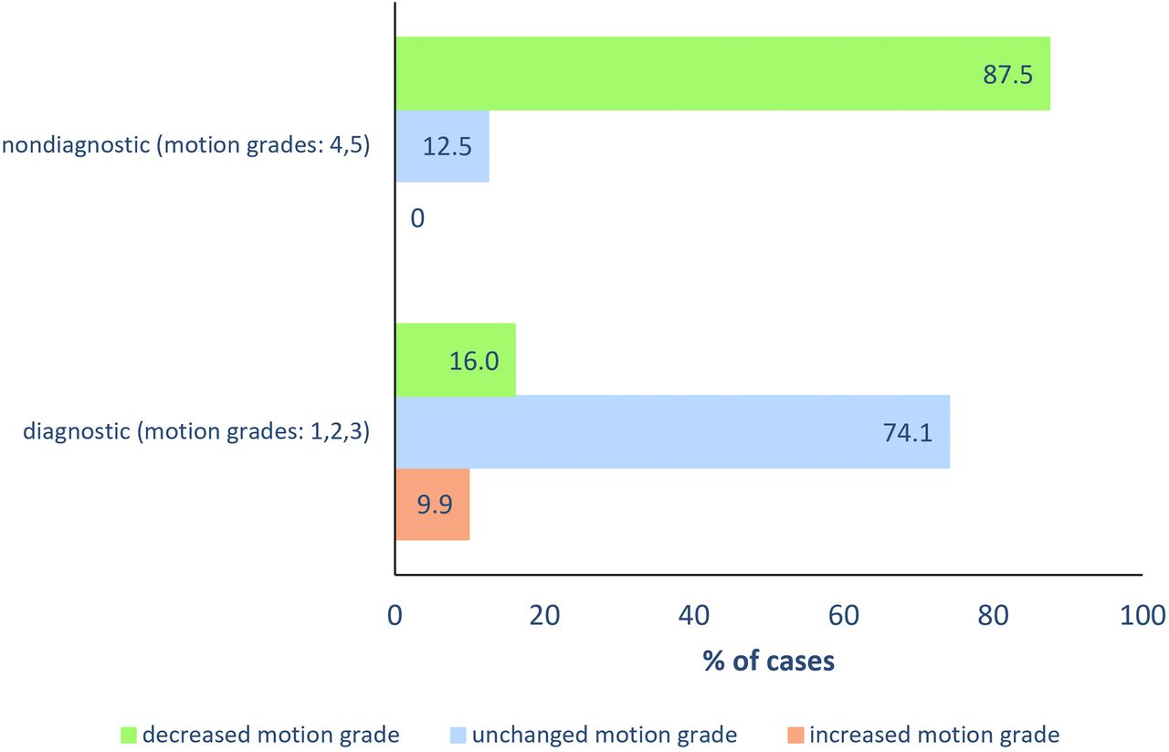
  • FIG 3.
    • Download figure
    • Open in new tab
    • Download powerpoint
    FIG 3.

    Proportion of nondiagnostic and diagnostic examinations that demonstrated worsening, no change, and improvement of motion grade after SAMER correction. Motion grades 1 (no motion), 2 (minimal), and 3 (mild) are considered diagnostic in terms of motion artifacts, whereas motion grades 4 (moderate) and 5 (severe) are considered nondiagnostic in terms of motion artifacts.

  • FIG 4.
    • Download figure
    • Open in new tab
    • Download powerpoint
    FIG 4.

    Axial MR images of 2 cases with severe motion artifacts (grade 5) in which SAMER motion correction restored diagnostic value (by reducing motion grade to 3). A, The extent of cortical/gyral enhancement (arrows) and edematous expansion of the left temporoparietal region with a mild rightward mass effect is better visualized on the motion-corrected image of a 67-year-old man with traumatic brain injury. B, Motion-corrected image shows better visualization of cortical laminar necrosis in the left occipital lobe (arrows) and better evaluation of left temporal lobe volume loss (arrows) in an 86-year-old woman with history of stroke.
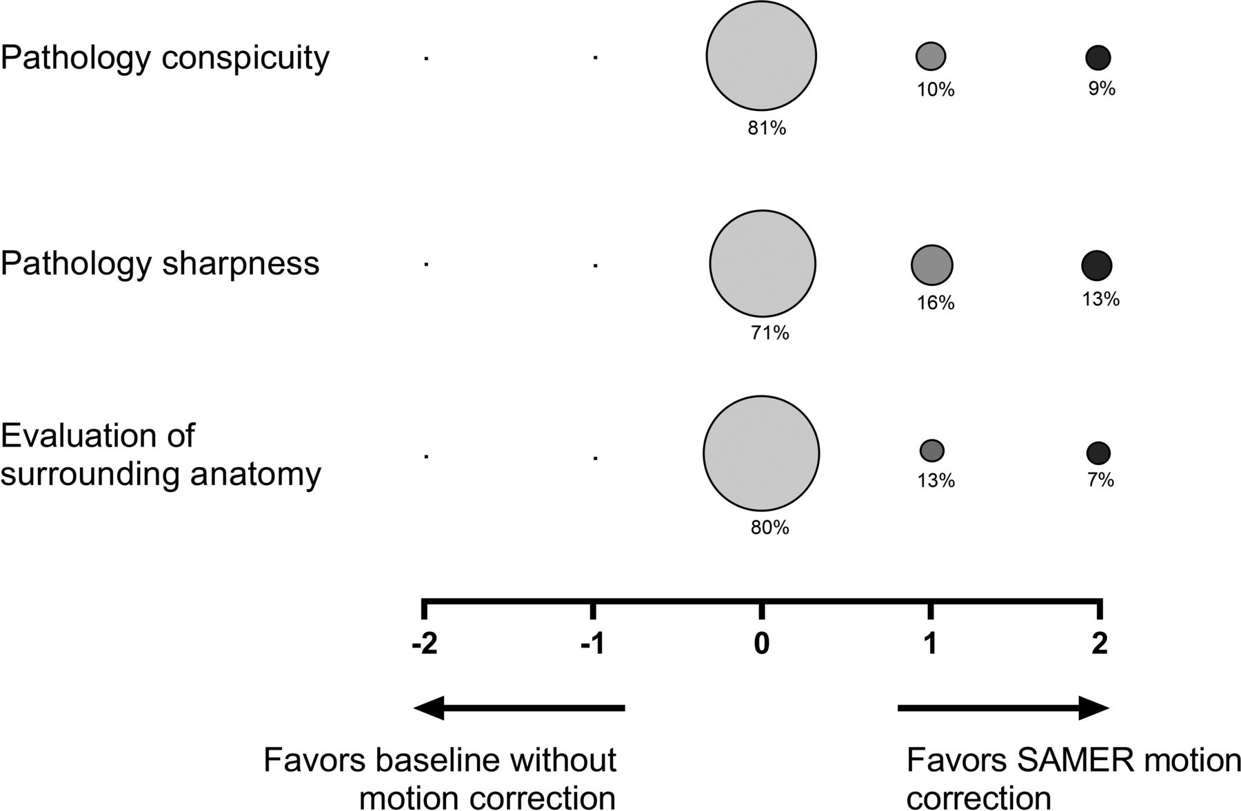
  • FIG 5.
    • Download figure
    • Open in new tab
    • Download powerpoint
    FIG 5.

    Balloon plot showing the results of the head-to-head comparison of baseline MPRAGE and SAMER motion-corrected MPRAGE studies for pathology conspicuity, pathology sharpness, and evaluation of surrounding anatomy. A total 79 of 97 cases had pathology on imaging and were included in this assessment. The size of the circle correlates to the percentage of cases assigned a given score, and the percentage of cases receiving a given score is indicated below each circle. A zero score indicates equivalency, negative scores (left) favor baseline MPRAGE images, and positive scores (right) favor SAMER motion-corrected MPRAGE images.

  • FIG 6.
    • Download figure
    • Open in new tab
    • Download powerpoint
    FIG 6.

    Boxplot charts demonstrating the distribution of CNR and SNR in the gray matter and white matter in MPRAGE without and with SAMER motion correction. CNRs and SNRs are significantly greater (P < .001) in the MPRAGE SAMER motion-corrected images compared with baseline MPRAGE images. Triple asterisks indicate P < .001.

Tables

  • Figures
    • View popup
    Table 1:

    Clinical characteristics of the patients

    Value
    No. of subjects97
    Mean age (yr)60.2 (SD, 16)
    Sex (F/M)48:49
    Clinical indication for MR imaging (No.) (%)
     Tumor32 (33%)
     Stroke17 (17.5%)
     AMS15 (15.4%)
     Neurologic deficit11 (11.3%)
     Abscess3 (3%)
     Dementia2 (2%)
     Headache2 (2%)
     Seizure2 (2%)
     TBI2 (2%)
     TIA2 (2%)
     Other9 (9.2%)
    MR imaging protocol (No.) (%)
     SAMER MPRAGE without contrast34 (35%)
     Routine brain without contrast33 (34%)
     Memory loss without contrast1 (1%)
     SAMER MPRAGE with contrast63 (65%)
     Routine brain without and with contrast61 (63%)
     Memory loss without and with contrast2 (2%)
    • Note:—AMS indicates altered mental status; TBI, traumatic brain injury.

    • View popup
    Table 2:

    Overall change in motion grade after SAMER implementation

    Before Motion Correction, Motion Grade (No. of cases)After Motion, Correction Motion Grade (No. of cases)Change in Motion Grade (mean)
    Grade 1 (5)Grade 1 (5)0
    Grade 2 (46)Grade 1 (2)0.1 (SD, 0.7)
    Grade 2 (36)
    Grade 3 (8)
    Grade 3 (30)Grade 2 (11)–0.3 (SD, 0.5)
    Grade 3 (19)
    Grade 4 (8)Grade 2 (1)–1.1 (SD, 0.3)
    Grade 3 (7)
    Grade 5 (8)Grade 3 (3)–1.1 (SD, 0.8)
    Grade 4 (3)
    Grade 5 (2)
    Mean motion grade (2.7 [SD, 1.0])Mean motion grade (2.4 [SD, .08])Change in motion grade (–0.2 [SD, 0.7])
    • Note:—Grade 1 indicates no motion; grade 2, minimal motion; grade 3, mild motion; grade 4, moderate motion; grade 5, severe motion.

    • View popup
    Table 3:

    Change in motion grade after SAMER implementation for noncontrast examinations

    Before Motion Correction, Motion Grade (No. of Cases)After Motion Correction, Motion Grade (No. of Cases)Change in Motion Grade (mean)
    Grade 1 (3)Grade 1 (3)0
    Grade 2 (18)Grade 2 (16)0.1 (SD, 0.3)
    Grade 3 (2)
    Grade 3 (7)Grade 2 (5)–0.7 (SD, 0.4)
    Grade 3 (2)
    Grade 4 (2)Grade 2 (1)–1.5 (SD, 0.7)
    Grade 3 (1)
    Grade 5 (4)Grade 3 (3)–1.7 (SD, 0.5)
    Grade 4 (1)
    Mean motion grade (2.6 [SD, 1.1])Mean motion grade (2.2 [SD, 0.6])Change in motion grade (–0.4 [SD, 0.8])
    • View popup
    Table 4:

    Change in motion grade after SAMER implementation for contrast-enhanced examinations

    Before Motion Correction, Motion Grade (No. of Cases)After Motion Correction, Motion Grade (No. of Cases)Change in Motion Grade (mean)
    Grade 1 (2)Grade 1 (2)0
    Grade 2 (28)Grade 1 (2)0.1 (SD, 0.5)
    Grade 2 (20)
    Grade 3 (6)
    Grade 3 (23)Grade 2 (6)–0.2 (SD, 0.4)
    Grade 3 (17)
    Grade 4 (6)Grade 3 (6)–1 (SD, 0)
    Grade 5 (4)Grade 4 (2)–0.5 (SD, 0.5)
    Grade 5 (2)
    Mean motion grade (2.7 [SD, 0.9])Mean motion grade (2.6 [SD, 0.8])Change in motion grade (–0.2 [SD, 0.6])
    Mean motion grade (2.6 [SD, 1.1])Mean motion grade (2.2 [SD, 0.6])Change in motion grade (–0.4 [SD, 0.8])
PreviousNext
Back to top

In this issue

American Journal of Neuroradiology: 44 (2)
American Journal of Neuroradiology
Vol. 44, Issue 2
1 Feb 2023
  • Table of Contents
  • Index by author
  • Complete Issue (PDF)
Advertisement
Print
Download PDF
Email Article

Thank you for your interest in spreading the word on American Journal of Neuroradiology.

NOTE: We only request your email address so that the person you are recommending the page to knows that you wanted them to see it, and that it is not junk mail. We do not capture any email address.

Enter multiple addresses on separate lines or separate them with commas.
Clinical Evaluation of Scout Accelerated Motion Estimation and Reduction Technique for 3D MR Imaging in the Inpatient and Emergency Department Settings
(Your Name) has sent you a message from American Journal of Neuroradiology
(Your Name) thought you would like to see the American Journal of Neuroradiology web site.
CAPTCHA
This question is for testing whether or not you are a human visitor and to prevent automated spam submissions.
Cite this article
M. Lang, A. Tabari, D. Polak, J. Ford, B. Clifford, W.-C. Lo, K. Manzoor, D.N. Splitthoff, L.L. Wald, O. Rapalino, P. Schaefer, J. Conklin, S. Cauley, S.Y. Huang
Clinical Evaluation of Scout Accelerated Motion Estimation and Reduction Technique for 3D MR Imaging in the Inpatient and Emergency Department Settings
American Journal of Neuroradiology Feb 2023, 44 (2) 125-133; DOI: 10.3174/ajnr.A7777

Citation Manager Formats

  • BibTeX
  • Bookends
  • EasyBib
  • EndNote (tagged)
  • EndNote 8 (xml)
  • Medlars
  • Mendeley
  • Papers
  • RefWorks Tagged
  • Ref Manager
  • RIS
  • Zotero
0 Responses
Respond to this article
Share
Bookmark this article
Scout Accelerated Motion Estimation in MR Imaging
M. Lang, A. Tabari, D. Polak, J. Ford, B. Clifford, W.-C. Lo, K. Manzoor, D.N. Splitthoff, L.L. Wald, O. Rapalino, P. Schaefer, J. Conklin, S. Cauley, S.Y. Huang
American Journal of Neuroradiology Feb 2023, 44 (2) 125-133; DOI: 10.3174/ajnr.A7777
del.icio.us logo Twitter logo Facebook logo Mendeley logo
  • Tweet Widget
  • Facebook Like
  • Google Plus One
Purchase

Jump to section

  • Article
    • Abstract
    • ABBREVIATIONS:
    • MATERIALS AND METHODS
    • RESULTS
    • DISCUSSION
    • CONCLUSIONS
    • Footnotes
    • References
  • Figures & Data
  • Info & Metrics
  • Responses
  • References
  • PDF

Related Articles

  • No related articles found.
  • PubMed
  • Google Scholar

Cited By...

  • Clinical Evaluation of a 2-Minute Ultrafast Brain MR Protocol for Evaluation of Acute Pathology in the Emergency and Inpatient Settings
  • Crossref (5)
  • Google Scholar

This article has been cited by the following articles in journals that are participating in Crossref Cited-by Linking.

  • Clinical Evaluation of a 2-Minute Ultrafast Brain MR Protocol for Evaluation of Acute Pathology in the Emergency and Inpatient Settings
    Min Lang, Bryan Clifford, Wei-Ching Lo, Brooks P. Applewhite, Azadeh Tabari, Augusto Lio M. Goncalves Filho, Zahra Hosseini, Maria Gabriela Figueiro Longo, Stephen F. Cauley, Kawin Setsompop, Berkin Bilgic, Thorsten Feiweier, Michael H. Lev, Pamela W. Schaefer, Otto Rapalino, Susie Y. Huang, John Conklin
    American Journal of Neuroradiology 2024 45 4
  • Clinical Evaluation of 3D Motion-Correction Via Scout Accelerated Motion Estimation and Reduction Framework Versus Conventional T1-Weighted MRI at 1.5 T in Brain Imaging
    Laura S. Leukert, Katya Hoffmannbeck Heitkötter, Andrea Kronfeld, Roman H. Paul, Daniel Polak, Daniel Nicolas Splitthoff, Marc A. Brockmann, Sebastian Altmann, Ahmed E. Othman
    Investigative Radiology 2025
  • Clinical evaluation of scout accelerated motion estimation and reduction (SAMER) for brain MRI in non-sedated children: initial clinical experience
    Azadeh Tabari, Komal Manzoor, Min Lang, Daniel Polak, Daniel Nicolas Splitthoff, Bryan Clifford, Wei-Ching Lo, Stephen Cauley, John Conklin, Michael S. Gee, Susie Y. Huang
    Pediatric Radiology 2025
  • Quantitative evaluation of Scout Accelerated Motion Estimation and Reduction (SAMER) MPRAGE for morphometric analysis of brain tissue in patients undergoing evaluation for memory loss
    Nelson Gil, Azadeh Tabari, Wei-Ching Lo, Bryan Clifford, Min Lang, Komal Awan, Kyla Gaudet, Daniel Nicolas Splitthoff, Daniel Polak, Stephen Cauley, Susie Y. Huang
    NeuroImage 2024 300
  • Sequence‐agnostic motion‐correction leveraging efficiently calibrated Pilot Tone signals
    Yannick Brackenier, Lucilio Cordero‐Grande, Sarah McElroy, Raphael Tomi‐Tricot, Hugo Barbaroux, Philippa Bridgen, Shaihan J. Malik, Joseph V. Hajnal
    Magnetic Resonance in Medicine 2024 92 5

More in this TOC Section

  • Diagnostic Neuroradiology of Monoclonal Antibodies
  • ML for Glioma Molecular Subtype Prediction
  • Segmentation of Brain Metastases with BLAST
Show more Adult Brain

Similar Articles

Advertisement

Indexed Content

  • Current Issue
  • Accepted Manuscripts
  • Article Preview
  • Past Issues
  • Editorials
  • Editors Choice
  • Fellow Journal Club
  • Letters to the Editor

Cases

  • Case Collection
  • Archive - Case of the Week
  • Archive - Case of the Month
  • Archive - Classic Case

Special Collections

  • Special Collections

Resources

  • News and Updates
  • Turn around Times
  • Submit a Manuscript
  • Author Policies
  • Manuscript Submission Guidelines
  • Evidence-Based Medicine Level Guide
  • Publishing Checklists
  • Graphical Abstract Preparation
  • Imaging Protocol Submission
  • Submit a Case
  • Become a Reviewer/Academy of Reviewers
  • Get Peer Review Credit from Publons

Multimedia

  • AJNR Podcast
  • AJNR SCANtastic
  • Video Articles

About Us

  • About AJNR
  • Editorial Board
  • Not an AJNR Subscriber? Join Now
  • Alerts
  • Feedback
  • Advertise with us
  • Librarian Resources
  • Permissions
  • Terms and Conditions

American Society of Neuroradiology

  • Not an ASNR Member? Join Now

© 2025 by the American Society of Neuroradiology All rights, including for text and data mining, AI training, and similar technologies, are reserved.
Print ISSN: 0195-6108 Online ISSN: 1936-959X

Powered by HighWire