Advertisement

Main menu

  • Home
  • Content
    • Current Issue
    • Accepted Manuscripts
    • Article Preview
    • Past Issue Archive
    • AJNR Case Collection
    • Case of the Week Archive
    • Classic Case Archive
    • Case of the Month Archive
  • Special Collections
    • Spinal CSF Leak Articles (Jan 2020-June 2024)
    • 2024 AJNR Journal Awards
    • Most Impactful AJNR Articles
  • Multimedia
    • AJNR Podcast
    • AJNR Scantastics
    • Video Articles
  • For Authors
    • Submit a Manuscript
    • Author Policies
    • Fast publishing of Accepted Manuscripts
    • Graphical Abstract Preparation
    • Manuscript Submission Guidelines
    • Imaging Protocol Submission
    • Submit a Case for the Case Collection
  • About Us
    • About AJNR
    • Editorial Board
  • More
    • Become a Reviewer/Academy of Reviewers
    • Subscribers
    • Permissions
    • Alerts
    • Feedback
    • Advertisers
    • ASNR Home
  • Other Publications
    • ajnr

User menu

  • Alerts
  • Log in

Search

  • Advanced search
American Journal of Neuroradiology

American Journal of Neuroradiology

ASHNR American Society of Functional Neuroradiology ASHNR American Society of Pediatric Neuroradiology ASSR
  • Alerts
  • Log in

Advanced Search

  • Home
  • Content
    • Current Issue
    • Accepted Manuscripts
    • Article Preview
    • Past Issue Archive
    • AJNR Case Collection
    • Case of the Week Archive
    • Classic Case Archive
    • Case of the Month Archive
  • Special Collections
    • Spinal CSF Leak Articles (Jan 2020-June 2024)
    • 2024 AJNR Journal Awards
    • Most Impactful AJNR Articles
  • Multimedia
    • AJNR Podcast
    • AJNR Scantastics
    • Video Articles
  • For Authors
    • Submit a Manuscript
    • Author Policies
    • Fast publishing of Accepted Manuscripts
    • Graphical Abstract Preparation
    • Manuscript Submission Guidelines
    • Imaging Protocol Submission
    • Submit a Case for the Case Collection
  • About Us
    • About AJNR
    • Editorial Board
  • More
    • Become a Reviewer/Academy of Reviewers
    • Subscribers
    • Permissions
    • Alerts
    • Feedback
    • Advertisers
    • ASNR Home
  • Follow AJNR on Twitter
  • Visit AJNR on Facebook
  • Follow AJNR on Instagram
  • Join AJNR on LinkedIn
  • RSS Feeds

Welcome to the new AJNR, Updated Hall of Fame, and more. Read the full announcements.


AJNR is seeking candidates for the position of Associate Section Editor, AJNR Case Collection. Read the full announcement.


Research ArticleHead & Neck

Ecchordosis Physaliphora: Does It Even Exist?

A.R. Stevens, B.F. Branstetter, P. Gardner, T.M. Pearce, G.A. Zenonos and K. Arani
American Journal of Neuroradiology August 2023, 44 (8) 889-893; DOI: https://doi.org/10.3174/ajnr.A7932
A.R. Stevens
aFrom the Lake Erie College of Osteopathic Medicine (A.R.S.), Erie, Pennsylvania
  • Find this author on Google Scholar
  • Find this author on PubMed
  • Search for this author on this site
  • ORCID record for A.R. Stevens
B.F. Branstetter IV
bDepartments of Radiology (B.F.B., K.A.)
cOtolaryngology (B.F.B.)
  • Find this author on Google Scholar
  • Find this author on PubMed
  • Search for this author on this site
  • ORCID record for B.F. Branstetter IV
P. Gardner
dNeurosurgery (P.G., G.A.Z.)
  • Find this author on Google Scholar
  • Find this author on PubMed
  • Search for this author on this site
  • ORCID record for P. Gardner
T.M. Pearce
ePathology (T.M.P.), University of Pittsburgh School of Medicine, Pittsburgh, Pennsylvania
  • Find this author on Google Scholar
  • Find this author on PubMed
  • Search for this author on this site
  • ORCID record for T.M. Pearce
G.A. Zenonos
dNeurosurgery (P.G., G.A.Z.)
  • Find this author on Google Scholar
  • Find this author on PubMed
  • Search for this author on this site
  • ORCID record for G.A. Zenonos
K. Arani
bDepartments of Radiology (B.F.B., K.A.)
  • Find this author on Google Scholar
  • Find this author on PubMed
  • Search for this author on this site
  • ORCID record for K. Arani
  • Article
  • Figures & Data
  • Info & Metrics
  • Responses
  • References
  • PDF
Loading

Abstract

SUMMARY: The term ecchordosis physaliphora (EP) has been used historically to describe a benign notochordal remnant with no growth potential, most commonly occuring in the central clivus. Unfortunately, the radiologic appearance of EP overlaps considerably with the appearance of low-grade chordomas, which do have the potential for growth. In this article, we review new pathologic terminology that better describes this family of diseases, and we propose new radiologic terms that better address the uncertainty of the radiologic diagnosis. The surgical importance of accurate terminology and the implications for patient care are discussed.

ABBREVIATIONS:

BNCT
benign notochordal cell tumor
EP
ecchordosis physaliphora

Ecchordosis physaliphora (EP) is a term that has been traditionally used for a notochordal remnant that arises from ectopic rests, most commonly at the spheno-occipital synchondrosis of the clivus. It is characterized by a low proliferative index, a lack of bony invasion, small size, and no clinical symptoms.1 It is usually discovered incidentally on neuroimaging performed for other reasons. Since the establishment of the defining histopathologic criteria, the pathology nomenclature has been revised from EP to benign notochordal cell tumor (BNCT).2 Unfortunately, the radiologic and pathologic features of BNCT overlap those of clival chordoma, the malignant counterpart of EP, which similarly derives from notochordal remnants.3 Although attempts have been made to differentiate EP from chordoma, no definitive radiographic parameters have been validated. Furthermore, histologic features of these entities overlap and can be subjective in borderline cases. Therefore, we suggest that radiologists abandon the potentially misleading term EP in favor of the more modern terminology. Notochordal remnants are best understood as a spectrum of disease ranging from benign to low-grade to frankly malignant.

Case Presentation

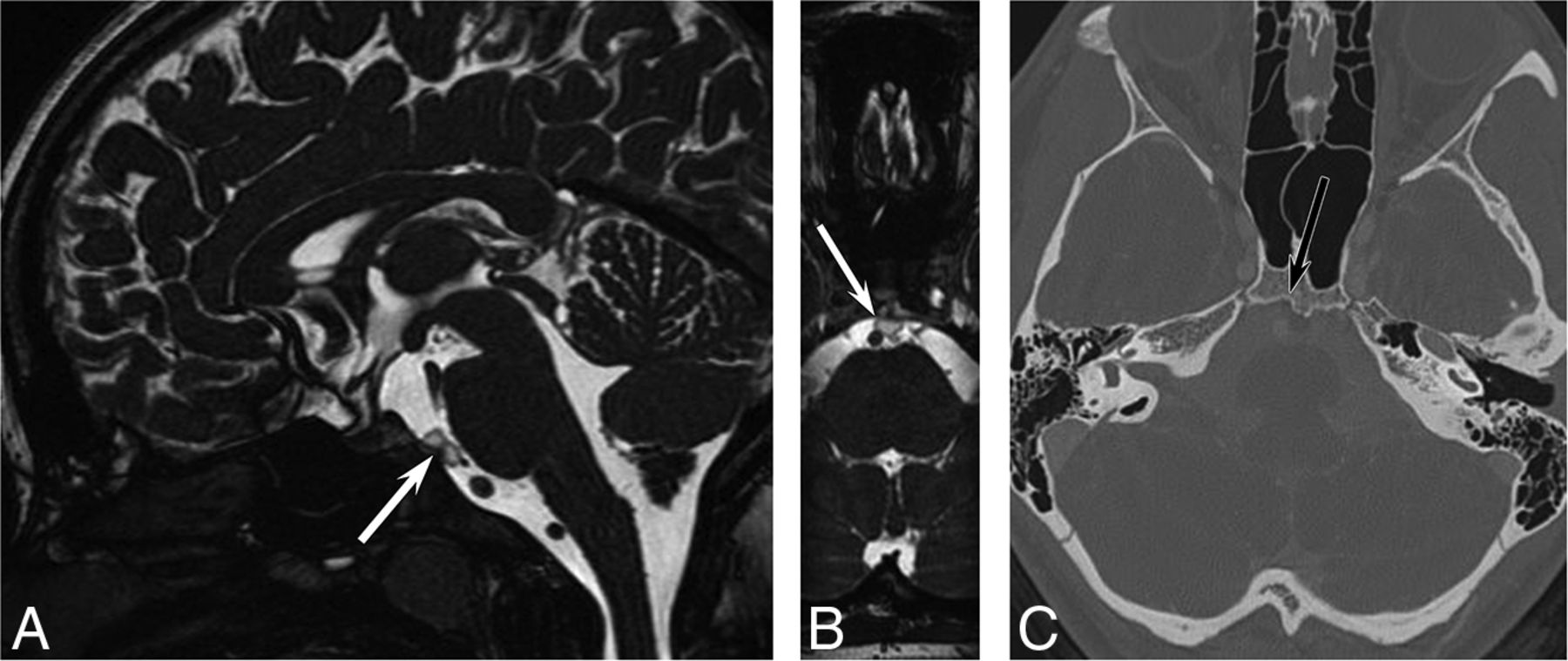
A previously healthy 16-year-old girl underwent MR imaging for headaches. The examination findings were normal except for a well-defined 5-mm T2-hyperintense prepontine mass with a dark rim and clival extension (Fig 1). A CT was performed for further characterization, showing a well-defined sclerotic margin to the clival component. There was no associated enhancement or mass effect. The interpreting radiologist stated that the lesion presumably represents ecchordosis physaliphora, and long-interval follow-up imaging was recommended.

Fig 1.
  • Download figure
  • Open in new tab
  • Download powerpoint
Fig 1.

A 16-year-old girl with headaches. A, Sagittal steady-state free precession MR imaging shows a lobular, well-defined T2-hyperintense lesion (arrow) in the prepontine cistern. Axial reformatted image (B) shows a benign-appearing abutment to the clivus. Axial CT (C) shows slight remodeling of the posterior clivus without erosion.

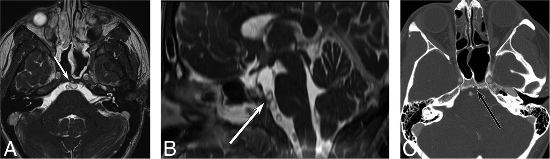
The patient returned 4 years later with recurrent headaches and again underwent MR imaging (Fig 2). The lesion had marginally enlarged but retained all of its benign features. Because of the change in size, the neurosurgery team advised resection, and the patient agreed. The resection was accomplished with an endoscopic endonasal transclival resection.4 Histopathologic analysis (Fig 3) revealed a neoplasm composed of cells with multivacuolated (physaliphorous)-to-eosinophilic cytoplasm embedded in a myxoid background, with a chronic lymphocytic inflammatory infiltrate seen in fibrous septa separating the lobules of the tumor. There was a variable degree of nuclear enlargement, hyperchromasia, and nuclear membrane irregularities. Mitotic activity was sparse, and the Ki-67 proliferation index was very low (<1%), indicating indolent or benign disease. However, no amplification of 1p36 or 9p21 was detected with fluorescence in site hybridization, so the tumor could not be classified as having minimal risk. Lobules of the tumor were identified intradurally as well as extending into the clivus, indicating some degree of aggressive features. Immunohistochemical staining for Brachyury showed strong, diffuse nuclear staining, confirming notochordal origin. When all of the histopathologic, genetic, and fluorescence stain data were taken into consideration, a diagnosis of low-grade chordoma was made, with a description of the relatively bland histologic features displayed by the tumor. Follow-up MR imaging 6 months later revealed no evidence of residual disease.

Fig 2.
  • Download figure
  • Open in new tab
  • Download powerpoint
Fig 2.

Follow-up imaging 4 years later. Axial steady-state free precession MR imaging (A) shows increased lesion size (arrow) with new involvement of the clivus. Sagittal reformat (B) shows increased superoinferior extent. The clival involvement is confirmed on axial contrast-enhanced CT (C).

Fig 3.
  • Download figure
  • Open in new tab
  • Download powerpoint
Fig 3.

H&E-stained sections demonstrate a lobulated neoplasm composed of predominantly physaliphorous cells in a myxoid extracellular matrix, with a chronic inflammatory infiltrate centered in the fibrous septa (A) and extending to involve clival bone (B). Higher-power view shows cells with nuclear enlargement and atypia (C). An immunohistochemical stain for Brachyury shows strong diffuse nuclear staining in tumor cells (D).

On a follow-up visit, the patient asked whether the lesion should have been resected when she first presented at age 16 and, in particular, whether the extent of surgery would have been less at that time.

Notochordal Embryology

Notogenesis is the formation of the embryologic notochord from precursor epiblasts during the third week of fetal development. The notochord initiates the process of neurulation, which results in the development of the CNS. On the formation of the brain and spine, the notochord regresses into the nucleus pulposus. Incomplete regression can result in persistence of isolated notochordal remnants, which are a potential source of neoplastic disease. Notochordal remnants usually remain near the midline, with a predilection for the sacrococcygeal region and the spheno-occipital synchondrosis.5⇓-7 Both benign entities (BNCT) and malignant entities (chordoma) can occur in these locations, presumably originating from these remnants.

EP/BNCT

BNCTs (a term that encompasses both extraosseous lesions [EP] as well as intraosseous notochordal remnants) are typically asymptomatic, so the lesions are often discovered incidentally when imaging for other reasons. A radiologic hallmark of BNCT that can be detected on CT or MR imaging is a small bony stalk or pedicle projecting from the basal portion of the clivus.8 Like other radiologic features, however, this parameter may only be used to suggest BNCT over other retroclival pathologies such as epidermoid or neurenteric cysts.9 Other nonspecific MR imaging findings include T1-hypointensity, T2-hyperintensity, lack of gadolinium enhancement, and lack of bony erosion.10 A diagnosis of BNCT is often made presumptively on imaging without histopathologic proof, because excisional biopsy is considered overly invasive for a benign disease.

Histologically, BNCTs display sheets of physaliphorous cells, which are characterized by mucin-containing intracytoplasmic vacuoles and lack lobular architecture, fibrous septa, necrosis, and nuclear atypia that characterize chordomas. The lesions are often hypocellular, have bland nuclear features, and lack mitoses. BNCTs also have a low Ki-67 proliferation index, indicating low growth potential.11,12 However, there can be substantial overlap in the histology and immunophenotype of BNCT and chordoma, and the architectural features that help distinguish these entities may be difficult to appreciate on surgical samples of small lesions. Similar challenges can arise in distinguishing a BNCT from a chordoma in the lumbosacral spine, and the term “atypical notochordal cell tumor” has been recently proposed for cases that defy definitive classification.13

Chordoma

Chordomas are typically locally invasive, aggressive tumors that are presumed to arise from persistent notochordal cells. They can arise anywhere along the craniospinal axis, with a predilection for the base of the skull and sacrococcygeal region. Histologically, conventional chordomas are composed of the same physaliphorous cells seen in benign notochordal cell tumors, along with eosinophilic cytoplasm and intracytoplasmic vacuoles.14 Interspersed within the physaliphorous cells are variable amounts of myxoid material, which results in variable imaging characteristics for this tumor.15 The tumors typically have a capsule and grow in lobules separated by fibrous septa, often accompanied by an inflammatory infiltrate. The degree of nuclear pleomorphism and mitotic activity is highly variable, with conventional chordomas ranging from histologically bland tumors with mild nuclear atypia, sparse mitotic activity, and low proliferative activity, up to more overtly atypical, highly proliferative lesions. Poorly-differentiated chordomas are defined by loss of nuclear INI-1 (SMARCB1) expression and often have increased cellularity and mitotic activity. A dedifferentiated chordoma consists of a frankly sarcomatous component juxtaposed with conventional chordomas. Poorly differentiated and dedifferentiated chordomas are quite rare, with conventional chordomas (including the chondroid subtype) accounting for ∼95% of all tumors. Within conventional chordomas, histologic grading criteria to stratify tumor behavior do not exist, though molecular panels to improve prognostication have been recently developed.16,17 By definition, chordomas are considered malignant and are thus capable of locally destructive behavior and metastasis.18

The radiologic appearance of conventional chordoma depends on location, aggressivity, and molecular makeup. On MR imaging, chordomas are typically lobular, septate, and heterogeneous, showing hypointensity on T1-weighted images (but frequently containing high-signal foci) and hyperintensity on T2-weighted images. Enhancement after gadolinium administration is also heterogeneous and often septal, with myxoid areas enhancing poorly.19

Due to the infiltrative nature of these tumors, they often present clinically with pain and site-related neurologic symptoms. Cranial and skull base chordomas may present with headache or cranial nerve dysfunction. The most frequently affected cranial nerve is the abducens nerve (CN VI) because it is exposed to clival tumors while traveling through the Dorello canal along the posterior clivus in the clival venous plexus.20 In rare cases, clival chordomas can present with rhinorrhea due to CSF leak.21 Craniocervical and cervical chordomas tend to present with nonspecific neck and upper-extremity pain. Dysphagia may be present due to mass effect. Thoracic, lumbar, and sacral chordomas tend to present with nonspecific dull pain.22

Although chordomas have a slow rate of progression, they are associated with substantial morbidity from mass effect.23 If a biopsy confirms the diagnosis of chordoma, en bloc resection with wide margins is performed.24 Chordomas have a high rate of reoccurrence, so adjuvant radiation and yearly MR imaging surveillance are often recommended.25

Recent data suggest that homozygous 9p21 deletions (p176/CDKN2A) and 1p36 deletions can further risk-stratify chordomas, even within the “conventional chordoma” category, and are predictive of progression-free survival.16,17 These markers were also found to be relevant to seeding.26 Depending on the prevalence of these deletions within the tumor cells, skull base chordomas can be further classified into an indolent group A, a highly aggressive group C, and an intermediate group B. According to this data set, radiation therapy did not confer any benefit after complete resections in groups A and B.17 Tumors in group A were more likely to be found in younger patients, were smaller, and were more likely to be asymptomatic on presentation. This group may encompass cases that would more accurately be described as EP or BNCT. Whether circulating tumor DNA would have a role in differentiating EP/BNCT from chordomas remains to be determined.27

Spectrum of Disease

Unfortunately, low-grade chordomas may have imaging features that overlap with the classic imaging appearance of BNCT. Imaging differentiation relies on sequential imaging, with BNCT remaining stable indefinitely and low-grade chordoma slowly enlarging. However, there is no established minimum rate of growth that indicates chordoma. Thus, it is difficult to recommend imaging intervals that would properly assess growth rates of a tumor with uncertain malignant potential. Furthermore, there are cases of presumed BNCTs that, on later excisional biopsy, are better classified as chordomas (as exemplified in the above case presentation). Several cases of BNCT have been published that displayed atypical characteristics, with resemblance to chordomas clinically or radiologically.28⇓-30 Similarly, there are documented cases of chordoma that display a low proliferative index, a diameter of <2 cm, and a lack of any clinical symptoms, with detection occurring only incidentally.3,31⇓-33

Given the overlap in clinical, radiologic, and histopathologic findings for EP/BNCT and low-grade chordoma, the distinction between these entities may be arbitrary. Until histopathologic analysis is completed, radiologically characteristic lesions in typical locations are best described as “benign-appearing notochordal lesions,” to avoid ascribing expected behavior before repeat imaging. Repeat imaging, first after 6 months, then with larger intervals (doubling each time) as stability is established, is a more clinically relevant method for evaluating the malignant potential of these lesions and will allow more nuanced prognostic discussions with patients. Although many of these tumors will never require surgical intervention, a patient-centered approach can be used that factors in patient age, the presence of attributable symptoms, rate of tumor growth, changes in imaging characteristics, and patient preferences (Table). The potential for malignant transformation of benign lesions should be considered, and continued imaging surveillance should be pursued even in cases that do not demonstrate appreciable growth or development of symptoms. More research is needed to establish evidence-based criteria for surgical intervention.

View this table:
  • View inline
  • View popup

Considerations for excisional biopsy of benign-appearing notochordal lesions

CONCLUSIONS

The classic dichotomy of a benign notochordal remnant (ecchordosis physaliphora) and a malignant notochordal tumor (chordoma) is outdated. Notochordal lesions fall on a spectrum of disease from completely benign to aggressively malignant. Although aggressive disease can be identified radiologically, low-grade malignancies cannot be reliably distinguished from benign disease, and even benign disease has the occasional potential for malignant transformation. Repeat imaging, beginning at a 6-month interval, is recommended even for lesions that appear completely benign on MR imaging. The term benign-appearing notochordal lesion is a more accurate descriptive term that does not make undue assumptions about the clinical behavior of incidentally discovered lesions in the central clivus and skull base.

Footnotes

  • Disclosure forms provided by the authors are available with the full text and PDF of this article at www.ajnr.org.

References

  1. 1.↵
    1. Park HH,
    2. Lee KS,
    3. Ahn SJ, et al
    . Ecchordosis physaliphora: typical and atypical radiologic features. Neurosurg Rev 2017;40:87–94 doi:10.1007/s10143-016-0753-4 pmid:27206421
    CrossRefPubMed
  2. 2.↵
    1. Yamaguchi T,
    2. Suzuki S,
    3. Ishiiwa H, et al
    . Benign notochordal cell tumors: a comparative histological study of benign notochordal cell tumors, classic chordomas, and notochordal vestiges of fetal intervertebral discs. Am J Surg Pathol 2004;28:756–61 doi:10.1097/01.pas.0000126058.18669.5d pmid:15166667
    CrossRefPubMed
  3. 3.↵
    1. Rodriguez L,
    2. Colina J,
    3. Lopez J, et al
    . Intradural prepontine growth: giant ecchordosis physaliphora or extraosseous chordoma? Neuropathology 1999;19:336–40 doi:10.1046/j.1440-1789.1999.00241.x
    CrossRef
  4. 4.↵
    1. Stippler M,
    2. Gardner PA,
    3. Snyderman CH, et al
    . Endoscopic endonasal approach for clival chordomas. Neurosurgery 2009;64:268–77; discussion 277–78 doi:10.1227/01.NEU.0000338071.01241.E2 pmid:19190456
    CrossRefPubMed
  5. 5.↵
    1. de Bree K,
    2. de Bakker BS,
    3. Oostra RJ
    . The development of the human notochord. PLoS One 2018;13:e0205752 doi:10.1371/journal.pone.0205752 pmid:30346967
    CrossRefPubMed
  6. 6.↵
    1. Ramesh T,
    2. Nagula SV,
    3. Tardieu GG, et al
    . Update on the notochord including its embryology, molecular development, and pathology: a primer for the clinician. Cureus 2017;9:e1137 doi:10.7759/cureus.1137 pmid:28480155
    CrossRefPubMed
  7. 7.↵
    1. Lawson L,
    2. Harfe BD
    . Notochord to nucleus pulposus transition. Curr Osteoporos Rep 2015;13:336–41 doi:10.1007/s11914-015-0284-x pmid:26231139
    CrossRefPubMed
  8. 8.↵
    1. Dias LA,
    2. Nakanishi M,
    3. Mangussi-Gomes J, et al
    . Successful endoscopic endonasal management of a transclival cerebrospinal fluid fistula secondary to ecchordosis physaliphora: an ectopic remnant of primitive notochord tissue in the clivus. Clin Neurol Neurosurg 2014;117:116–19 doi:10.1016/j.clineuro.2013.11.026 pmid:24438817
    CrossRefPubMed
  9. 9.↵
    1. Baek WK,
    2. Lachkar S,
    3. Iwanaga J, et al
    . Comprehensive review of spinal neurenteric cysts with a focus on histopathological findings. Cureus 2018;10:e3379 doi:10.7759/cureus.3379 pmid:30519518
    CrossRefPubMed
  10. 10.↵
    1. Mehnert F,
    2. Beschorner R,
    3. Küker W, et al
    . Retroclival ecchordosis physaliphora: MR imaging and review of the literature. AJNR Am J Neuroradiol 2004;25:1851–55 pmid:15569763
    PubMed
  11. 11.↵
    1. Choudhri O,
    2. Feroze A,
    3. Hwang P, et al
    . Endoscopic resection of a giant intradural retroclival ecchordosis physaliphora: surgical technique and literature review. World Neurosurg 2014;82:912.e21–26 doi:10.1016/j.wneu.2014.06.019 pmid:24937599
    CrossRefPubMed
  12. 12.↵
    1. Kandukuri SR,
    2. Lin F,
    3. Gui L, et al
    . Application of immunohistochemistry in undifferentiated neoplasms: a practical approach. Arch Pathol Lab Med 2017;141:1014–32 doi:10.5858/arpa.2016-0518-RA pmid:28745568
    CrossRefPubMed
  13. 13.↵
    1. Carter JM,
    2. Wenger DE,
    3. Rose PS, et al
    . Atypical notochordal cell tumors: a series of notochordal-derived tumors that defy current classification schemes. Am J Surg Pathol 2017;41:39–48 doi:10.1097/PAS.0000000000000766 pmid:27819871
    CrossRefPubMed
  14. 14.↵
    1. Wasserman JK,
    2. Gravel D,
    3. Purgina B
    . Chordoma of the head and neck: a review. Head Neck Pathol 2018;12:261–68 doi:10.1007/s12105-017-0860-8 pmid:28980142
    CrossRefPubMed
  15. 15.↵
    1. Egerbacher M,
    2. Böck P
    . Myxoid tissue: its morphology, histochemistry, and relationship with other supporting tissues. Arch Histol Cytol 1997;60:121–31 doi:10.1679/aohc.60.121 pmid:9232176
    CrossRefPubMed
  16. 16.↵
    1. Zenonos GA,
    2. Fernandez-Miranda JC,
    3. Mukherjee D, et al
    . Prospective validation of a molecular prognostication panel for clival chordoma. J Neurosurg 2019;130:1528–37 doi:10.3171/2018.3.JNS172321 pmid:29905508
    CrossRefPubMed
  17. 17.↵
    1. Abdallah HM,
    2. Gersey ZC,
    3. Muthiah N, et al
    . An integrated management paradigm for skull base chordoma based on clinical and molecular characteristics. J Neurol Surg B Skull Base 2021;82:601–07 doi:10.1055/s-0041-1730958 pmid:34745826
    CrossRefPubMed
  18. 18.↵
    1. Fernandez-Miranda JC,
    2. Gardner PA,
    3. Snyderman CH, et al
    . Clival chordomas: a pathological, surgical and radiotherapeutic review. Head Neck 2014;36:892–906 doi:10.1002/hed.23415 pmid:23804541
    CrossRefPubMed
  19. 19.↵
    1. Sung MS,
    2. Lee GK,
    3. Kang HS, et al
    . Sacrococcygeal chordoma: MR imaging in 30 patients. Skeletal Radiol 2005;34:87–94 doi:10.1007/s00256-004-0840-4 pmid:15480648
    CrossRefPubMed
  20. 20.↵
    1. Martin JJ,
    2. Niranjan A,
    3. Kondziolka D, et al
    . Radiosurgery for chordomas and chondrosarcomas of the skull base. J Neurosurg 2007;107:758–64 doi:10.3171/JNS-07/10/0758 pmid:17937220
    CrossRefPubMed
  21. 21.↵
    1. Alshammari J,
    2. Monnier P,
    3. Daniel RT, et al
    . Clival chordoma with an atypical presentation: a case report. J Med Case Rep 2012;6:410 doi:10.1186/1752-1947-6-410 pmid:23194159
    CrossRefPubMed
  22. 22.↵
    1. Young VA,
    2. Curtis KM,
    3. Temple HT, et al
    . Characteristics and patterns of metastatic disease from chordoma. Sarcoma 2015;2015:517657 doi:10.1155/2015/517657 pmid:26843835
    CrossRefPubMed
  23. 23.↵
    1. Smoll NR,
    2. Gautschi OP,
    3. Radovanovic I, et al
    . Incidence and relative survival of chordomas: the standardized mortality ratio and the impact of chordomas on a population. Cancer 2013;119:2029–37 doi:10.1002/cncr.28032 pmid:23504991
    CrossRefPubMed
  24. 24.↵
    1. Walcott BP,
    2. Nahed BV,
    3. Mohyeldin A, et al
    . Chordoma: current concepts, management, and future directions. Lancet Oncol 2012;13:e69–76 doi:10.1016/S1470-2045(11)70337-0 pmid:22300861
    CrossRefPubMed
  25. 25.↵
    1. Rohatgi S,
    2. Ramaiya NH,
    3. Jagannathan JP, et al
    . Metastatic chordoma: report of the two cases and review of the literature. Eurasian J Med 2015;47:151–54 doi:10.5152/eurasianjmed.2015.52 pmid:26180502
    CrossRefPubMed
  26. 26.↵
    1. Fernandes Cabral DT,
    2. Zenonos GA,
    3. Fernandez-Miranda JC, et al
    . Iatrogenic seeding of skull base chordoma following endoscopic endonasal surgery. J Neurosurg 2018;129:947–53 doi:10.3171/2017.6.JNS17111 pmid:29271711
    CrossRefPubMed
  27. 27.↵
    1. Frederico SC,
    2. Darling C,
    3. Zhang X, et al
    . Circulating tumor DNA: A potential aid in the management of chordomas. Front Oncol 2022;12:1016385 doi:10.3389/fonc.2022.1016385 pmid:36338734
    CrossRefPubMed
  28. 28.↵
    1. Ling SS,
    2. Sader C,
    3. Robbins P, et al
    . A case of giant ecchordosis physaliphora: a case report and literature review. Otol Neurotol 2007;28:931–33 doi:10.1097/MAO.0b013e318068b2c8 pmid:17592399
    CrossRefPubMed
  29. 29.↵
    1. Sun R,
    2. Ajam Y,
    3. Campbell G, et al
    . A rare case of ecchordosis physaliphora presenting with headache, abducens nerve palsy, and intracranial hypertension. Cureus 2020;12:e8843 doi:10.7759/cureus.8843 pmid:32754387
    CrossRefPubMed
  30. 30.↵
    1. Golden LD,
    2. Small JE
    . Benign notochordal lesions of the posterior clivus: retrospective review of prevalence and imaging characteristics. J Neuroimaging 2014;24:245–49 doi:10.1111/jon.12013 pmid:23464492
    CrossRefPubMed
  31. 31.↵
    1. Gonzalez-Martinez JA,
    2. Guthikonda M,
    3. Vellutini E, et al
    . Intradural invasion of chordoma: two case reports. Skull Base 2002;12:155–61 doi:10.1055/s-2002-33462 pmid:17167671
    CrossRefPubMed
  32. 32.↵
    1. Ciarpaglini R,
    2. Pasquini E,
    3. Mazzatenta D, et al
    . Intradural clival chordoma and ecchordosis physaliphora: a challenging differential diagnosis: case report. Neurosurgery 2009;64:E387–88; discussion E388 doi:10.1227/01.NEU.0000337064.57270.F0 pmid:19190444
    CrossRefPubMed
  33. 33.↵
    1. Yuh SJ,
    2. Woulfe J,
    3. Corsten MJ, et al
    . Diagnostic imaging dilemma of a clival lesion and its clinical management implications. J Neurol Surg B Skull Base 2014;75:177–82 doi:10.1055/s-0033-1363171 pmid:24967152
    CrossRefPubMed
  • Received January 9, 2023.
  • Accepted after revision June 8, 2023.
  • © 2023 by American Journal of Neuroradiology
PreviousNext
Back to top

In this issue

American Journal of Neuroradiology: 44 (8)
American Journal of Neuroradiology
Vol. 44, Issue 8
1 Aug 2023
  • Table of Contents
  • Index by author
  • Complete Issue (PDF)
Advertisement
Print
Download PDF
Email Article

Thank you for your interest in spreading the word on American Journal of Neuroradiology.

NOTE: We only request your email address so that the person you are recommending the page to knows that you wanted them to see it, and that it is not junk mail. We do not capture any email address.

Enter multiple addresses on separate lines or separate them with commas.
Ecchordosis Physaliphora: Does It Even Exist?
(Your Name) has sent you a message from American Journal of Neuroradiology
(Your Name) thought you would like to see the American Journal of Neuroradiology web site.
CAPTCHA
This question is for testing whether or not you are a human visitor and to prevent automated spam submissions.
Cite this article
A.R. Stevens, B.F. Branstetter, P. Gardner, T.M. Pearce, G.A. Zenonos, K. Arani
Ecchordosis Physaliphora: Does It Even Exist?
American Journal of Neuroradiology Aug 2023, 44 (8) 889-893; DOI: 10.3174/ajnr.A7932

Citation Manager Formats

  • BibTeX
  • Bookends
  • EasyBib
  • EndNote (tagged)
  • EndNote 8 (xml)
  • Medlars
  • Mendeley
  • Papers
  • RefWorks Tagged
  • Ref Manager
  • RIS
  • Zotero
0 Responses
Respond to this article
Share
Bookmark this article
Ecchordosis Physaliphora: Fact or Fiction?
A.R. Stevens, B.F. Branstetter, P. Gardner, T.M. Pearce, G.A. Zenonos, K. Arani
American Journal of Neuroradiology Aug 2023, 44 (8) 889-893; DOI: 10.3174/ajnr.A7932
del.icio.us logo Twitter logo Facebook logo Mendeley logo
  • Tweet Widget
  • Facebook Like
  • Google Plus One
Purchase

Jump to section

  • Article
    • Abstract
    • ABBREVIATIONS:
    • CONCLUSIONS
    • Footnotes
    • References
  • Figures & Data
  • Info & Metrics
  • Responses
  • References
  • PDF

Related Articles

  • No related articles found.
  • PubMed
  • Google Scholar

Cited By...

  • Benign Notochordal Cell Tumor: An Entity Distinct from Ecchordosis Physaliphora and Chordoma
  • Crossref (6)
  • Google Scholar

This article has been cited by the following articles in journals that are participating in Crossref Cited-by Linking.

  • Skull Base Chordoma and Chondrosarcoma: Neuroradiologist’s Guide to Diagnosis, Surgical Management, and Proton Beam Therapy
    Gillian M. Potter, Rekha Siripurapu, Amit Herwadkar, Sarah Abdulla, Oluwaseun Ikotun, Philip Broadhurst, Mark Woodward, Rajiv K. Bhalla, Laurence J. Glancz, Charlotte L. Hammerbeck-Ward, Scott A. Rutherford, Omar N. Pathmanaban, Federico Roncaroli, Rovel J. Colaco, Shermaine Pan, Gillian A. Whitfield
    RadioGraphics 2024 44 10
  • Delayed FLAIR-enhancement of benign notochordal remnant (ecchordosis physaliphora)
    Jeroen Peels, Christof Vulsteke, Michael Boedts, Eline Marin, Laurens J L De Cocker
    The Neuroradiology Journal 2025 38 2
  • Skull Base Tumors: Therapeutic Challenges and Multi-Disciplinary Care
    Garret Choby
    Cancers 2024 16 3
  • Benign Notochordal Cell Tumor: An Entity Distinct from Ecchordosis Physaliphora and Chordoma
    Carrie M. Carr, Jamie Van Gompel, Aditya Raghunathan
    American Journal of Neuroradiology 2024 45 8
  • Current understanding of brachyury in chordoma
    Tianshun Ma, Jiwei Bai, Yazhuo Zhang
    Biochimica et Biophysica Acta (BBA) - Reviews on Cancer 2023 1878 6
  • Ecchordosis Physaliphora and Posttraumatic Headache: A Diagnostic Challenge in Neurotrauma
    Carlos Enrique Hernández Borroto, Orquidia Reyes de Hernández, Yuri Medrano Plana
    Indian Journal of Neurotrauma 2025

More in this TOC Section

  • Chondrosarcoma vs Synovial Chondromatosis: Imaging
  • NI-RADS for HEAD&NECK Cancer Recurrence
  • WHO Classification Update: Nasal&Skull Base Tumors
Show more HEAD & NECK

Similar Articles

Advertisement

Indexed Content

  • Current Issue
  • Accepted Manuscripts
  • Article Preview
  • Past Issues
  • Editorials
  • Editors Choice
  • Fellow Journal Club
  • Letters to the Editor

Cases

  • Case Collection
  • Archive - Case of the Week
  • Archive - Case of the Month
  • Archive - Classic Case

Special Collections

  • Special Collections

Resources

  • News and Updates
  • Turn around Times
  • Submit a Manuscript
  • Author Policies
  • Manuscript Submission Guidelines
  • Evidence-Based Medicine Level Guide
  • Publishing Checklists
  • Graphical Abstract Preparation
  • Imaging Protocol Submission
  • Submit a Case
  • Become a Reviewer/Academy of Reviewers
  • Get Peer Review Credit from Publons

Multimedia

  • AJNR Podcast
  • AJNR SCANtastic
  • Video Articles

About Us

  • About AJNR
  • Editorial Board
  • Not an AJNR Subscriber? Join Now
  • Alerts
  • Feedback
  • Advertise with us
  • Librarian Resources
  • Permissions
  • Terms and Conditions

American Society of Neuroradiology

  • Not an ASNR Member? Join Now

© 2025 by the American Society of Neuroradiology All rights, including for text and data mining, AI training, and similar technologies, are reserved.
Print ISSN: 0195-6108 Online ISSN: 1936-959X

Powered by HighWire