
Article Figures & Data
Figures
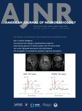
- FIG 1.
A 53-year-old patient with a history of bilateral sinonasal inverted papillomas and left nasopharyngeal papillary squamous cell carcinoma. Axial T2-weighted MR image (A) shows material of intermediate-to-hyperintense signal in the left posterior Eustachian tube (short arrow), middle ear (long arrow), and external auditory canal (arrowhead). Consecutive axial postcontrast T1-weighted MR images from caudal to cranial (B, C, D) show corresponding enhancement of this abnormal soft tissue in the posterior Eustachian tube (short arrow in B), middle ear (long arrow in B, C, and D), and external auditory canal (arrowhead in B), representing tumor. There is an extension to the mastoid antrum (arrowhead in D). Ear biopsy showed squamous cell carcinoma possibly arising from an inverted papilloma.
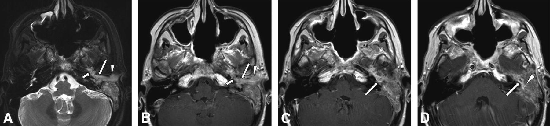
- FIG 2.
A 65-year-old patient with a history of recurrent, right sinonasal, inverted papilloma and carcinoma. Axial T2-weighted (A) and postcontrast T1-weighted (B) MR images show a large, bulky, lobulated septate enhancing mass in the right sinonasal region. Note the absence of abnormal soft tissue at the right Eustachian tube opening (short arrow), middle ear (long arrow), and external auditory canal (arrowhead). The patient underwent partial resection, chemoradiation, and immunotherapy. Two years later, he presented with otologic symptoms, and MR imaging was performed. Axial precontrast (C) and postcontrast (D) T1-weighted MR images show bulky enhancing soft tissue at the right Eustachian tube opening (short arrow), middle ear (long arrow), and external auditory canal (small arrowheads), with invasion into the mastoid and petrous portions of the temporal bone (large arrowheads). Also note tumor involvement in the skull base foramina such as the right pterygopalatine fossa (short broad arrows).
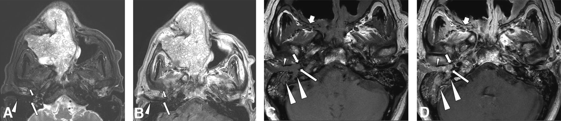
- FIG 3.
A 60-year-old patient with a right nasal cavity inverted papilloma. Axial precontrast (A) and postcontrast (B) T1-weighted images show the initial right sinonasal papilloma (arrow) 2 years prior. On presentation to otology, CT in soft-tissue (C) and bone (D) windows shows an expansile bulky mass in the right middle ear (long arrow in C), external auditory canal (arrowhead in C), and mastoid (large arrowhead in C) with bone destruction (arrows in D). PET (E) shows avid FDG uptake by the mass (arrow). Axial precontrast (F) and postcontrast (G) T1-weighted images show enhancing tumor along the right Eustachian tube (small arrow), in the middle ear (long arrow), and in the mastoid (large arrowhead). Pathology of the ear mass proved to be inverted papilloma.
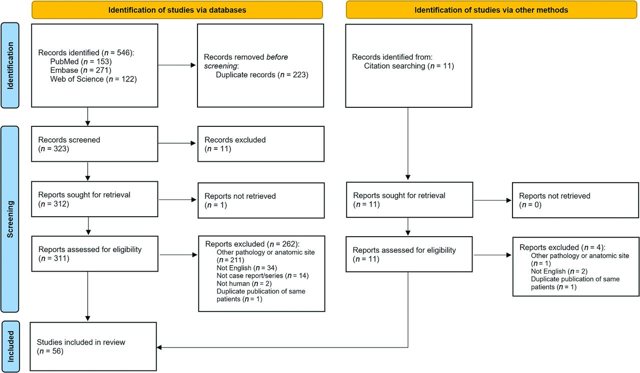
- FIG 4.
PRISMA flow diagram showing search algorithm used for systematic review.


Tables
- Table 1:
Summary of patient characteristics in systematic review and case series of temporal bone Schneiderian neoplasmsa
Characteristic Demographics (n = 76) Age at presentation (median) (IQR) 53 (44–63) Sex ratio (male/female) 1.5 (45:31) Main presenting otologic signs/symptoms (n = 76)b Hearing loss 51 (67%) Otorrhea or otorrhagia 36 (47%) Aural fullness 14 (18%) Otalgia 13 (17%) Tinnitus or pulsatile tinnitus 12 (16%) Acute or chronic otitis media and/or mastoiditis 9 (12%) Visible ear mass 9 (12%) Facial paralysis 7 (9%) Dizziness or vertigo 2 (3%) Association of temporal bone tumor with sinonasal tumor (n = 76) Primary (isolated) 37 (49%) Secondary 39 (51%) Temporal association of secondary tumor diagnosis with sinonasal tumor (n = 39) Synchronous 9 (23%) Metachronous 30 (77%) Temporal bone tumor pathology (n = 74) Benign 48 (65%) Malignant 26 (35%) Sinonasal tumor pathology (n = 39) Benign 24 (62%) Malignant/high-risk 15 (38%) - Table 2:
Contingency table on the association of temporal bone Schneiderian tumors that are primary (isolated) or secondary (synchronous or metachronous to a similar sinonasal tumor) and pathology that is malignant/high-risk (carcinoma, carcinoma in situ, or papilloma with severe/high-grade dysplasia) or benign (papilloma without severe/high-grade dysplasia)
Primary Secondary Malignant 6 22 Benign 31 17


Jump to section
Related Articles
Cited By...
- No citing articles found.